Review
. 2025 Jul 14;15(30):24624-24638.
doi: 10.1039/d5ra02939b. eCollection 2025 Jul 10.

Challenges and opportunities in the application of carbon nanotubes as membrane channels to improve mass transfer to cells

Affiliations
Review

Challenges and opportunities in the application of carbon nanotubes as membrane channels to improve mass transfer to cells

Sara Yazdani et al. RSC Adv. .

Abstract

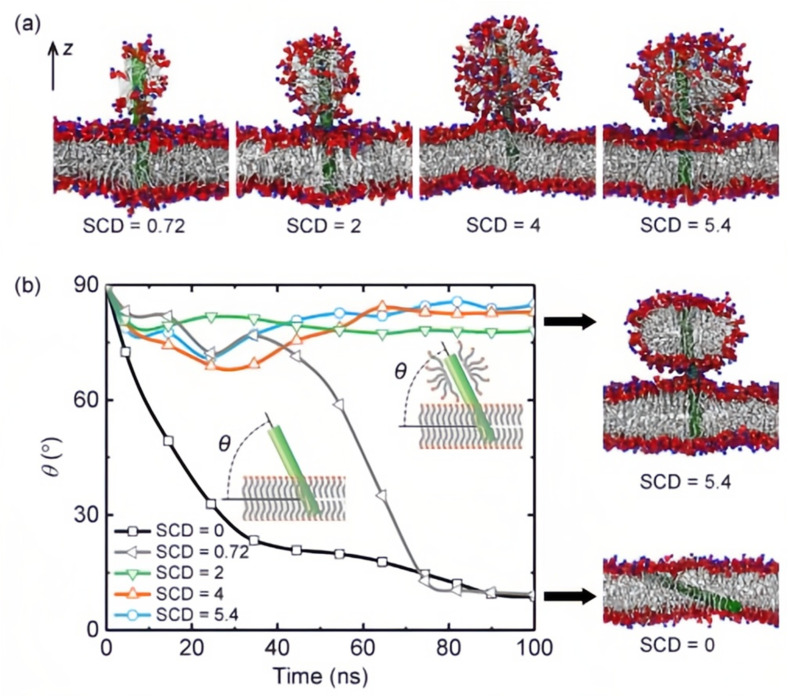
The regulation and improvement of mass transfer through the living cell's membrane is of great importance in various industrial, environmental and medical applications. Designing membrane channels based on carbon nanotubes (CNTs) has been considered as a promising approach to this end because of the geometry of CNTs, their physical properties, high chemical stability, and excellent transport features. Despite their advantages, CNTs have a few problems such as their toxicity to living cells, low bioavailability in an aqueous medium and difficulties with managing their orientation within the cell membrane which should be addressed in the first place. Here, we tried to review recent studies on overcoming these challenges and critically evaluate their advances and suggestions for future research. Functionalization of CNTs with biocompatible materials has been recommended as the main solution which decreases the inherent cytotoxicity of the pristine CNTs, enhances their solubility and dispersibility in aqueous solution, and affects their orientation in the cell membrane. Molecular dynamics simulation results for the interactions of the functionalized CNTs and the cell membrane have been reviewed as well to demonstrate the effectiveness of functionalizing CNTs for membrane channel applications. Finally, we highlighted that modified CNTs with appropriate functional groups and favorable physical and geometrical conditions can be considered as an effective tool to make artificial channels in the cell membrane.


Conflict of interest statement

There are no conflicts to declare.

Figures

Fig. 1
Fig. 1. Natural size rules and gatekeepers within a mammalian cell. The thickness of membrane bilayer is typically between 4 to 10 nm. The diameter of nuclear pore complex is approximately 80–120 nm. The sizes of endocytic vesicles in both phagocytosis and pinocytosis pathways for nanoparticle internalization were also introduced. Phagocytes could take up large particles (or nanoparticle aggragates), opsonized nanoparticles, or nanoparticles with certain liagnd modification via phagocytosis. Nanoparticle internalization in a nonphagocytic mammalian cell is mainly through pinocytosis or direct penetration. With various surface modifications, nanoparticles may be taken up via specific or nonspecific endocytosis. MR, mannose receptor; PRRs, pattern-recognition receptors; FcγR, immunoglobulin Fcγ receptor; CR, complement receptor; CPPs, cell-penetrating peptides; IgG, immunoglobulin G; ER, endoplasmic reticulum; Golgi, Golgi apparatus. Figure reprinted with permission from ref. . Copyright 2013, American Chemical Society.
Fig. 2
Fig. 2. Different cellular uptake mechanisms for CNTs (reproduced from ref. 40).
Fig. 3
Fig. 3. Incorporation of CNTs coated with lipid into a POPC bilayer membrane. (a) Snapshots of the configurations at starting time with different SCD. (b) Time evolution of the insertion angle θ of CNTs at five SCD values (schematically shown in the insets). Right panel indicates snapshots of balance configurations for two SCD after 100 ns simulations. Figure reproduced with permission from ref. . Copyright 2020, Elsevier.
Fig. 4
Fig. 4. (a) Diameter histogram of p-MWCNTs@GUVs at a 2 μg mL−1 concentration of CNTs, (b) confocal images of p-MWCNTs@GUVs at a 2 μg mL−1 concentration, (c) cartoon of the interaction in the p-MWCNTs@GUVs systems, (d) diameter histogram of oxMWCNTs@GUVs at the concentration of 2 μg mL−1 of CNTs after one hour of incubation, (e) confocal images of ox-MWCNTs@GUVs at 2 μg mL−1, (f) cartoon of the interaction of the system ox-MWCNTs@GUVs, (g) diameter histogram of alk-MWCNTs@GUVs at a 2 μg mL−1 concentration of alk-MWCNTs, (h) confocal images of alk-MWCNTs@GUVs at a 2 μg mL−1 CNTs concentration, colocalization of alk-MWCNTs GUVs on “ghost-like” structures, and (i) confocal images of alk-MWCNTs@GUVs at a 2 μg mL−1 CNTs concentration, colocalization of alk-MWCNTs GUVs on “ghost-like” structures. Figure reprinted with permission from ref. . Copyright 2018, Springer Nature.
Fig. 5
Fig. 5. An entering route of pure CNT and functionalized CNT situated in the membrane vertically and horizontally. (a) Snapshots at critical times for pure CNT. (b and c) Time evolvement of the center of mass (COM) distance in the z-direction, the tilt angle, and the interaction energy between pure CNT and membrane. (d and e) Snapshots at t = 0 ns and 100 ns for functionalized CNT, (f) the tilt angle, and (g) the hydrogen bonds between functionalized CNT and lipid bilayer membrane. Figure reproduce with permission from ref. . Copyright 2019, Multidisciplinary Digital Publishing Institute (MDPI).

References

    1. Korn E. D. Annu. Rev. Biochem. 1969;38:263–288. - PubMed
    1. Gatenby R. A. Int. J. Mol. Sci. 2019;20:3609. - PMC - PubMed
    1. Zargar S. M. Hafshejani D. K. Eskandarinia A. Rafienia M. Kharazi A. Z. J. Med. Signals Sens. 2019;9:181–189. - PMC - PubMed
    1. Tan S. Wu T. Zhang D. Zhang Z. Theranostics. 2015;5:863. - PMC - PubMed
    1. Dai C. Xu Q. Li L. Liu Y. Qu S. ACS Biomater. Sci. Eng. 2024;10:1988–2000. - PubMed

LinkOut - more resources