[Preprint]. 2025 Jun 15:2025.06.13.659541.
doi: 10.1101/2025.06.13.659541.

Hibernation improves neural performance during energy stress in regions across the central nervous system in the American bullfrog

Affiliations

Hibernation improves neural performance during energy stress in regions across the central nervous system in the American bullfrog

Nikolaus Bueschke et al. bioRxiv. .

Abstract

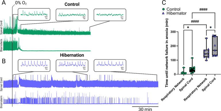
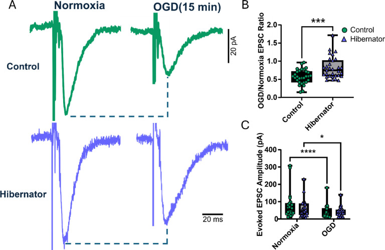
Neuronal signaling requires high rates of ATP production via the oxidative metabolism of glucose. The American bullfrog is intriguing, as this species has typical brain energy requirements for an average vertebrate but modifies synaptic physiology and metabolism after hibernation to maintain function during hypoxia and ischemia. Given the importance of the respiratory system in restoring metabolic homeostasis during emergence from underwater hibernation, work to date has addressed this response in the brainstem respiratory network. Thus, metabolic plasticity has been interpreted as an adaptation used to restart respiratory motor behavior under hypoxic conditions during the transition from skin breathing to air breathing. It remains unclear whether these improvements are specific to the brainstem regions critical for breathing versus a global response within the central nervous system (CNS). To address this question, we recorded neural activity from the spinal cord, forebrain, and brainstem respiratory network in vitro. As expected, hypoxia disrupted the function of each network in control animals. After hibernation, each network improved its activity in hypoxia compared to controls. These results suggest that plasticity that improves neural function during energy stress following hibernation reflects a global response that may impact many behaviors controlled by the CNS and is not limited to regions involved in metabolic homeostasis.


Conflict of interest statement

Conflict of Interests: The authors declare no conflicts of interest.

Figures

Figure 1.
Figure 1.. Experimental Approach.
(A) Image of an isolated brainstem and spinal cord, ventral side up with nerve rootlets for recordings labeled where respiratory-associated rootlets in the brainstem are shown on the left, and rootlets that output spontaneous spinal cord activity are shown on the right. (B) Representative trace of a simultaneous recording of both spinal cord (top) and brainstem (bottom). Gray lines indicate brainstem bursts to demonstrate that network activity is occurring asynchronously to the spinal cord activity. (C) Image (10X magnification) of a transverse forebrain slice with a stimulator placed over thalamic projections that innervate dorsal pallium neurons, recorded from patch clamp electrodes. (C1) Example trace of a stimulator-evoked EPSC in a recorded dorsal pallium neuron. (C2) Example trace of an mEPSC recording in the dorsal pallium neuron.
Figure 2.
Figure 2.. Metabolic plasticity phenotype in the brainstem is present in the spinal cord.
(A-B) Representative traces of spontaneous rhythmic output from the brainstem and spinal cord rootlets in the presence of anoxia (0% O2) with timing on the same scale between groups. (A) Anoxic response in control conditions shows a rapid cessation of activity in the brainstem. Failure of spinal cord activity was slightly delayed, and was shown as a steady decrease in rhythmic burst amplitude as shown in inlets. (B) Anoxic response post-hibernation shows prolonged brainstem function and spinal cord function that persists for the entire anoxic duration. (C) Time until final duration in anoxia between brainstem and spinal cord activity before and after hibernation. After hibernation, both brainstem and spinal cords persisted longer in anoxia (n = 9) compared to pre-hibernated controls (n = 7), as shown from results following a Holm-Sidak’s multiple comparisons test. Additionally, spinal cord activity persisted longer in anoxia than brainstems before and after hibernation.
Figure 3.
Figure 3.. Hibernation decreases sensitivity to oxygen and glucose deprivation in dorsal pallium synapses.
OGD onset results in attenuated evoked EPSC amplitudes that are less severe after hibernation. (A) Example traces of individual evoked currents comparing amplitudes before and after 15 minutes of OGD exposure in control (top) and after hibernation (bottom). (B) Evoked EPSC amplitude in OGD relative to baseline in controls (n = 30 neurons, N = 12 animals) and hibernators (n = 31 neurons, N = 15 animals). Significance in amplitude changes was shown with a Mann-Whitney U test. (C) Recorded evoked EPSC amplitudes to compare differences in absolute amplitudes between groups. Holm-Sidak’s multiple comparisons show significance in both controls and hibernators.
Figure 4.
Figure 4.. Hibernation does not alter mEPSC amplitude or frequency.
(A) Change in mEPSC amplitude in 15 minutes of OGD relative to normoxic conditions. Mann-Whitney U results showing no difference in mEPSC amplitude suggest that OGD has no influence on changes in postsynaptic response to excitatory quanta. (B) Difference in mEPSC frequency in OGD relative to normoxic conditions. Mann-Whitney U test results showing no changes in altered frequency after hibernation suggest no change in presynaptic release sites.

Similar articles

  • Short-Term Memory Impairment.
    Cascella M, Al Khalili Y. Cascella M, et al. 2024 Jun 8. In: StatPearls [Internet]. Treasure Island (FL): StatPearls Publishing; 2025 Jan–. 2024 Jun 8. In: StatPearls [Internet]. Treasure Island (FL): StatPearls Publishing; 2025 Jan–. PMID: 31424720 Free Books & Documents.
  • The Black Book of Psychotropic Dosing and Monitoring.
    DeBattista C, Schatzberg AF. DeBattista C, et al. Psychopharmacol Bull. 2024 Jul 8;54(3):8-59. Psychopharmacol Bull. 2024. PMID: 38993656 Free PMC article. Review.
  • Ventilator Management(Archived).
    Mora Carpio AL, Mora JI. Mora Carpio AL, et al. 2023 Mar 27. In: StatPearls [Internet]. Treasure Island (FL): StatPearls Publishing; 2025 Jan–. 2023 Mar 27. In: StatPearls [Internet]. Treasure Island (FL): StatPearls Publishing; 2025 Jan–. PMID: 28846232 Free Books & Documents.
  • Systemic Inflammatory Response Syndrome.
    Baddam S, Burns B. Baddam S, et al. 2025 Jun 20. In: StatPearls [Internet]. Treasure Island (FL): StatPearls Publishing; 2025 Jan–. 2025 Jun 20. In: StatPearls [Internet]. Treasure Island (FL): StatPearls Publishing; 2025 Jan–. PMID: 31613449 Free Books & Documents.
  • Physical interventions to interrupt or reduce the spread of respiratory viruses.
    Jefferson T, Dooley L, Ferroni E, Al-Ansary LA, van Driel ML, Bawazeer GA, Jones MA, Hoffmann TC, Clark J, Beller EM, Glasziou PP, Conly JM. Jefferson T, et al. Cochrane Database Syst Rev. 2023 Jan 30;1(1):CD006207. doi: 10.1002/14651858.CD006207.pub6. Cochrane Database Syst Rev. 2023. PMID: 36715243 Free PMC article.

References

    1. Adams S., Zubov T., Bueschke N., & Santin J. M. (2021). Neuromodulation or energy failure? Metabolic limitations silence network output in the hypoxic amphibian brainstem. American Journal of Physiology-Regulatory, Integrative and Comparative Physiology, 320(2), R105–R116. 10.1152/ajpregu.00209.2020 - DOI - PMC - PubMed
    1. Amaral-Silva L., & Santin J. (2024). Neural Processing without O 2 and Glucose Delivery: Lessons from the Pond to the Clinic. Physiology, 39(6), 000–000. 10.1152/physiol.00030.2023 - DOI - PMC - PubMed
    1. Amaral-Silva L., & Santin J. M. (2023). Synaptic modifications transform neural networks to function without oxygen. BMC Biology, 21(1), 54. 10.1186/s12915-023-01518-0 - DOI - PMC - PubMed
    1. Ashton D., Van Reempts J., Haseldonckx M., & Willems R. (1989). Dorsal-ventral gradient in vulnerability of CA1 hippocampus to ischemia: A combined histological and electrophysiological study. Brain Research, 487(2), 368–372. 10.1016/0006-8993(89)90842-1 - DOI - PubMed
    1. Baghdadwala M. I. (2016). Diving into the mammalian swamp of respiratory rhythm generation with the bullfrog. Respiratory Physiology. - PubMed

Publication types