[Preprint]. 2025 Jun 11:2025.06.10.658880.
doi: 10.1101/2025.06.10.658880.

Structural basis of RNA-guided transcription by a dCas12f-σE-RNAP complex

Affiliations

Structural basis of RNA-guided transcription by a dCas12f-σE-RNAP complex

Renjian Xiao et al. bioRxiv. .

Abstract

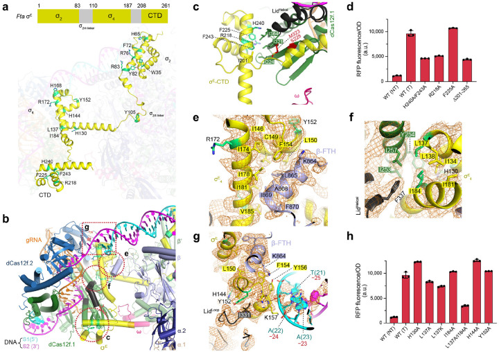
RNA-guided proteins have emerged as critical transcriptional regulators in both natural and engineered biological systems by modulating RNA polymerase (RNAP) and its associated factors1-5. In bacteria, diverse clades of repurposed TnpB and CRISPR-associated proteins repress gene expression by blocking transcription initiation or elongation, enabling non-canonical modes of regulatory control and adaptive immunity1,6,7. Intriguingly, a distinct class of nuclease-dead Cas12f homologs (dCas12f) instead activates gene expression through its association with unique extracytoplasmic function sigma factors (σE)8, though the molecular basis has remained elusive. Here we reveal a novel mode of RNA-guided transcription initiation by determining cryo-electron microscopy structures of the dCas12f-σE system from Flagellimonas taeanensis. We captured multiple conformational and compositional states, including the DNA-bound dCas12f-σE-RNAP holoenzyme complex, revealing how RNA-guided DNA binding leads to σE-RNAP recruitment and nascent mRNA synthesis at a precisely defined distance downstream of the R-loop. Rather than following the classical paradigm of σE-dependent promoter recognition, these studies show that recognition of the -35 element is largely supplanted by CRISPR-Cas targeting, while the melted -10 element is stabilized through unusual stacking interactions rather than insertion into the typical recognition pocket. Collectively, this work provides high-resolution insights into an unexpected mechanism of RNA-guided transcription, expanding our understanding of bacterial gene regulation and opening new avenues for programmable transcriptional control.


Conflict of interest statement

COMPETING INTERESTS S.H.S. is a co-founder and scientific advisor to Dahlia Biosciences, a scientific advisor to CrisprBits and Prime Medicine, and an equity holder in Dahlia Biosciences and CrisprBits. S.H.S., F.T.H., and T.W. are inventors on patents related to CRISPR-Cas-like systems and uses thereof. The other authors declare no competing interests.

Figures

Figure 1 |
Figure 1 |. Biochemical characterization of the dCas12f-σE system and structure of dCas12f-gRNA.
a, Genomic organization of the dCas12f-σE operon in F. taeanensis (left), and the gRNA-matching DNA target site upstream of susC/susD membrane transport genes (right). b, Schematic showing base-pairing between the gRNA and target DNA, which includes native mismatches at positions 11–12. The downstream transcription start site (TSS) is indicated. c, OD-normalized RFP fluorescence from cellular transcriptional reporter assays using the indicated Fta σE homologs. dCas12f-gRNA complexes were presented in all conditions, and contained a targeting (T) or non-targeting (NT) guide. Data are shown as mean ± s.d. for n = 3 biologically independent samples. d, SDS-PAGE and urea-PAGE analysis of purified dCas12f-gRNA, RNAP, and σE-RNAP complexes. e, Cryo-EM density map of the dCas12f-gRNA complex at 3.28 Å. f, Domain organization of dCas12f.1 and dCas12f.2, showing the REC, WED, RuvC, and Lid motif domains. g, Structures of the Lid motif in ‘Loop’ (left) and ‘Helical’ (right) conformations. h, Cartoon representation of the experimental gRNA structure. i, Secondary structure diagram of the gRNA, highlighting key structural elements. Stem-loops are labeled P1–P5.
Figure 2 |
Figure 2 |. Structure of the dCas12f-gRNA-target DNA complex.
a, Cryo-EM density map of dCas12f-gRNA-DNA in a partial R-loop state. b, Cryo-EM density map of dCas12f-gRNA-DNA in a full R-loop state. c, Interaction between the TAM duplex and dCas12f.1. d, Schematic of interactions between the TAM duplex and dCas12f.1. e, OD-normalized RFP fluorescence from cellular transcriptional reporter assays using the indicated dCas12f mutants that perturb TAM duplex recognition. Data are shown as mean ± s.d. for n = 3 biologically independent samples. f, Recognition of the 5’-G TAM, highlighting hydrogen bonding between the base of C(−1) and the backbone carbonyl oxygen of S106. g, Lid motif conformation in the partial R-loop state. h, Lid motif conformation in the full R-loop state. Insets in g and h show focused views of key residues in the Lid motif undergoing conformational rearrangement, along with the corresponding cryo-EM density. i, Structural superimposition of RNA-DNA duplex in partial and full R-loop states. j, Lid motif conformation in UnCas12f-gRNA-DNA complex for comparison. k, Lid motif conformation in the dCas12f-gRNA-DNA-σE-RNAP complex.
Figure 3 |
Figure 3 |. Structures of the DNA-bound dCas12f-σE-RNAP complex and apo RNAP.
a, Schematic showing the target DNA and gRNA in the DNA-bound dCas12f-σE-RNAP structure. The target DNA features two DNA bubbles, one mediated by the dCas12f-gRNA R-loop and the other mediated by the RNAP transcription bubble; the TSS is located at position 46 of the template strand. b, Two representative 2D class averages of cryo-EM images of the DNA-bound dCas12f-σE-RNAP complex. c, Cryo-EM density map of the DNA-bound dCas12f-σE-RNAP complex. d, Cartoon representation of the atomic model. e, Schematic of the DNA-bound dCas12f-σE-RNAP structure. f, Contact between dCas12f.1 and the RNAP ω subunit, showing the cryo-EM map displayed as a surface representation (top) and the surface potential of dCas12f.1 and the ω subunit (bottom). g, Cryo-EM map of apo RNAP. h, Atomic model of apo RNAP.
Figure 4 |
Figure 4 |. Structure of Fta RNAP core subunits.
a, Domain organization of the RNAP core subunits. b, Structure of the β’ subunit with each domain color-coded. c, Structure of β’ in the context of σE and DNA. d, Structure of the β subunits with each domain color-coded. e, Structure of β in the context of σE and DNA. f, Structure of the Clamp domain alone (left) and in the context of σE and DNA (right); the conformational change between both states is indicated. g, Structure of the ‘Cleft’ formed by the β-cleft, β’-cleft, and β-protrusion. h, View of the ‘Funnel’ structure formed by the β’-pore, β’-funnel, and β’-cleft domains.
Figure 5 |
Figure 5 |. Interactions between dCas12f and σE.
a, Structure of σE in cartoon representation, with the remainder of the DNA-bound dCas12f-RNAP complex shown in transparency, for clarity. The σE domain organization is shown at the top. b, Overall view showing the interactions between σE and other components, with magnified inset regions indicated with dotted lines. c, Interactions between the σE CTD and dCas12f.1. d, OD-normalized RFP fluorescence from cellular transcriptional reporter assays using the indicated σE CTD mutants. e, Interactions between the σE4 domain and β-flip-tip-helix (β-FTH). f, Interactions between the σE4 domain and dCas12f.1. g, The σE4 domain is encircled by β-FTH, the lid motif of dCas12f.2, and DNA. h, OD-normalized RFP fluorescence from cellular transcriptional reporter assays using the indicated σE4 mutants. Data in d and h are shown as mean ± s.d. for n = 3 biologically independent samples.
Figure 6 |
Figure 6 |. Target DNA interactions and comparison with canonical σE factors.
a, Interactions between Fta σE and the −10 region of DNA; the cryo-EM map is shown in mesh. b, Interactions between Fta σE and the −10 region of DNA, with the σE surface displayed. c, Interactions between the Eco σE and the −10 region of DNA, shown as in b, highlighting the pocket formed by the specificity loop. d, Interactions between Fta σE and the target DNA. e, Interactions between Eco σE and the target DNA. f, Superimposed structure comparing dCas12f-associated Fta σE (yellow) and non-dCas12f-associated Eco σE (brown).

Similar articles

References

    1. Wiegand T., Hoffmann F. T., Walker M. W. G., Tang S., Richard E., Le H. C., Meers C. & Sternberg S. H. TnpB homologues exapted from transposons are RNA-guided transcription factors. Nature 631, 439–448, doi: 10.1038/s41586-024-07598-4 (2024). - DOI - PMC - PubMed
    1. Qi L. S., Larson M. H., Gilbert L. A., Doudna J. A., Weissman J. S., Arkin A. P. & Lim W. A. Repurposing CRISPR as an RNA-guided platform for sequence-specific control of gene expression. Cell 152, 1173–1183, doi: 10.1016/j.cell.2013.02.022 (2013). - DOI - PMC - PubMed
    1. Gilbert L. A., Horlbeck M. A., Adamson B., Villalta J. E., Chen Y., Whitehead E. H., Guimaraes C., Panning B., Ploegh H. L., Bassik M. C., Qi L. S., Kampmann M. & Weissman J. S. Genome-Scale CRISPR-Mediated Control of Gene Repression and Activation. Cell 159, 647661, doi: 10.1016/j.cell.2014.09.029 (2014). - DOI - PMC - PubMed
    1. Bikard D., Jiang W., Samai P., Hochschild A., Zhang F. & Marraffini L. A. Programmable repression and activation of bacterial gene expression using an engineered CRISPR-Cas system. Nucleic Acids Res 41, 7429–7437, doi: 10.1093/nar/gkt520 (2013). - DOI - PMC - PubMed
    1. Zalatan J. G., Lee M. E., Almeida R., Gilbert L. A., Whitehead E. H., La Russa M., Tsai J. C., Weissman J. S., Dueber J. E., Qi L. S. & Lim W. A. Engineering complex synthetic transcriptional programs with CRISPR RNA scaffolds. Cell 160, 339–350, doi: 10.1016/j.cell.2014.11.052 (2015). - DOI - PMC - PubMed

Publication types

LinkOut - more resources