[Preprint]. 2025 May 29:2025.05.28.656686.
doi: 10.1101/2025.05.28.656686.

Filopodia numbers impact chemotactic migration speed

Affiliations

Filopodia numbers impact chemotactic migration speed

Annika L Schroder et al. bioRxiv. .

Abstract

Migrating cells sense and respond to external chemical and physical cues, enabling them to efficiently reach their destinations. Filopodia are slender actin-filled membrane protrusions implicated in interacting with the extracellular environment in many contexts, such as neuronal growth cone guidance and the capture of prey by immune cells and unicellular organisms. The role of filopodia in chemotactic guidance in fast-moving amoeboid cells has not been well-studied. The social amoeba Dictyostelium relies on chemotaxis for development and finding food, making it an excellent system for investigating the role of filopodia in amoeboid chemotaxis. Stimulation of amoebae with the chemoattractant cAMP activates a transient increase of filopodia formation by recruiting the filopodial myosin DdMyo7 to the cell cortex. Filopodia formation is biased towards the source of chemoattractant, yet myo7 null cells that lack filopodia exhibit normal directional migration. However, cells either lacking filopodia or having increased numbers of filopodia move more slowly than those with wildtype numbers of filopodia. Thus, while filopodia are dispensable for detection of chemical gradients by amoeboid cells, changes in filopodia number can impact their migration speed possibly due to altering cell-substrate adhesion.

Keywords: actin; amoeboid migration; chemotaxis; directed migration; filopodia; myosin.


Figures
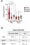
Figure 1.
Figure 1.. Filopodia number impact the speed of randomly moving cells.
A. Average speed of vegetative wild type (Ax3), myo7 null or myo7 nulls expressing the KKAA autoinhibition mutant when assayed under agarose. Shown is the speed of cells when placed in phosphate starvation buffer (PB) or in nutrient media (HL5). N= 3, n = 16 (minimum) for each assay. Kruskal-Wallis ANOVA and multiple comparisons tests (Ax3: ****p<0.0001, n.s.= not significant, p=0.067; Ax2, n.s., p=0.14) B. Summary of means values and significance measured for each line ± SEM.)
Figure 2.
Figure 2.. Global stimulation with chemoattractant cAMP results in cortical recruitment of DdMyo7 and stimulates filopodia formation.
A. Representative images of wild type D. discoideumcells overexpressing actin marker RFP-LifeAct, GFP-DdMyo7, or GFP-CRAC immediately prior to (0s), and 4 seconds after global cAMP stimulation. Yellow arrows indicate regions of cortical enrichment and white arrows indicate nascent filopodia tipped with GFP-DdMyo7. Max cortex/cytoplasm intensities and peak times ± SEM are shown to the right. Scale bar = 10 μm. B. RFP-LifeAct (n = 38 cells, 3 experiments), GFP-DdMyo7 (n = 55 cells, 2 experiments), and GFP-CRAC (n = 32 cells, 6 experiments) are rapidly recruited to the cortex following global cAMP stimulation. The cortical intensity It= (cortex - background) / (cytoplasm - background). Itwas normalized to time 0s (I0), immediately before cAMP stimulation. Error bars = SEM for all cells. C. The number of GFP-DdMyo7 tipped filopodia per cell over time after global stimulation with cAMP. Vertical lines indicate stimulation time and peak time for GFP-DdMyo7 cortical recruitment from panels A and B. Error bars = SEM. D. Model of DdMyo7 and VASP recruitment following cAMP stimulation and consequent extension of DdMyo7 tipped filopodia (light green = pre-existing filopodia and cortical actin; dark green = stimulation-induced filopodia and increased cortical actin).
Figure 3.
Figure 3.. Filopodia formation is biased towards a chemotactic source.
A. Representative micrographs of myo7 null cells expressing GFP-DdMyo7 chemotaxing towards 1μM cAMP under agarose. Scale bar = 10 μm. B. Example of the arc length analysis. The closest pixel of the cell mask (ii, blue) to the tip mask center (ii,yellow) is assigned as the filopodia shaft. Arc length is measured from the right most pixel (ii, white arrow for tip 1). Arc lengths are then visualized as circumference fraction (iii). Scale bar = 10 μm. C and D. Arc length rose plot of all filopodia events where degrees are normalized arc lengths. C. Rose plot of all filopodia events detected during chemotaxis of myo7 null cells expressing GFP-DdMyo7 (n = 74 cells, 6 experiments). The 0° position points towards higher cAMP concentrations, and the radial axis is the number of filopodia events in each bin (9.7° per bin). Dotted lines indicate the quadrant relative to the gradient. The relation between quadrant and total filopodia was significant, χ2 (DF = 3, N = 2364 filopodia) = 366.5, p< 0.0001. D. Subset of panel C, plotting only filopodia initiation arc lengths, where initiation events are defined as the first time point a filopodium appears. The relation between quadrant and initiation events was significant, χ2 (DF = 3, N = 458 initiation events) = 81.83, **** p< 0.0001.
Figure 4.
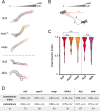
Figure 4.. Filopodia are dispensable for directional and persistent chemotaxis.
A. Representative perimeter plots for different genotypes migrating under agarose in a 1μM cAMP gradient for 5 minutes. B. Depiction of chemotaxis parameter calculations. Total distance traveled = line a. Displacement = line b. Chemotactic gradient vector = line c. Chemotactic index = cosine of the angle between b and c. Persistence = b / a. C. Average chemotactic index for each mutant. Dashed line separates genetic backgrounds Ax3 and Ax2 for the different mutant lines. Violin plots display kernel density distributions of individual cells; dashed line = median; dotted lines = quartiles. Black circles represent means of individual experiments. n.s. = no statistical significance (Ax3: Kruskal-Wallis ANOVA and multiple comparisons test, p > 0.99; Ax2: Mann-Whitney U test, p = 0.32). D. Summary of chemotaxis parameters. n = number of cells per genotype, combined from multiple individual experiments. One way ANOVA and multiple comparisons test (Ax3: * p < 0.05; **** p < 0.0001), and unpaired t test (Ax2; * p < 0.05).
Figure 5.
Figure 5.. Filopodia number tunes the speed of chemotactic migration.
A. Perimeter plots of Ax3 and DdMyo7 mutant KKAA migrating under agarose in a 1μM cAMP gradient over 5 minutes (n=30, 3 experiments). B. Average filopodia made per chemotaxing cell. C. Speed = total distance traveled (see Fig 3B, line a) / total time. Kruskal-Wallis ANOVA and multiple comparisons test (Ax3: *** p = 0.0001; **** p < 0.0001), and unpaired t test (Ax2; n.s. p = 0.76). Dashed line separates genetic backgrounds Ax3 and Ax2 for the different mutant lines. D. Average speed of all cells of a given genotype: myo7-, Ax3, KKAA plotted as a function of filopodia number in filopodia-positive cell subset. The line drawn on the plot is to illustrate the trend of the data.

Similar articles

  • The Black Book of Psychotropic Dosing and Monitoring.
    DeBattista C, Schatzberg AF. DeBattista C, et al. Psychopharmacol Bull. 2024 Jul 8;54(3):8-59. Psychopharmacol Bull. 2024. PMID: 38993656 Free PMC article. Review.
  • Sexual Harassment and Prevention Training.
    Cedeno R, Bohlen J. Cedeno R, et al. 2024 Mar 29. In: StatPearls [Internet]. Treasure Island (FL): StatPearls Publishing; 2025 Jan–. 2024 Mar 29. In: StatPearls [Internet]. Treasure Island (FL): StatPearls Publishing; 2025 Jan–. PMID: 36508513 Free Books & Documents.
  • Modulation of Filopodial Myosin Function.
    Crawford A, Schwartz JK, Hall E, Houdusse A, Titus MA. Crawford A, et al. bioRxiv [Preprint]. 2025 May 29:2025.05.29.656896. doi: 10.1101/2025.05.29.656896. bioRxiv. 2025. PMID: 40501999 Free PMC article. Preprint.
  • Management of urinary stones by experts in stone disease (ESD 2025).
    Papatsoris A, Geavlete B, Radavoi GD, Alameedee M, Almusafer M, Ather MH, Budia A, Cumpanas AA, Kiremi MC, Dellis A, Elhowairis M, Galán-Llopis JA, Geavlete P, Guimerà Garcia J, Isern B, Jinga V, Lopez JM, Mainez JA, Mitsogiannis I, Mora Christian J, Moussa M, Multescu R, Oguz Acar Y, Petkova K, Piñero A, Popov E, Ramos Cebrian M, Rascu S, Siener R, Sountoulides P, Stamatelou K, Syed J, Trinchieri A. Papatsoris A, et al. Arch Ital Urol Androl. 2025 Jun 30;97(2):14085. doi: 10.4081/aiua.2025.14085. Epub 2025 Jun 30. Arch Ital Urol Androl. 2025. PMID: 40583613 Review.
  • Short-Term Memory Impairment.
    Cascella M, Al Khalili Y. Cascella M, et al. 2024 Jun 8. In: StatPearls [Internet]. Treasure Island (FL): StatPearls Publishing; 2025 Jan–. 2024 Jun 8. In: StatPearls [Internet]. Treasure Island (FL): StatPearls Publishing; 2025 Jan–. PMID: 31424720 Free Books & Documents.

References

    1. Artemenko Y, Lampert TJ, Devreotes PN (2014). Moving towards a paradigm: common mechanisms of chemotactic signaling in Dictyostelium and mammalian leukocytes. Cell Mol. Life Sci. 71, 3711–3747. - PMC - PubMed
    1. Arthur AL, Songster LD, Sirkia H, Bhattacharya A, Kikuti C, Borrega FP, Houdusse A, Titus MA (2019). Optimized filopodia formation requires myosin tail domain cooperation. PNAS 116, 22196–22204. - PMC - PubMed
    1. Bentley D, Toroian-Raymond A (1986). Disoriented pathfinding by pioneer neurone growth cones deprived of filopodia by cytochalasin treatment. Nature 323, 712–715. - PubMed
    1. Berg S, Kutra D, Kroeger T, Straehle CN, Kausler BX, Haubold C, Schiegg M, Ales J, Beier T, Rudy M et al. (2019). ilastik: interactive machine learning for (bio)image analysis. Nat Meth 16, 1226–1232. - PubMed
    1. Brazill D (2016). Chemotaxis: Under Agarose Assay. In Cytoskeleton: Methods and Protocols, Gavin RH, ed. (New York: Springer Science + Business Media; ), pp. 339–346.

Publication types

LinkOut - more resources