[Preprint]. 2025 Jun 12:2025.06.10.658684.
doi: 10.1101/2025.06.10.658684.

An ultrasensitive and modular platform to detect Siglec ligands and control immune cell function

Affiliations

An ultrasensitive and modular platform to detect Siglec ligands and control immune cell function

Zeinab Jame-Chenarboo et al. bioRxiv. .

Abstract

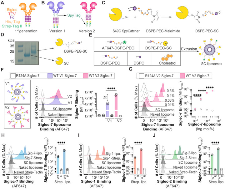
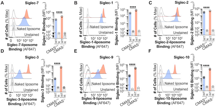
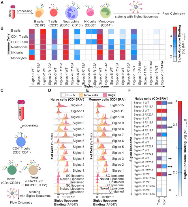
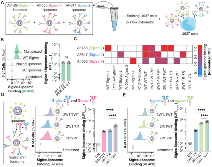
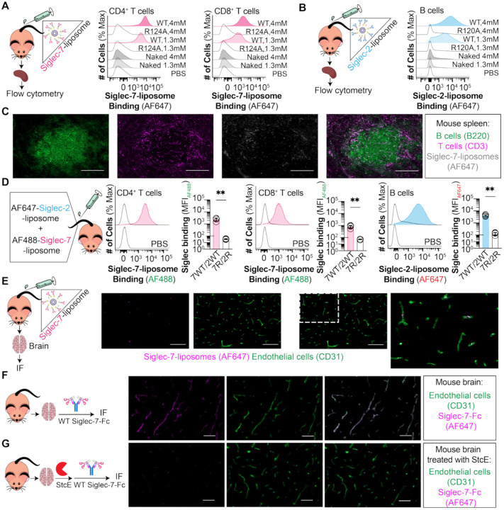
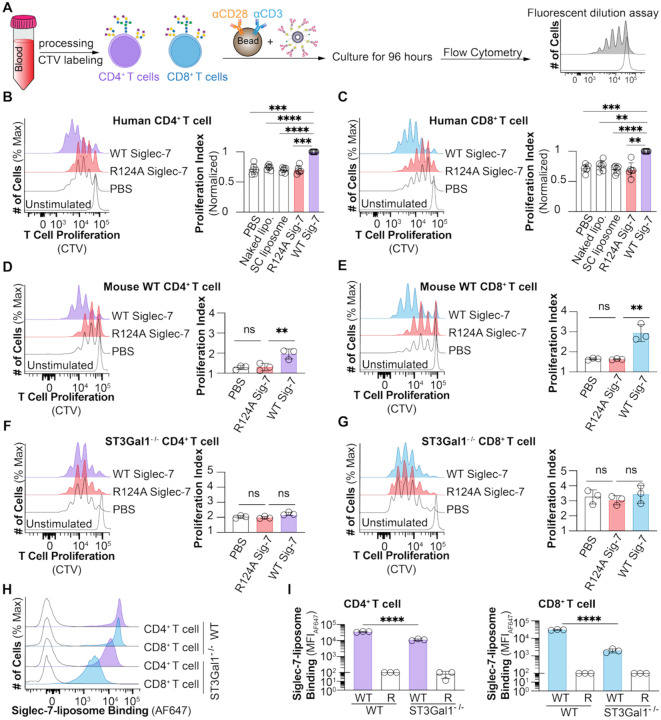
Siglecs are immunomodulatory receptors that regulate immune cell function. A fundamental challenge in studying Siglec-ligand interactions is the low affinity of Siglecs for their ligands. Inspired by how nature uses multivalency, we developed Siglec-liposomes as a highly multivalent and versatile platform for detecting Siglec glycan ligands in which recombinant Siglecs were conjugated to liposomes using the SpyCatcher-SpyTag system. Siglec-liposomes offer tunable multivalency and a modular assembly, enabling presentation of different Siglecs on the same liposome. Using Siglec-liposomes, we profiled Siglec ligands on human leukocytes, revealing new insights into Siglec ligands. Moreover, Siglec-liposomes are in vivo compatible, where we demonstrated that Siglec-7-liposomes bind to the brain vasculature in a mucin-dependent manner. Given the abundance of Siglec ligands on T cells, we investigated whether Siglec-liposomes modulate T cell function and find that Siglec-7-liposomes increase T cell proliferation in a ST3Gal1-dependent and CD43-independent manner. Taken together, Siglec-liposomes are a versatile and sensitive tool for detecting Siglec ligands and immunomodulation.


Conflict of interest statement

Competing interests: The authors declare that they have no conflicts of interest with the contents of this article.

Figures

Fig. 1.
Fig. 1.. Development and validation of Siglec-liposomes.
(A) First generation Siglec-Fc. (B) V1 and V2 constructs of Siglec-Fc with a genetically-encoded SpyTag. (C) Schematic of site-specific conjugation of SC to DSPE-PEG-Maleimide. (D) SDS-PAGE of SC and DSPE-PEG-SC. (E) Schematic of making SC-liposomes. (F) Binding of V1 and V2 WT Siglec-7- and R124A Siglec-7-liposomes to U937 cells. Data is represented as flow cytometry histograms and median fluorescent intensity (MFI) of Siglec-7 binding. (G) Binding of V2 of WT Siglec-7 and R124A Siglec-7 in different densities on liposomes to U937 cells. Data is represented as flow cytometry histograms and MFI of Siglec-7 binding. Binding of (H) WT and R124A V2 Siglec-7, (I) WT and R116A V2 Siglec-1, and (J) WT and R120A V2 Siglec-2 pre-complexed with Strep-Tactin (Strep.) and conjugated to liposomes (lipo.) to U937 cells. Data is presented as flow cytometry histograms and MFI of Siglec binding. The p value for three technical replicates was calculated using an unpaired one-way ANOVA. ****p<0.0001.
Fig. 2.
Fig. 2.. Siglecs expressed in sialic acid deficient CHO cells show improved binding toward their ligands.
Binding of WT and arginine mutated of (A) Siglec-7-, (B) Siglec-1-, (C) Siglec-2-, (D) Siglec-3-, (E) Siglec-9-, and (F) Siglec-10-liposomes expressed in CMAS+/+ and CMAS−/− CHO cells to U937 cells. Data is presented as flow cytometry histograms and MFI of Siglec binding. The p value for three technical replicates was calculated using an un-paired one-way ANOVA. ****p<0.0001.
Fig. 3.
Fig. 3.. Profiling Siglec ligands on primary cells.
(A) Schematic of staining immune cells isolated from human peripheral blood with Siglec-liposomes. (B) Siglec-liposomes binding to B cells, T cells, mature NK cells, monocytes, and neutrophils isolated from PBMCs. (C) Schematic of staining Treg and Tconv cells isolated from human peripheral blood with Siglec-liposomes. Binding of Siglec-liposomes to (D) naïve and (E) memory Treg and Tconv cells isolated from human peripheral blood. Data is presented as flow cytometry histograms. (F) Binding of Siglec-liposomes to naïve Treg and Tconv cells isolated from human peripheral blood. Data is presented as a heat map. The p value for four (panel B) and five (panel F) biological replicates was calculated using a paired one-way ANOVA. *0.05>p 0.01, **0.01>p0.001, ***0.001>p0.0001.
Fig. 4.
Fig. 4.. Siglec-liposomes as a multiplexing platform.
(A) Schematic for multiplexing liposomes with different fluorophores. (B) Binding of single color and multiplexed of AF488-Siglec-1-liposome to U937 cells. Data is presented as flow cytometry histograms and MFI of Siglec binding. (C) Binding of multiplexed and single color of AF488-Siglec-1-, AF555-Siglec-7-, and AF647-Siglec-2-liposomes in different combinations to U937 cells. Data is presented as a heat map. Binding of (D) Siglec-2/Siglec-7, and (E) Siglec-1/Siglec-2 on the same liposomes to U937 cells. Data is presented as flow cytometry histograms and MFI of Siglec binding. The p value for three technique replicates was calculated using an unpaired one-way ANOVA. Not significant (ns) p>0.05, ****p<0.0001.
Fig. 5.
Fig. 5.. In vivo and ex vivo staining of cells and tissues using Siglec-liposomes.
In vivo binding of WT and arginine mutated of (A) Siglec-7-liposome to CD4+ and CD8+ T cells and (B) Siglec-2-liposome to B cells isolated from mouse splenocytes. Data is presented as flow cytometry histograms. (C) IF microscopy images from mouse spleen representing in vivo binding of WT Siglec-7-liposomes to T cells. scale bar: 100 μm. (D) In vivo binding of multiplexed AF488-Siglec-7-liposomes to CD4+ and CD8+ T cells and AF647-Siglec-2-liposomes to B cells. Data is presented as flow cytometry histograms and MFI of Siglec binding. (E) IF microscopy images of mouse brain tissues representing in vivo binding of Siglec-7-liposomes to blood vessels. Scale bar: 100 μm. IF microscopy images of ex vivo staining of mouse brain tissues with (F) WT Siglec-7-Fc and αCD31, and (G) StcE mucinase enzyme treatment followed by WT Siglec-7-Fc and αCD31. Scale bar: 50 μm. The p value for four biological replicates was calculated using a paired Student’s t test. **0.01>p0.001.
Fig. 6.
Fig. 6.. Modulation of T cell proliferation by Siglec-7-liposomes.
(A) Schematic of T cell isolation from human blood and co-culturing with αCD3/28 beads and Siglec-liposomes. T cell proliferation of human (B) CD4+ and (C) CD8+ T cells co-cultured with WT and R124A Siglec-7-liposomes. Data is presented as flow cytometry histograms and proliferation index. T cell proliferation of (D) WT CD4+, (E) WT CD8+, (F) ST3Gal1−/− CD4+, and (G) ST3Gal1−/− CD8+ T cells isolated from mouse solenocytes. Data is presented as flow cytometry histograms and proliferation index. Binding of WT and R124A Siglec-7-liposomes to WT and ST3Gal1−/− T cells. Data is presented as (H) flow cytometry histograms and (I) MFI of Siglec-7 binding. The p value for six (panel B,C) and three (panel D-G) biological replicates was calculated using a paired one-way ANOVA. The p value for three technical replicates was calculated using an unpaired ANOVA. Not significant (ns) p>0.05, **0.01>p0.001, ***0.001>p0.0001, ****p<0.0001.

Similar articles

References

    1. Lin S. Y., Schmidt E. N., Takahashi-Yamashiro K., Macauley M. S., Roles for Siglec-glycan interactions in regulating immune cells. Semin Immunol 77, 101925 (2024). - PubMed
    1. Stanczak M. A., Laubli H., Siglec receptors as new immune checkpoints in cancer. Mol Aspects Med 90, 101112 (2023). - PubMed
    1. Jame-Chenarboo Z., Gray T. E., Macauley M. S., Advances in understanding and exploiting Siglec-glycan interactions. Curr Opin Chem Biol 80, 102454 (2024). - PubMed
    1. Lin S. Y., Schmidt E. N., Takahashi-Yamashiro K., Macauley M. S., Roles for Siglec-glycan interactions in regulating immune cells. Semin Immunol 77, 101925 (2025). - PubMed
    1. Rodrigues E. et al. , A versatile soluble siglec scaffold for sensitive and quantitative detection of glycan ligands. Nat Commun 11, 5091 (2020). - PMC - PubMed

Publication types