[Preprint]. 2025 Jun 14:2025.06.10.658786.
doi: 10.1101/2025.06.10.658786.

Multi-selective RAS(ON) Inhibition Targets Oncogenic RAS Mutations and Overcomes RAS/MAPK-Mediated Resistance to FLT3 and BCL2 Inhibitors in Acute Myeloid Leukemia

Affiliations

Multi-selective RAS(ON) Inhibition Targets Oncogenic RAS Mutations and Overcomes RAS/MAPK-Mediated Resistance to FLT3 and BCL2 Inhibitors in Acute Myeloid Leukemia

Bogdan Popescu et al. bioRxiv. .

Abstract

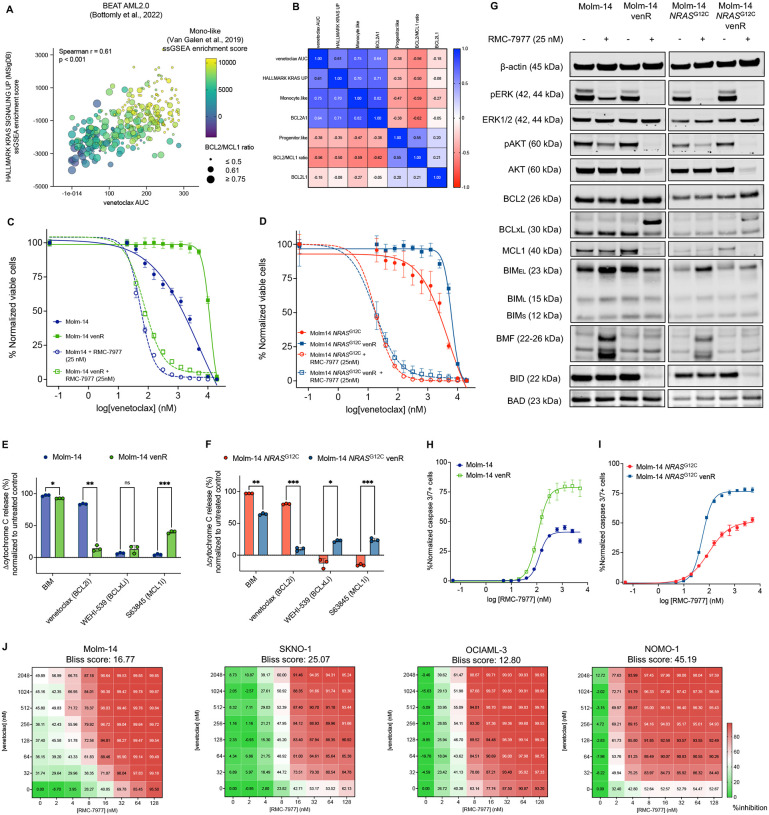
Aberrant activation of the RAS/MAPK signaling limits the clinical efficacy of several targeted therapies in acute myeloid leukemia (AML). In FLT3-mutant AML, the selection of clones harboring heterogeneous RAS mutations drives resistance to FLT3 inhibitors (FLT3i). RAS activation is also associated with resistance to other AML targeted therapies, including the BCL2 inhibitor venetoclax. Despite the critical need to inhibit RAS/MAPK signaling in AML, no targeted therapies have demonstrated clinical benefit in RAS-driven AML. To address this unmet need, we investigated the preclinical activity of RMC-7977, a multi-selective inhibitor of GTP-bound active [RAS(ON)] isoforms of mutant and wild-type RAS in AML models. RMC-7977 exhibited potent antiproliferative and pro-apoptotic activity across AML cell lines with MAPK-activating signaling mutations. In cell line models with acquired FLT3i resistance due to secondary RAS mutations, treatment with RMC-7977 restored sensitivity to FLT3i. Similarly, RMC-7977 effectively reversed resistance to venetoclax in RAS-addicted cell line models with both RAS wild-type and mutant genetic backgrounds. In murine patient-derived xenograft models of RAS-mutant AML, RMC-7977 was well tolerated and significantly suppressed leukemic burden in combination with gilteritinib or venetoclax. Our findings strongly support clinical investigation of broad-spectrum RAS(ON) inhibition in AML to treat and potentially prevent drug resistance due to activated RAS signaling.


Conflict of interest statement

C.C.S. reports research funding from Revolution Medicines, ERASCA, Abbvie; funding for clinical trials from Zentalis and Biomea; has served on advisory boards for Abbvie, Genentech, Servier and Biomea, and as a consultant for Astellas.

Figures

Figure 1 –
Figure 1 –. Activity of RAS(ON) multi-selective inhibitor RMC-7977 in AML cell lines.
A-C: Concentration-response curves representing relative proliferation of AML cell lines driven by FLT3 (A), KIT (B), NRAS or KRAS mutations (C) after 48 h of exposure to serial doses of RMC-7977. Data represent the mean ± SD of three replicates. D: Western blot analysis of seven AML cell lines exposed for 2 hours to indicated concentrations of RMC-7977; β-actin was used as loading control. E-F: Bubble plots showing enrichment of GSEA Hallmark gene sets expression in Molm-14 and OCIAML-3 cell lines after exposure for 24 hours to RMC-7977 (25 nM). The size of the bubbles represents statistical significance, and the colors indicate upregulated (red) or downregulated (blue) expression. G-H: PROGENy pathway activity scores in Molm-14 and OCIAML-3 cells after exposure for for 24 hours to RMC-7977 (25 nM).
Figure 2 –
Figure 2 –. RAS(ON) inhibition has a higher pro-apoptotic activity than downstream MAPK inhibition in AML cell lines.
A: Concentration-response curves showing activated caspase 3/7 expression in AML cell lines after 24 h of exposure to increasing doses of RMC-7977, normalized to untreated controls. Data represent the means ± SD of three replicates. B-C: Comparative caspase 3/7 activity in Molm-14 (B) and OCIAML-3 (C) cells exposed to RMC-7977 (RASi), naporafenib (RAFi), trametinib (MEKi) and ulixertinib (ERKi). D-E: Western blot analysis of Molm-14 (D) and OCIAML-3 (E) cells treated for 2 hours and 24 hours with the indicated inhibitors at concentrations approximately equivalent to IC50 for each compound; β-actin was used as loading control. F: Comparative caspase 3/7 activity in Molm-14 cells expressing either PIK3CAWT or PIK3CAE542K/PIK3CAH1047R after 24 hours of exposure to RMC-7977. Data represent the means ± SD of three replicates.
Figure 3 –
Figure 3 –. RMC-7977 re-sensitizes FLT3 and RAS co-mutant AML cells to FLT3 inhibitors.
A-B: Concentration-response curves representing relative proliferation of Molm-14 parental cells or Molm-14 expressing secondary NRASG12C or NRASQ61K mutations after 48 h of exposure to serial doses of gilteritinib relative to un-treated controls. In (B), the proliferation assay was conducted in the presence of RMC-7977 (25 nM). Data represent the mean ± SD of three replicates. C: Western blot analysis of parental or NRAS-mutant Molm-14 cells treated with either gilteritinib, RMC-7977 or both inhibitors; β-actin was used as loading control. D: Fluorescently-tagged Molm-14 (blue) or Molm-14 NRASQ61K (green) cells were mixed at a 1:1 ratio, then treated with gilteritinib, RMC-7977 or their combination. Scatter plots showing percentages of each cell population after 96 hours of indicated treatments and a staining with a live/dead dye (Sytox AADvanced). Plots representative of three replicates.
Figure 4 –
Figure 4 –. RMC-7977 inhibits RAS activation driving venetoclax resistance in both RASmut and RASWT AML.
A: Bubble plot showing correlation between ex vivo sensitivity to venetoclax (AUC) and RAS transcriptional signature (Hallmark KRAS Signaling Up, ssGSEA enrichment score) in 335 RASWT and RASmut primary AML samples from the BEAT AML2.0 trial. Size of bubbles represent BCL2/MCL1 normalized expression ratio and color gradient represents transcriptional scores of monocytic differentiation (Monocyte-like, ssGSEA enrichment score). B: Correlation matrix of nonparametric Spearman r correlation coefficients between venetoclax ex vivo sensitivity and indicated transcriptional parameters. C-D: Comparative concentration-response curves representing relative proliferation of Molm-14 and venetoclax-resistant Molm-14 cells (C) or Molm-14 NRASG12C and venetoclax-resistant Molm-14 NRASG12C cells (D) after 48 h of exposure to serial doses of venetoclax with or without RMC-7977 (25 nM). E-F: Dynamic iBH3 profiling showing relative differences in cytochrome c release upon exposure to BH3-mimetic peptide (BIM) or inhibitors (venetoclax, WEHI-539 and S63845) in Molm-14 (E) or Molm-14 NRASG12C (F) cells, compared to their venetoclax-resistant counterparts. Data represent the mean ± SD of three replicates; unpaired Welch t-test with Holm-Sidak’s correction for multiple comparisons was used for statistical significance (****p ≤ 0.0001, ***p ≤ 0.001, **p ≤ 0.01, *p ≤ 0.05, ns, non-significant). H-I: Comparative activated caspase 3/7 expression between venetoclax-sensitive and resistant Molm-14 (H) or Molm-14 NRASG12C cells after 24 hours of exposure to RMC-7977. Data represent the means ± SD of three replicates. J: Concentration-response matrices representing normalized cell viability inhibition following 48 h of treatment with increasing doses of RMC-7977 and venetoclax in the indicated cell lines. Synergy scores were computed using the Bliss method within SynergyFinder v.3.0 software.
Figure 5 –
Figure 5 –. Ex vivo activity of RMC-7977 in primary AML samples.
A-E: Differences in CFU-normalized counts in primary samples derived from individuals with AML with signaling mutations (A-C) or healthy donors (D-E). Data represent three replicates per sample; one-way ANOVA with Dunnet correction for multiple comparisons was used for statistical significance analysis (****p ≤ 0.0001, ***p ≤ 0.001, **p ≤ 0.01, *p ≤ 0.05, ns, non-significant). F: Western blot analysis of Molm-14 and OCIAML-3 cell lines exposed to either regular medium or plasma collected from a healthy donor in the presence or absence of RMC-7977 and cells exposed to plasma collected from a patient receiving daraxonrasib (RMC-6236, 200 mg daily).
Figure 6 –
Figure 6 –. In vivo activity of RMC-7977 and therapeutic combinations.
A and C: Schematic representations of PDX in vivo study designs. B-D and F-H: Quantification of hCD45+ cells in target organs for mice in each treatment group at study termination (n=6 mice/group for both trials). Data represent the mean percentage of hCD45+ cells ± SD; one-way ANOVA with Tukey correction for multiple comparisons was used for statistical significance analysis (****p ≤ 0.0001, ***p ≤ 0.001, **p ≤ 0.01, *p ≤ 0.05, ns, non-significant).

References

    1. Kantarjian H, Borthakur G, Daver N, et al. Current status and research directions in acute myeloid leukemia. Blood Cancer J. 2024;14(1):163. doi: 10.1038/s41408-024-01143-2 - DOI - PMC - PubMed
    1. Stone RM, Mandrekar SJ, Sanford BL, et al. Midostaurin plus Chemotherapy for Acute Myeloid Leukemia with a FLT3 Mutation. New England Journal of Medicine. 2017;377(5):454–464. doi: 10.1056/NEJMoa1614359 - DOI - PMC - PubMed
    1. Cortes JE, Khaled S, Martinelli G, et al. Quizartinib versus salvage chemotherapy in relapsed or refractory FLT3-ITD acute myeloid leukaemia (QuANTUM-R): a multicentre, randomised, controlled, open-label, phase 3 trial. Lancet Oncol. 2019;20(7):984–997. doi: 10.1016/S1470-2045(19)30150-0 - DOI - PubMed
    1. Perl AE, Martinelli G, Cortes JE, et al. Gilteritinib or Chemotherapy for Relapsed or Refractory FLT3 -Mutated AML. New England Journal of Medicine. 2019;381(18):1728–1740. doi: 10.1056/NEJMoa1902688 - DOI - PubMed
    1. Stein EM, DiNardo CD, Fathi AT, et al. Ivosidenib or enasidenib combined with intensive chemotherapy in patients with newly diagnosed AML: a phase 1 study. Blood. 2021;137(13):1792–1803. doi: 10.1182/blood.2020007233 - DOI - PMC - PubMed

Publication types

LinkOut - more resources