[Preprint]. 2025 Jun 15:2025.06.11.659167.
doi: 10.1101/2025.06.11.659167.

A lipid plug affects K2P6.1(TWIK-2) function

Affiliations

A lipid plug affects K2P6.1(TWIK-2) function

Abhisek Mondal et al. bioRxiv. .

Abstract

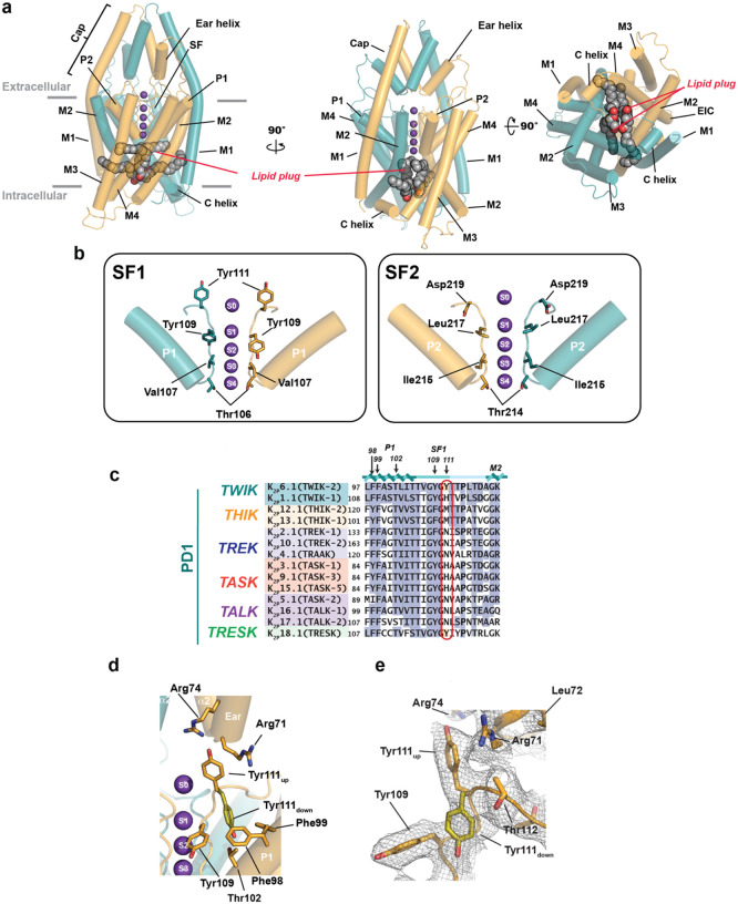
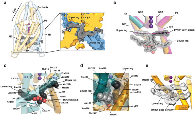
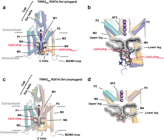
Lipids are integral to ion channel function yet delineating mechanisms by which they affect function remains challenging. Within the K2P family of leak potassium channels1-3, observation of tubular densities interpreted as alkyl chains occupying lateral fenestrations linking the pore and bilayer4-8 raised the possibility that lipid access from the bilayer acts as a regulatory mechanism4-7. Here, we present cryo-electron microscopy (cryo-EM) structures of the human leak potassium channel K2P6.1 (TWIK2)9-11 and mutants in nanodisc and detergent environments that reveal an unusual conformation in the first selectivity filter (SF1) and a pair of two-chain lipids within the channel cavity (denoted the 'lipid plug'). The chains of each plug lipid occupy separate binding sites that laterally extend to the bilayer from the channel cavity. One, the upper leg, matches the previously identified alkyl chain binding site4-8,12. The second, the lower leg, occupies a fenestration common with K2P1.1 (TWIK1)13. Together, they demonstrate a bidentate means to coordinate each plug lipid that offers a reinterpretation of previous observations. Structures of a K2P6.1 (TWIK2) mutant that directs the channel to the plasma membrane14 and an R257A mutant that increases function yield plugged and unplugged forms. Notably, the R257A plugged form shows a change in lipid plug position, indicating a key role for this residue in lipid binding. Together, our data suggest that occupation of the central cavity by the lipid plug serves as a mechanism to render the TWIK channels inactive and points to the importance of lipid plug removal to create an ion permeable pore. Such a mechanism could provide a potent way for limiting the leak function of K2Ps based on cellular location or other contextual factors.


Conflict of interest statement

Competing interests The authors declare no competing interests.

Figures

Figure 1
Figure 1. K2P6.1(TWIK-2) structural features.
a, K2P6.1(TWIK-2) cartoon diagram (deep teal and bright orange) showing side (left and center) and intracellular (right) views. Lipid plug (grey) is shown in space filling. Grey bars indicate membrane. Potassium ions are shown as purple spheres. b, Comparison of K2P6.1(TWIK-2) SF1 (left) and SF2 (right) filter conformations. Select matching positions are shown as sticks. c, Sequence comparison P1, SF1, and initial segment of M2 from PD1 for K2P6.1 (TWIK-2) with other K2Ps. Red oval indicates Tyr111 site. Labels indicate residues that interact with Tyr111 in the ‘down’ state. Conservation is indicated in blue. Tyr111 and similar residues are shaded red. Sequences are for human: K2P6.1(TWIK-2) (GENBANK 4758624); K2P 1.1(TWIK-1) (GENBANK 4504847); K2P13.1(THIK-1) (GENBANK 16306555); K2P12.1(THIK-2) (GENBANK 11545761); K2P2.1(TREK-1) (GENBANK 14589851); K2P10.1(TREK-2) (GENBANK 20143944); K2P4.1(TRAAK) (GENBANK 15718767); K2P3.1(TASK-1) (GENBANK 4504849); K2P9.1 (TASK-3) (GENBANK 542133161); K2P15.1(TASK-5) (GENBANK 333440483); K2P5.1(TASK-2) (GENBANK 333440483); K2P16.1(TALK-1) (GENBANK 14149764); K2P17.1(TALK-2) (GENBANK 17025230); and K2P18.1(TRESK) (GENBANK 32469495). d, K2P13.1(THIK-1) Try111 environment and interactions. ‘up’ (bright orange) and ‘down’ (olive) Tyr111 conformations are indicated. e, Cryo-EM density for the region including Tyr111 (σ=2.5).
Figure 2
Figure 2. K2P6.1(TWIK-2) lipid plug blocks the central cavity.
a, K2P6.1(TWIK-2) cartoon diagram (deep teal and bright orange). Cryo-EM density for lipid plug (grey) is shown (σ =3.0). Inset shows slice through space-filling model of the channel, lipid plug cryo-EM density and stick representation of lipid plug. b, Superposition of K2P6.1(TWIK-2) (deep teal and bright orange) and K2P1.1(TWIK-1) (magenta) (PDB:3UKM). K2P6.1(TWIK-2) lipid plug (grey and black) is shown as sticks and semi-transparent space filling. Upper and lower legs for one lipid are indicated. TWIK1 Alkyl chain is shown as sticks. c, K2P6.1(TWIK-2) lipid plug interactions. One lipid plug chain is shown (black, space filling). Contacting residues from the two K2P6.1(TWIK-2) subunits (deep teal and bright orange) are shown as sticks. d, View from the center of the bilayer towards the upper and lower leg binding sites. Lipid plug chains are grey and black and shown in space filling. Interacting residues are shown as sticks. e, Lipid plug density (1.9σ) from K2P1.1 (TWIK-1) at pH5.5 (EMDB 25169). Model shows a single K2P6.1(TWIK-2) subunit and lipid plug (grey).
Figure 3
Figure 3. Comparison of plugged and unplugged K2P6.1 (TWIK2) structures.
a, Superposition of TWIK2:ND (deep teal and bright orange) and TWIK2RM:Det (plugged) (deep purple) structures. b, Close up view of the central cavity from ‘a’. Lipid plugs from TWIK2:ND and TWIK2RM:Det (plugged) are shown as spheres (grey) and sticks (deep purple), respectively. c, Intracellular view of a slice through the TWIK2RM:Det (plugged) density. Subunits are deep teal and bright orange, lipid plug density is grey. d, Superposition of TWIK2:ND (deep teal and bright orange) and TWIK2RM:Det (unplugged) (firebrick) structures. d, Close up view of the central cavity from ‘d’. Lipid plug from TWIK2:ND is shown as spheres (grey). TWIK2RM:Det (unplugged) cavity ion is labeled. f, Intracellular view of a slice through the TWIK2RM:Det (unplugged) density. Subunits are firebrick and light red. g, Pore profile of K2P6.1(TWIK2) (deep teal and bright orange) calculated using HOLE. Selectivity filter and key cavity positions are shown as sticks. h, K2P6.1(TWIK2) pore profiles for plugged (blue) and unplugged (magenta) forms. Selectivity filter (SF) (grey) and cavity (blue) are indicated. in ‘g’ and ‘h’.
Figure 4
Figure 4. K2P6.1(TWIK2) cavity and filter sites affect function.
a, View of the K2P6.1(TWIK2) lipid plug from center of the membrane. Lipid plug is shown as grey spheres. Key interacting residues are shown as sticks. Potassium ions are purple spheres. b, Exemplar current-voltage responses from TEVC Xenopus oocytes expressing the indicated channels and cavity mutants. Uninjected (grey), TWIK2 (sky blue), TWIK2RM (blue), TWIK2RM V131D (olive), TWIK2RM M135A (orange), TWIK2RM M135A (red), TWIK2RM R257A (yellow), and TWIK2RM R257E (lavender). Inset shows protocol. c, Effects of mutants on K2P6.1(TWIK2) basal activity at 0 mV. **** p <0.0001, *** p <0.001, ** p < 0.01, * p = 0.01 −0.05. Statistical analysis was performed using a One-Way ANOVA and multiple comparisons were performed with Tukey’s test. Error bars are S.E.M.. d, Exemplar current-voltage responses from TEVC Xenopus oocytes expressing the indicated channels and mutants. Uninjected (grey), TWIK2 (sky blue), TWIK2RM (blue), TWIK2RM Y111A (green), TWIK2RM Y111H (salmon), and TWIK2RM Y111N (maroon). Inset shows protocol. e, Effects of mutants on K2P6.1(TWIK2) basal activity at 0 mV. **** p <0.0001, *** p <0.001, ** p < 0.01, * p = 0.01 −0.05. Statistical analysis was performed using One-Way ANOVA and multiple comparisons were performed with Tukey’s test. Error bars are S.E.M..
Figure 5
Figure 5. Comparison of plugged and unplugged K2P6.1 (TWIK2) R257A structures.
a, Superposition of TWIK2:ND (deep teal and bright orange) and TWIK2RM R257A:Det (plugged) (deep blue) structures. b, Close up view of the central cavity from ‘a’. Lipid plugs from TWIK2:ND and TWIK2RM R257A:Det are shown as spheres (grey) and sticks (deep blue), respectively. d, Superposition of TWIK2:ND (deep teal and bright orange) and TWIK2RM R257A:Det (unplugged) (dirtyviolet) structures. d, Close up view of the central cavity from ‘c’. Lipid plug from TWIK2:ND is shown as spheres (grey).
Figure 6
Figure 6. Model of K2P6.1(TWIK2) regulation.
Lipid plug prevents ion passage. Removal of plug in response to cellular localization changes, changes in the membrane lipid composition, or other factors yields channels that can conduct ions.

References

    1. Mathie A., Veale E.L., Cunningham K.P., Holden R.G., and Wright P.D. (2021). Two-Pore Domain Potassium Channels as Drug Targets: Anesthesia and Beyond. Annu Rev Pharmacol Toxicol 61, 401–420. 10.1146/annurev-pharmtox-030920-111536. - DOI - PubMed
    1. Feliciangeli S., Chatelain F.C., Bichet D., and Lesage F. (2015). The family of K2P channels: salient structural and functional properties. J Physiol 593, 2587–2603. 10.1113/jphysiol.2014.287268. - DOI - PMC - PubMed
    1. Enyedi P., and Czirjak G. (2010). Molecular background of leak K+ currents: two-pore domain potassium channels. Physiological reviews 90, 559–605. 90/2/559 [pii] 10.1152/physrev.00029.2009. - DOI - PubMed
    1. Miller A.N., and Long S.B. (2012). Crystal structure of the human two-pore domain potassium channel K2P1. Science 335, 432–436. 335/6067/432 [pii] 10.1126/science.1213274. - DOI - PubMed
    1. Turney T.S., Li V., and Brohawn S.G. (2022). Structural Basis for pH-gating of the K(+) channel TWIK1 at the selectivity filter. Nat Commun 13, 3232. 10.1038/s41467-022-30853-z. - DOI - PMC - PubMed

Publication types

LinkOut - more resources