[Preprint]. 2025 Jun 8:2025.06.08.653722.
doi: 10.1101/2025.06.08.653722.

RetiGene, a comprehensive gene atlas for inherited retinal diseases (IRDs)

Affiliations

RetiGene, a comprehensive gene atlas for inherited retinal diseases (IRDs)

Mathieu Quinodoz et al. bioRxiv. .

Update in

  • RetiGene, a comprehensive gene atlas for inherited retinal diseases.
    Rivolta C, Celik E, Kamdar D, Cancellieri F, Kaminska K, Ullah M, Barberán-Martínez P, Bouckaert M, Cortón M, Delanote E, Fernández-Caballero L, García García G, Holtes LK, Karali M, Lopez I, Peter VG, Schneider N, Vincke L, Ayuso C, Banfi S, Bocquet B, Coppieters F, Cremers FPM, Inglehearn CF, Iwata T, Kalatzis V, Koenekoop RK, Millán JM, Sharon D, Toomes C, Quinodoz M. Rivolta C, et al. Am J Hum Genet. 2025 Oct 2;112(10):2253-2265. doi: 10.1016/j.ajhg.2025.08.017. Epub 2025 Sep 16. Am J Hum Genet. 2025. PMID: 40961941 Review.

Abstract

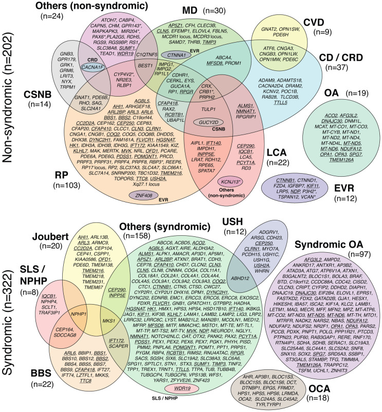
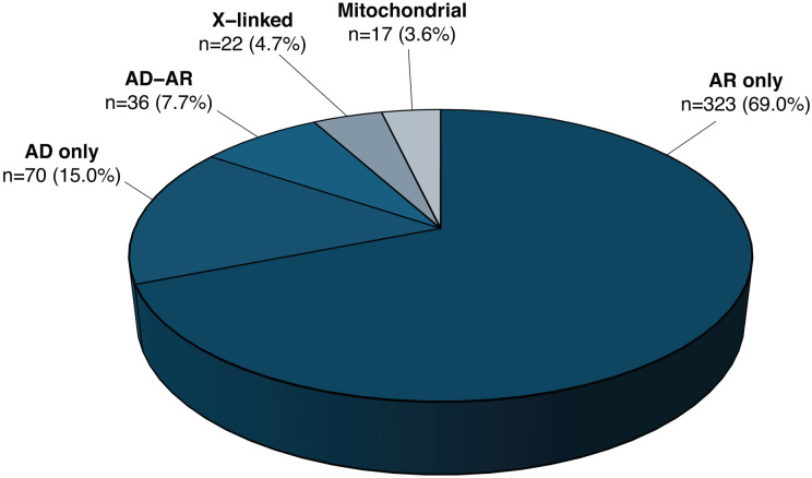
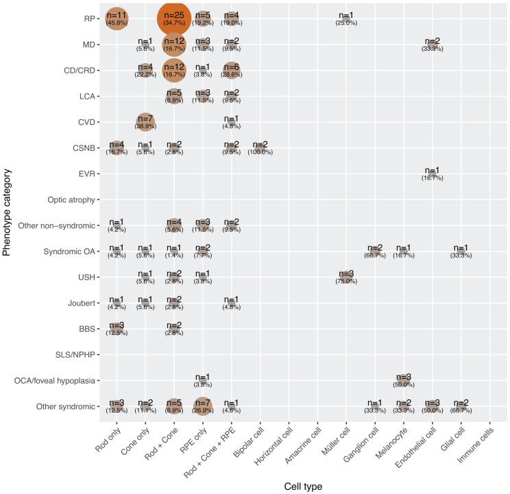
Inherited retinal diseases (IRDs) are rare disorders, typically presenting as Mendelian traits, that result in stationary or progressive visual impairment. They are characterized by extensive genetic heterogeneity, possibly the highest among all human genetic diseases, as well as diverse inheritance patterns. Despite advances in gene discovery, limited understanding of gene function and challenges in accurately interpreting variants continue to hinder both molecular diagnosis and genetic research in IRDs. One key problem is the absence of a comprehensive and widely accepted catalogue of disease genes, which would ensure consistent genetic testing and reliable molecular diagnoses. With the rapid pace of IRD gene discovery, gene catalogues require frequent validation and updates to remain clinically and scientifically useful. To address these gaps, we developed RetiGene, an expert-curated gene atlas that integrates variant data, bulk and single-cell RNA sequencing, and functional annotations. Through the integration of diverse data sources, RetiGene supports candidate gene prioritization, functional studies, and therapeutic development in IRDs.


Conflict of interest statement

Declaration of interests The authors declare no competing financial or non-financial interests.

Figures

Figure 1:
Figure 1:
Venn diagram of genes and loci associated with IRDs (total=468). Underlined genes are linked to both non-syndromic and syndromic phenotypes. Asterisks point to genes that can also be involved in non-retinal ocular diseases. n, number of genes.
Figure 2:
Figure 2:
Inheritance mode of diseases associated with all curated genes and loci. n, number of genes.
Figure 3:
Figure 3:
Discovery of IRD genes through time. (A) Cumulative number of genes identified, per year. (B) Annual count of new gene discoveries. (C) Cumulative percentage of genes discovered, stratified by inheritance mode. (D) Cumulative percentage of genes discovered, stratified by broad phenotypic categories.
Figure 4.
Figure 4.
Functional categorization of IRD-associated genes. The bar graph shows the number of genes assigned to each functional category relevant to IRDs. Bar segments indicate whether each gene is annotated in a unique category (blue) or appears in multiple categories (coral). The "Others" group includes genes with roles that could not be confidently assigned to the main categories.
Figure 5:
Figure 5:
Inheritance and expression features of IRD genes. Co-occurrence matrices between: (A) Inheritance mode and broad phenotypic categories, (B) Inheritance mode and type of pathogenic variants, (C) Expression in retina / other tissues and broad phenotypic categories, (D) Expression in retinal cell types and broad phenotypic categories. n, number of genes.
Figure 6:
Figure 6:
Co-occurrence matrix between phenotypes and retinal single-cell gene expression data. n, number of genes.

References

    1. Hussey K.A., Hadyniak S.E., and Johnston R.J. Jr. (2022). Patterning and Development of Photoreceptors in the Human Retina. Front Cell Dev Biol 10, 878350. - PMC - PubMed
    1. Strauss O. (2005). The retinal pigment epithelium in visual function. Physiol Rev 85, 845–881. - PubMed
    1. Intartaglia D., Giamundo G., and Conte I. (2022). Autophagy in the retinal pigment epithelium: a new vision and future challenges. FEBS J 289, 7199–7212. - PMC - PubMed
    1. Masland R.H. (2012). The neuronal organization of the retina. Neuron 76, 266–280. - PMC - PubMed
    1. Rashid K., Akhtar-Schaefer I., and Langmann T. (2019). Microglia in Retinal Degeneration. Front Immunol 10, 1975. - PMC - PubMed

Publication types

LinkOut - more resources