[Preprint]. 2025 Jun 12:2025.06.09.658633.
doi: 10.1101/2025.06.09.658633.

NDR kinase SAX-1 controls dendrite branch-specific elimination during neuronal remodeling in C. elegans

Affiliations

NDR kinase SAX-1 controls dendrite branch-specific elimination during neuronal remodeling in C. elegans

Paola V Figueroa-Delgado et al. bioRxiv. .

Abstract

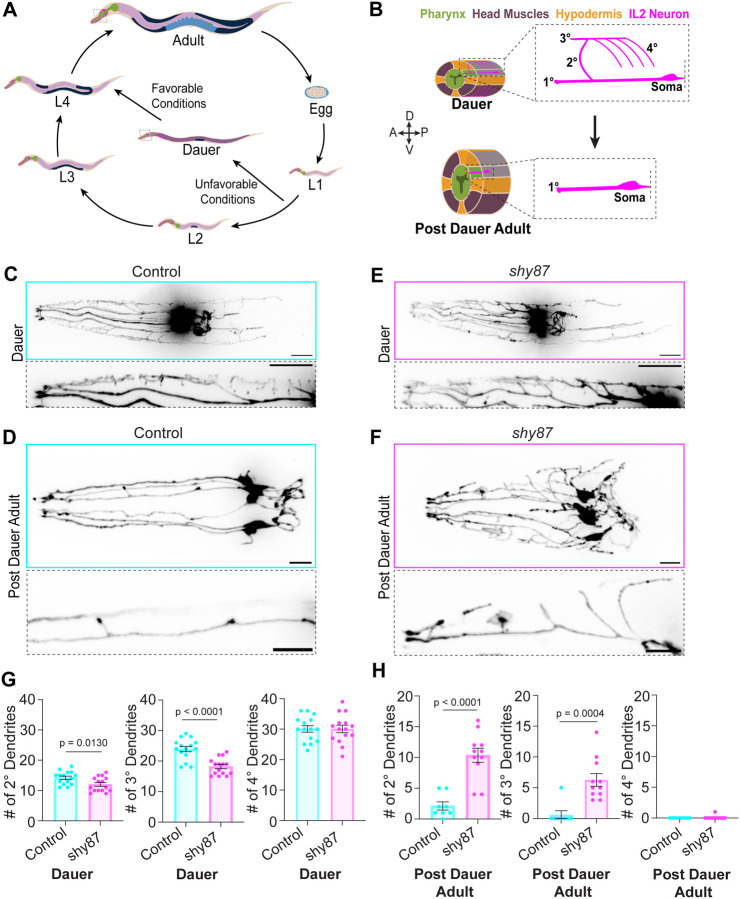
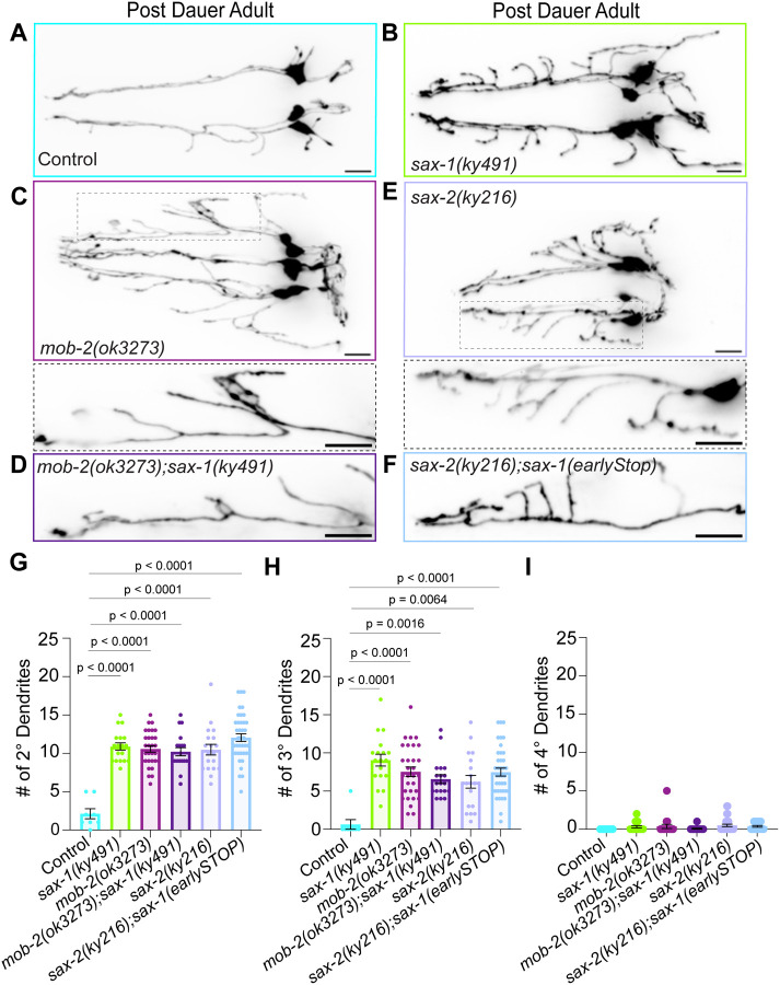
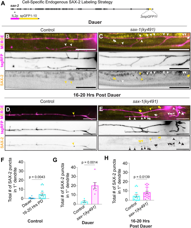
Neuronal remodeling is crucial for proper nervous system development and function, and can be initiated by developmental programs, activity-dependent mechanisms, or stress. Despite significant advances, the underlying mechanisms that govern this process remain poorly understood. Here, we adapted C. elegans IL2 sensory dendrites as a model system to study developmental and stress-mediated dendrite pruning. Upon entering a stress-induced developmental diapause, IL2 dendrites grow a complex dendritic arbor, which is later pruned when reproductive development resumes. We identified unexpected specificity in the pruning process, with distinct genetic requirements to direct branch-specific elimination of secondary, tertiary, and quaternary branches. The serine/threonine kinase SAX-1/NDR promotes elimination of secondary and tertiary, but not quaternary, dendrites. SAX-1 functions with its conserved interactors SAX-2/Furry and MOB-2 in the removal of both dendritic branches. The guanine-nucleotide exchange factor RABI-1/Rabin8 and the small GTPase RAB-11.2 mediate the elimination of secondary branches with SAX-1, but their effect on tertiary branches is minimal. Consistent with the known roles of RABI-1 and RAB-11.2 in regulating membrane dynamics, we find that SAX-1 promotes endocytosis during remodeling. Together, our findings reveal distinct mechanisms for branch-specific elimination under stress-induced and developmentally regulated neuronal remodeling.

Keywords: MOB-2/Mob; RABI-1/Rabin8; SAX-1/NDR; SAX-2/Fry; dauer; dendrite pruning; membrane trafficking; neuronal remodeling; small GTPase RAB-11.2.


Conflict of interest statement

Declaration of Interests The authors declare no competing interests.

Figures

Figure 1.
Figure 1.. shy87 mutants exhibit a remodeling defect in post dauer adults.
(A) Schematic depicting C. elegans life cycle. Adapted from WormAtlas. Under unfavorable conditions, the nematode arrests into an alternative developmental molt (dauer). Upon re-exposure to favorable conditions, reproductive development is resumed into adulthood. (B) Oblique transverse schematic of IL2Q neurons (magenta) at dauer arrest (top) and in a post dauer adult worm (bottom). Pharynx (green); Head muscles (purple); Hypodermis (yellow). At dauer, the IL2Q neurons extend a stereotypical dendritic arbor characterized by a 2° dendrite extending from a 1° dendrite towards the dorsal midline; 3° dendrites bifurcate from 2° dendrite towards the anterior and posterior in parallel to 1° dendrite; 4° dendrites extending into the head muscle quadrants (purple). When reproductive development is resumed, post dauer adult worms remove the higher-order dendrites, leaving the 1° dendrite intact. (C-D) Z-projection of control dauer (C) and post dauer adult (D) expressing tba-6p::tagRFP in IL2s (top). Zoomed inset of select IL2 dorsal neuron (bottom, dashed box). (E-F) Maximum intensity Z-projection of shy87 mutant dauer (E) and post dauer adult (F) expressing cytosolic tagRFP under tba-6p (top). Zoomed inset of select IL2 dorsal neuron (bottom, dashed box). Scale bar, 10μm. (G) Quantification of the number of IL2Q 2°-4° dendrites at dauer comparing shy87 mutants (n = 15) to control animals (n = 15). Unpaired t-test. (H) Quantification of the number of IL2Q higher-order dendrites in control (n = 8) versus shy87 mutants post dauer adults (n = 11). Unpaired t-test with Welch’s correction (2°) or Mann-Whitney U test (3°-4°). Error bars are ±SEM.
Figure 2.
Figure 2.. SAX-1/NDR promotes IL2Q remodeling in a cell-specific and kinase activity-dependent manner.
(A) AlphaFold predicted structure of SAX-1 with key functional domains indicated: N-terminal regulatory domain (1–79AA, orange), kinase catalytic domain I-VII (80–270AA, dark green), auto-inhibitory sequence (270–310AA, dark purple), kinase catalytic domain VIII-IX (310–370AA, light green), and the AGC family kinase C-terminal domain (370–446AA, magenta). Close-up view of glycine316 (red), which is conserved across species from yeast to human homologs of NDR1/2 kinases and mutated into an asparagine in shy87 mutants. (B-D) Maximum intensity Z-projection of control (B), early stop (C), and engineered repair of the shy87 allele (D) post dauer adult animals, demonstrating that loss of sax-1 function is responsible for the pruning defects in shy87 mutants. Zoomed inset of select IL2Q are shown for early stop (C) and shy87 engineered repair (D) with a dashed box. Scale bars, 10μm. (E, F) Quantifications of IL2Q 2° dendrite (E) and 3° dendrite number in post dauer adults of control and indicated sax-1 alleles. Brown-Forsythe and Welch ANOVA with Dunnett’s correction (E) or Kruskal-Wallis test with Dunn’s correction (F), all genotypes compared to control and shy87 mutants, n = 8–22. All sax-1 mutant alleles phenocopied shy87. Repair of the shy87 missense point mutation rescued the mutant phenotype back to control numbers. (G-H) Maximum intensity Z-projection of shy87 post dauer adult mutants expressing wildtype SAX-1 cDNA (G) and a kinase-dead (S279) construct (H) under an IL2-specific promoter (tba-6p). Scale bars, 10μm. (I, J) Quantifications comparing the total number of IL2Q 2° dendrites (I) and 3°dendrites (J) in post dauer adults of control, sax-1 mutants, and indicated rescue transgenes. Brown-Forsythe and Welch ANOVA with Dunnett’s multiple comparison (I) and Kruskal-Wallis with Dunn’s correction (J), n = 8–21. Error bars are ±SEM.
Figure 3.
Figure 3.. MOB-2 and SAX-2/Furry function with SAX-1/NDR to direct IL2Q dendrite elimination.
Post dauer adult confocal images of (A) control and (B) sax-1 loss-of-function, (C) mob-2, (D) sax-1;mob-2 double mutants, (E) sax-2, and (F) sax-1; sax-2 double-mutants. Zoomed inset of select IL2Q are shown for mob-2 and sax-2 single mutants with a dashed box. Scale bars, 10μm. (G-H) Quantification of the total number of 2° (G), 3° (H), and 4° dendrites (I) in post dauer adults for genotypes shown in A-F. Brown-Forsythe and Welch ANOVA with Dunnett’s correction (2°) or Kruskal-Wallis test with Dunn’s correction (3°-4°), n = 8–39. Error bars are ±SEM, with individual data points shown.
Figure 4.
Figure 4.. SAX-2/Furry localization depends on SAX-1 kinase.
(A) Schematic illustrating the strategy to endogenously label SAX-2 in IL2 neurons using split-GFP, with GFP1–10 expressed under IL2 specific promoter. (B-C) Representative images of endogenous SAX-2::GFP (pseudo-colored yellow) in IL2 neurons (pseudo-colored magenta) at dauer arrest in control (B) and sax-1 mutants (C). Scale bar, 10μm. (D-E) Representative IL2Q inset maximum intensity Z-projection of endogenous SAX-2::GFP in IL2 neurons 16–20 hours post dauer in control (D) and sax-1 mutants (E). Zoomed insets show a single IL2D 1° dendrite. Yellow arrowheads denote SAX-2::GFP puncta localized at 1° dendrites; white/black arrowheads point to SAX-2::GFP puncta localized to higher-order dendrites. Scale bar, 10μm. (F) Quantification of SAX-2::GFP puncta at 1° dendrites in control animals at dauer and 16–20 hours post dauer. Mann-Whitney U test, n = 17–20. Dendritic SAX-2 puncta increase 16–20 hours following dauer recovery. (G) Quantifications of total # of SAX-2 puncta at 1° dendrites in control versus sax-1 mutants at dauer. Unpaired t-test with Welch’s correction, n = 5–7. (H) Quantifications of total # of SAX-2 puncta at 1° dendrites in control versus sax-1 mutants at 16–20 hours post dauer. Mann-Whitney U test, n = 17–21. Error bars are ±SEM.
Figure 5.
Figure 5.. SAX-1 coordinates IL2Q remodeling with RABI-1/Rabin8 and RAB-11.2.
(A-I) Representative confocal images of select IL2Q insets for control (A), rab-11.1 (B), rab-8 (C), rab10 (D), rabi-1 (E), rab-11.2 (F), sax-1 (G), rabi-1;sax-1 (H), and rab-11.2;sax-1 (I) post dauer adult mutants. Scale bar, 10μm. (J-K) Quantification of total number of 2° (J) and 3° (K) dendrites for A-F. Brown-Forsythe and Welch ANOVA with Dunnett’s correction (J) or Kruskal-Wallis with Dunn’s correction (K), n = 8–62. (L) Quantification of total number of 2° dendrites in indicated genotypes. Brown-Forsythe and Welch ANOVA with Dunnett’s correction, n = 8–33. Error bars are ±SEM, with individual data points shown.
Figure 6.
Figure 6.. SAX-1/NDR promotes endocytic events during pruning.
(A) Schematic of generic endocytosis reporter. mCD8::tagRFP (blue) fused to a GFP-binding nanobody are expressed under an IL2-specific promoter. Secreted GFP (yellow) from muscle binds the nanobody, enabling visualization of endocytosed GFP-mCD8 complexes as blue/yellow colocalized puncta. (B) Schematic of a control endocytosis reporter chimera lacking the GFP-binding nanobody. (C) Zoomed insets of confocal image depicting a single IL2Q 1° dendrite in control dauer with the endocytosis reporter (C-D) and a control construct lacking GFP-binding nanobody (E-F). White arrowheads show blue-yellow colocalization, suggestive of endocytosed complexes. Scale bar, 10μm. (D) Normalized fluorescence linescan of endocytic reporter showing co-localization of blue-yellow puncta (white dashed box) at dauer. (F) Normalized fluorescence linescan of control construct lacking GFP-binding nanobody showing lack of co-localization of blue-yellow puncta at dauer. (G-H) Representative zoomed insets of confocal images of the endocytosis reporter in a single IL2Q neuron at 16–20 hours post dauer in control (G) and sax-1 mutants (H). White arrowheads depict blue-yellow co-localized puncta. Scale bar, 10μm. (I) Quantification of the total number of co-localized blue-yellow puncta at dauer and 16–20 hours post dauer in control animals and sax-1 mutants. Mann-Whitney U test, n = 5–7. Error bars are ±SEM. sax-1 is required for the endocytic puncta post dauer.

Similar articles

References

    1. Altun Z.F. and Hall D.H. 2009. “Introduction.” WormAtlas. 10.3908/wormatlas.1.1 - DOI
    1. Androwski Rebecca J., Flatt Kristen M., and Schroeder Nathan E.. 2017. “Phenotypic Plasticity and Remodeling in the Stress-Induced Caenorhabditis Elegans Dauer.” Wiley Interdisciplinary Reviews. Developmental Biology 6 (5). 10.1002/wdev.278. - DOI - PMC - PubMed
    1. Bichsel Samuel J., Tamaskovic Rastislav, Stegert Mario R., and Hemmings Brian A.. 2004. “Mechanism of Activation of NDR (Nuclear Dbf2-Related) Protein Kinase by the hMOB1 Protein*.” Journal of Biological Chemistry 279 (34): 35228–35. 10.1074/jbc.M404542200. - DOI - PubMed
    1. Brenner S. 1974. “THE GENETICS OF CAENORHABDITIS ELEGANS.” Genetics 77 (1): 71–94. 10.1093/genetics/77.1.71. - DOI - PMC - PubMed
    1. Brunson Kristen L., Enikö Kramár, Lin Bin, Chen Yuncai, Colgin Laura Lee, Yanagihara Theodore K., Lynch Gary, and Baram Tallie Z.. 2005. “Mechanisms of Late-Onset Cognitive Decline after Early-Life Stress.” Journal of Neuroscience 25 (41): 9328–38. 10.1523/JNEUROSCI.228-105.2005. - DOI - PMC - PubMed

Publication types

LinkOut - more resources