

This is a preprint.
In vivo characterization of Achilles subtendon function and morphology within the tendon cross section and along the free tendon
- PMID: 40661625
- PMCID: PMC12258896
- DOI: 10.1101/2025.05.29.656873
In vivo characterization of Achilles subtendon function and morphology within the tendon cross section and along the free tendon
Update in
-
In vivo characterization of Achilles subtendon function and morphology within the tendon cross section and along the free tendon.J Appl Physiol (1985). 2025 Sep 1;139(3):812-822. doi: 10.1152/japplphysiol.00479.2025. Epub 2025 Aug 19. J Appl Physiol (1985). 2025. PMID: 40828574 Free PMC article.
Abstract
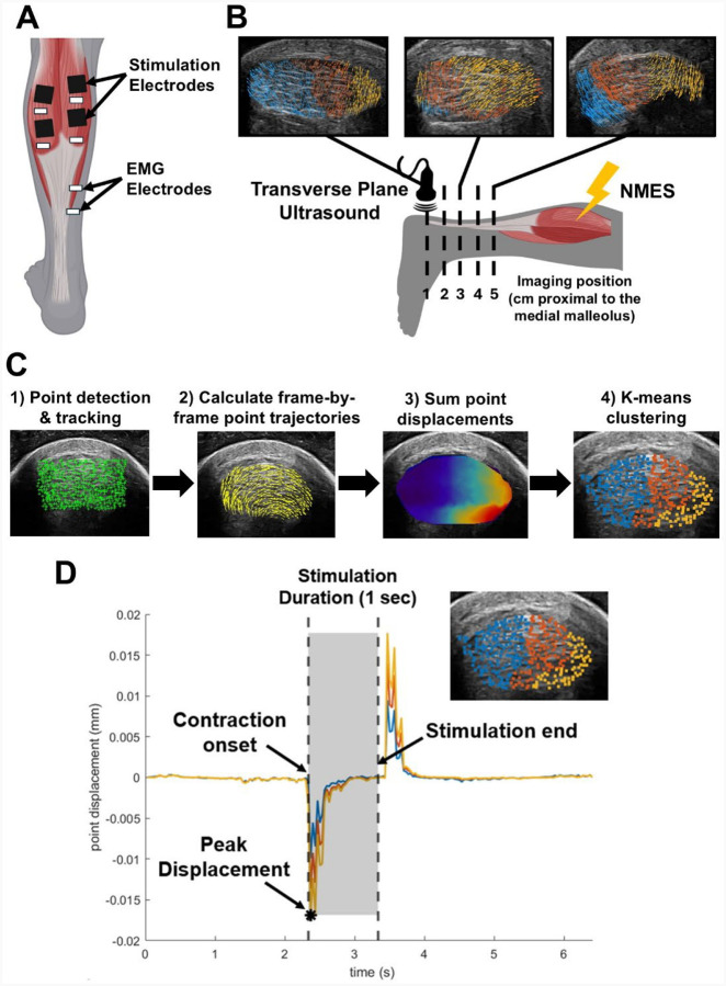
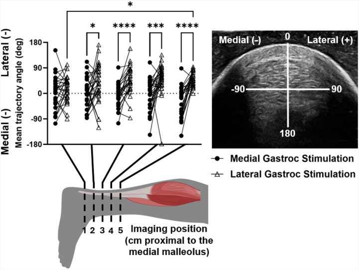
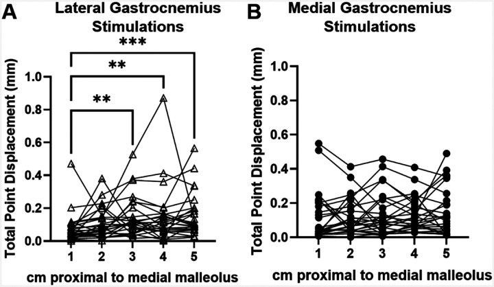
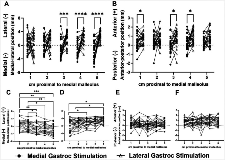
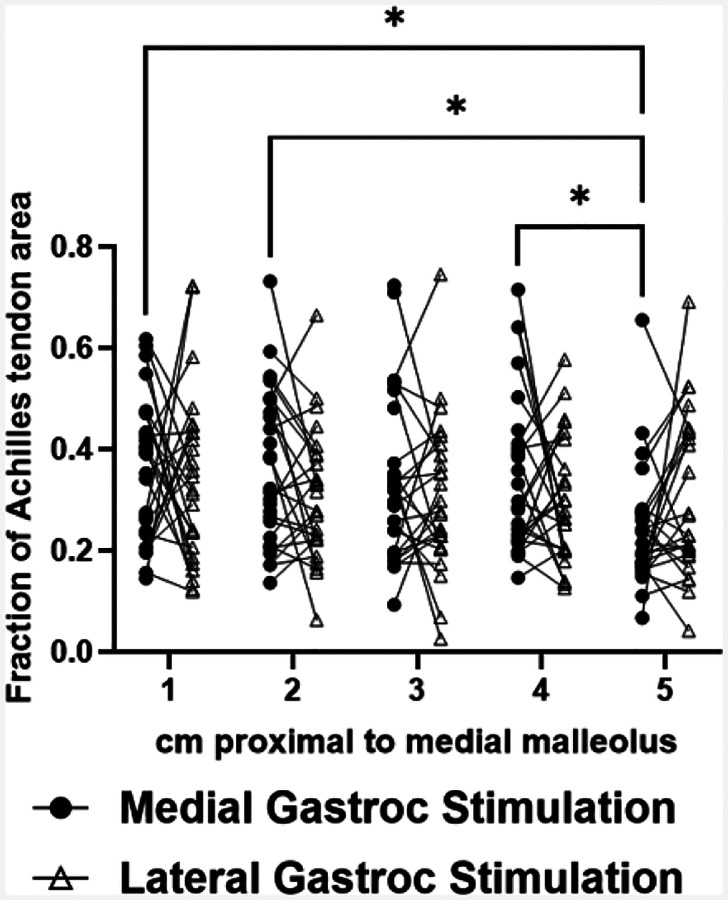
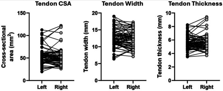
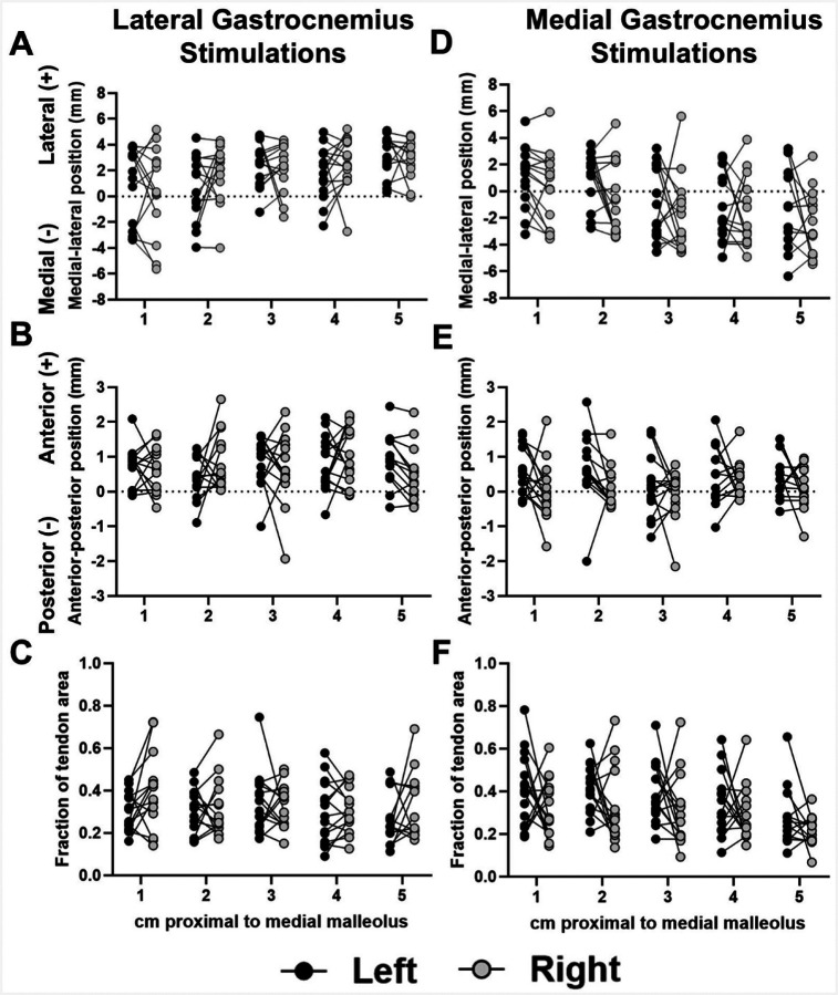
The Achilles tendon is composed of three distinct fascicle bundles, or "subtendons," each originating from the head of one of the three triceps surae muscles. In a healthy tendon, these subtendons slide relative to each other during muscle contractions. This subtendon sliding is reduced in older adults and younger adults who suffer an Achilles tendon injury. However, subtendon sliding is challenging to quantify in low-load scenarios that are critical for monitoring subtendon biomechanics in patients with mechanically compromised tendons, like following an Achilles tendon rupture and repair surgery. The purpose of this study was to develop a reliable method to characterize subtendon behavior in vivo using combined transverse plane ultrasound imaging and neuromuscular electrical stimulation of individual gastrocnemii. We used a Kanade-Lucas-Tomasi point tracking algorithm to quantify tendon displacement during isolated muscle stimulations. Next, we applied k-means clustering to characterize heterogeneous subtendon behavior within the tendon cross section. The tendon cross section displayed differential displacement patterns depending on the stimulated muscle (p<0.0001), and these displacements differed along the free tendon during lateral gastrocnemius stimulations (p=0.004). These results reflect possible differences in load-sharing between adjacent subtendons and differing muscle-tendon dynamics among the triceps surae muscles. Finally, this method confirmed bilaterally symmetric subtendon behavior and demonstrated high inter-session reliability (ICC>0.83). Overall, this study furthers our understanding of differential muscle-tendon dynamics of individual Achilles subtendons both within the tendon cross section and along the free tendon. Future work will apply this method to injured populations to develop biomarkers of altered subtendon function.
Keywords: Achilles Subtendons; muscular stimulation; ultrasound.
Conflict of interest statement
Disclosures The authors have no disclosures, financial or otherwise.
Figures
References
-
- Edama M, Kubo M, Onishi H, Takabayashi T, Inai T, Yokoyama E, et al. The twisted structure of the human Achilles tendon. Scand J Med Sci Sports. 2015. Oct 1;25(5):e497–503. - PubMed
-
- Pękala PA, Henry BM, Ochała A, Kopacz P, Tatoń G, Młyniec A, et al. The twisted structure of the Achilles tendon unraveled: A detailed quantitative and qualitative anatomical investigation. Scand J Med Sci Sports. 2017. Dec 1;27(12):1705–15. - PubMed
Publication types
Associated data
Grants and funding
LinkOut - more resources
Full Text Sources