[Preprint]. 2025 Jun 12:2025.06.09.658704.
doi: 10.1101/2025.06.09.658704.

Single-cell Analysis of Intracellular Transport and Expression of Cell Surface Proteins

Affiliations

Single-cell Analysis of Intracellular Transport and Expression of Cell Surface Proteins

Rekha Mudappathi et al. bioRxiv. .

Abstract

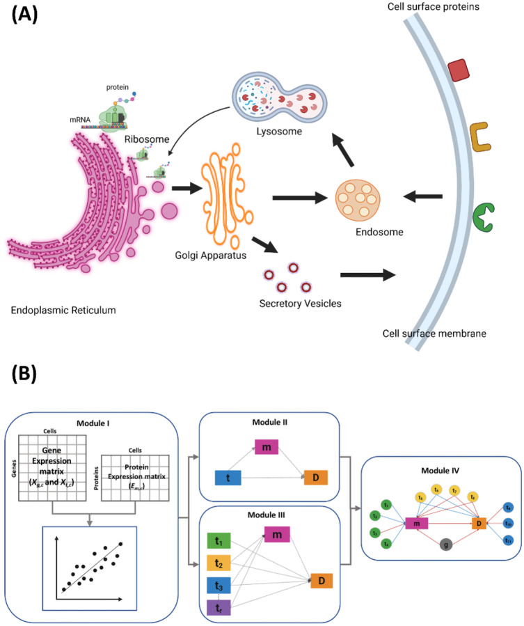
Intracellular protein transport (ICT) is a tightly regulated process that orchestrates protein localization and expression, ensuring proper cellular function. Dysregulated ICT can lead to aberrant expression of surface proteins involved in cell-cell communication, adhesion, and immune responses, contributing to disease progression and therapeutic resistance. Cellular Indexing of Transcriptomes and Epitopes by Sequencing (CITE-seq) enables the simultaneous measurement of mRNA and surface protein levels in the same cell, providing a powerful opportunity to investigate the molecular mechanisms underlying surface protein regulation. In this study, we introduce a novel computational frame for Modeling Protein Expression and Transport (MPET) that evaluates the contribution of ICT activity to differential surface protein expression using CITE-seq data. MPET comprises three modules for identification of ICT-surface protein regulatory circuits across biological scales and their contributions to phenotypic variation. We applied MPET to analyze single-cell data from COVID-19 patients with varying disease severity. Our analysis revealed context-dependent recruitment of ICT genes and pervasive rewiring of ICT pathways throughout the course of disease progression. Notably, we found that even when the transcriptional levels of key immune response proteins remained stable, their expression on cell surface were significantly altered due to dysregulated ICT. MPET provides a valuable new tool for dissecting complex regulatory networks and offers mechanistic insight into post-transcriptional regulation of cell surface proteins in diseases.

Keywords: CITE-seq; COVID-19; immune dysregulation; intracellular transport; mediation analysis; mixed-effects modeling; post-transcriptional regulation; single-cell multi-omics; surface protein expression.


Conflict of interest statement

Competing interests The authors declare no competing interests.

Figures

Figure 1.
Figure 1.
Overview of intracellular protein transport and MPET algorithm. (A) Ribosomes along with newly synthesized proteins attach onto endoplasmic reticulum (ER) membrane to enter ER lumen. After processing, membranous vesicles shuttle proteins from ER to Golgi apparatus. The proteins are then packed into secretory vesicles and transported to plasma membrane. Cells can internalize surface proteins via endocytosis. Endocytic vesicles fuse with recycling endosomes, from where they eventually move to lysosomes for degradation. Golgi cargo can also be sorted to endosomes, lysosomes, and ER for degradation and recycling. (B) CITE-seq data containing the gene expression matrix and protein expression matrix are input to MPET Module 1, in which mixed-effects regression analysis identifies ICT genes associated with surface protein abundance. The output from Module I can enter Module II for single-exposure mediation analysis or Module III for multiple-exposure mediation analysis. Module IV constructs a regulatory network to link surface proteins, their coding genes, and ICT genes to disease phenotype.
Figure 2.
Figure 2.
Mediation models in MPET. (A) Single-exposure mediation model. The transcription level of an ICT gene (t) may influence the disease phenotype (D) directly or indirectly via the expression level of a surface protein as a mediator (m). Coefficients in Eq. [2] and [3] are displayed along the corresponding edges. (B) Multiple exposure mediation model. Transcription levels of multiple ICT genes (t1,…,tr) are exposures. Coefficients in Eq. [4] and [5] are displayed along the corresponding edges. (C) Regulatory network represented as a directed acyclic graph. Nodes t1–t4 exhibit full mediation effects, Node t5–t8 show partial mediation. Nodes t9–t11 have null mediation. Red edges indicate positive associations, and blue edges indicate negative associations.
Figure 3.
Figure 3.
(A) UMAP visualization of CITE-seq cell clustering of cells from healthy control, and COVID-19 samples. (B) Scatter plot showing correlation between surface protein expression levels and the number of associated ICT genes in healthy cases, mild cases, and severe cases. (C) Venn diagram showing unique and shared PTTs between different phenotype groups.
Figure 4.
Figure 4.
Surface protein and ICT association revealed in Module I analysis. (A) Heatmap showing association between surface protein-coding gene pairs and ICTs in Healthy group. (B) Tanglegram comparing hierarchical clustering structures based on ICT associations (left) and on coding gene transcription (right). HLA-DR subunits are labeled in orange and CD16 subunits in green in both panels.
Figure 5.
Figure 5.
Modeling CD69 expression (A) Boxplot showing significantly increased surface protein expression of CD69 in CD16+ monocytes from patients with severe COVID-19 compared to mild and healthy controls (Wilcoxon rank-sum test, FDR < 0.05). (B) Boxplot showing no significant difference in CD69 mRNA expression across the phenotype groups (nominal p-value = 0.68). (C) Distribution of the mediation effects (α coefficients) of daICT genes for the CD69 protein. One-sample t-test supports mean α >0 with p = 5.7 × 10⁻¹³.
Figure 6.
Figure 6.
HLA-DR protein expression (A) Protein expression level is low in severe compared to healthy controls and mild. (B) Regulatory network with HLA-DR as the mediator, ICT genes as exposures, and disease severity (D) as the outcome. ICT genes are colored by mediation type - full mediation (green), partial mediation (yellow) or null mediation (blue).

References

    1. Alberts B., Johnson A., Lewis J., Raff M., Roberts K., and Walter P. (2002). Membrane Proteins.
    1. Xiong S., and Xu W. (2014). Human Immune System. Comprehensive Biomedical Physics 10, 91–114. 10.1016/B978-0-444-53632-7.01008-X. - DOI
    1. Engel P., Boumsell L., Balderas R., Bensussan A., Gattei V., Horejsi V., Jin B.-Q., Malavasi F., Mortari F., Schwartz-Albiez R., et al. (2015). CD Nomenclature 2015: Human Leukocyte Differentiation Antigen Workshops as a Driving Force in Immunology. The Journal of Immunology 195, 4555–4563. 10.4049/JIMMUNOL.1502033. - DOI - PubMed
    1. Dendrou C.A., Petersen J., Rossjohn J., and Fugger L. (2018). HLA variation and disease. Nature Reviews Immunology 2017 18:5 18, 325–339. 10.1038/nri.2017.143. - DOI - PubMed
    1. Pang X., He X., Qiu Z., Zhang H., Xie R., Liu Z., Gu Y., Zhao N., Xiang Q., and Cui Y. (2023). Targeting integrin pathways: mechanisms and advances in therapy. Signal Transduct Target Ther 8, 1. 10.1038/S41392-022-01259-6. - DOI - PMC - PubMed

Publication types

LinkOut - more resources