. 2025 Jul 13;13(7):e70632.
doi: 10.1002/fsn3.70632. eCollection 2025 Jul.

Proteostasis Disruption by Proteasome Inhibitor MG132 and Propolin G Induces ER Stress- and Autophagy-Mediated Apoptosis in Breast Cancer

Affiliations

Proteostasis Disruption by Proteasome Inhibitor MG132 and Propolin G Induces ER Stress- and Autophagy-Mediated Apoptosis in Breast Cancer

Jih-Tung Pai et al. Food Sci Nutr. .

Abstract

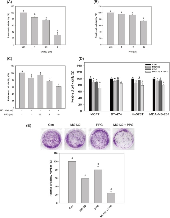
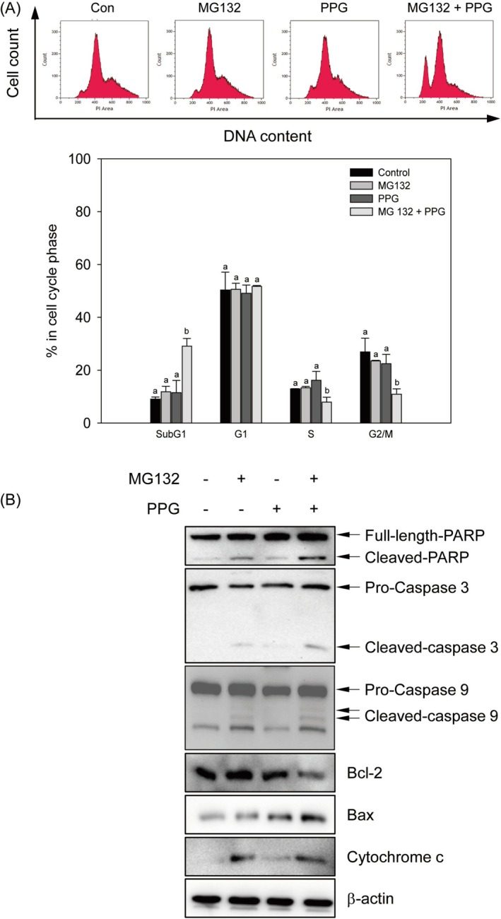
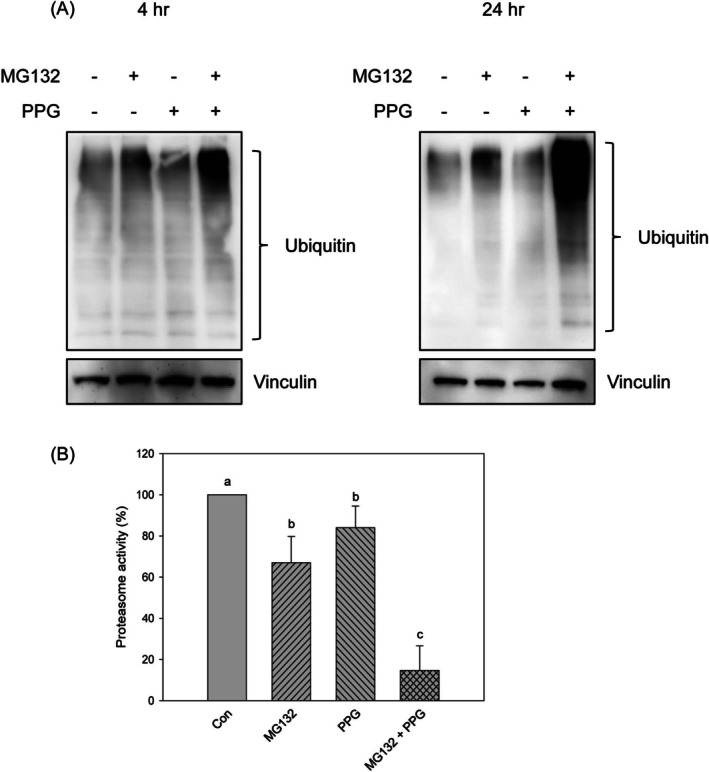
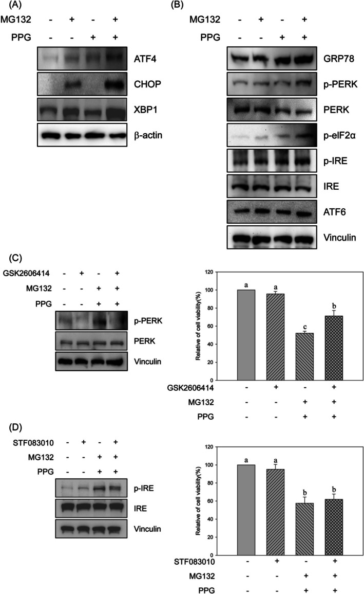
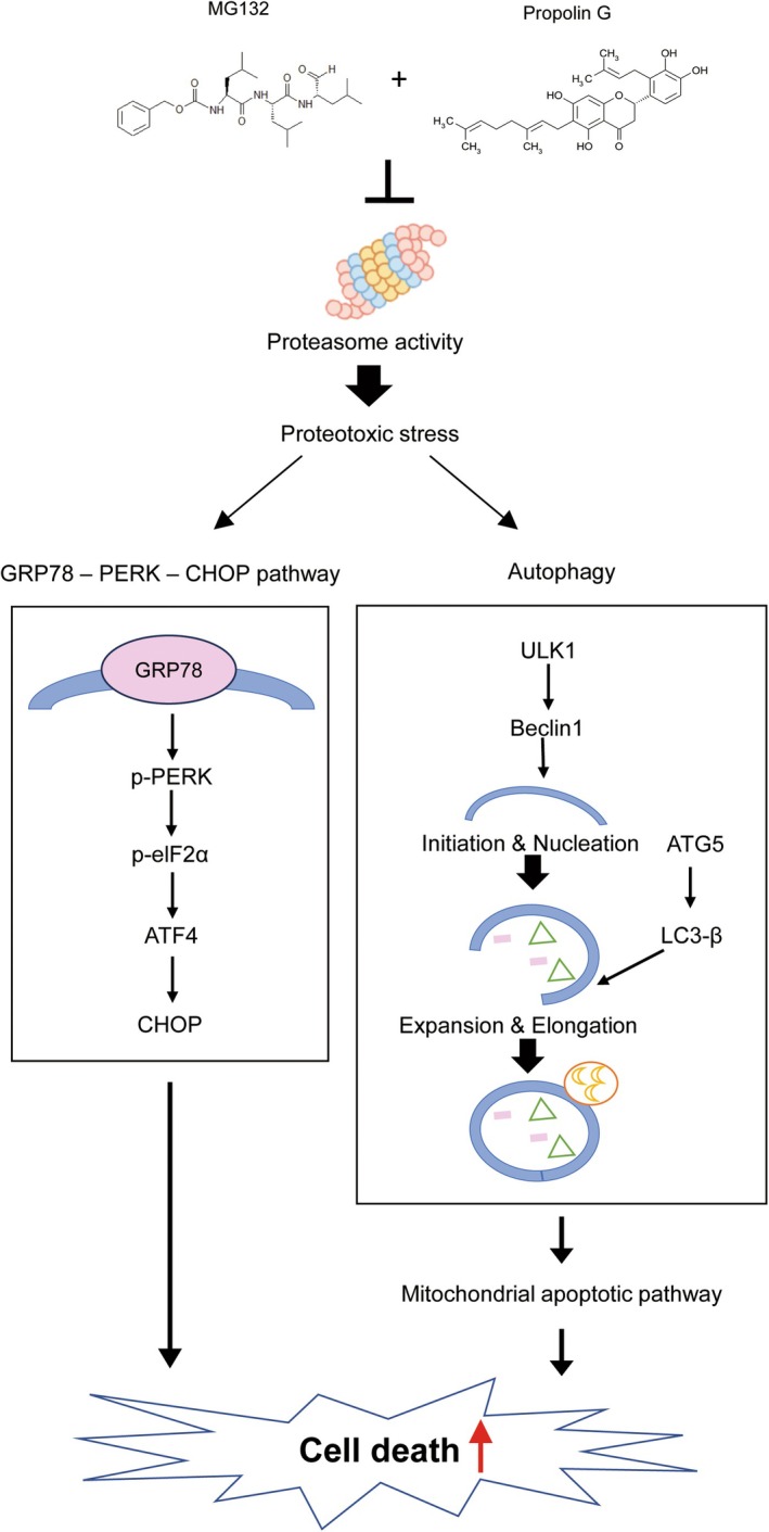
The maintenance of protein homeostasis, commonly referred to as proteostasis, is critical for the proper functioning of cells. Disruptions in protein degradation pathways can result in proteotoxic stress, which may ultimately lead to cellular apoptosis. Targeting the dysregulation of proteostasis has emerged as a promising approach in cancer therapy. Propolin G, a c-prenylflavanone derived from Taiwanese propolis, has demonstrated anticancer properties; however, its underlying mechanisms remain largely unexplored. In this study, we evaluated the combined effect of propolin G and the proteasome inhibitor MG132 on breast cancer cells. While individual treatments with MG132 (1 μM) or propolin G (10 μM) exhibited minimal effects on cell viability, their combination resulted in a synergistic suppression of proliferation and induction of apoptosis, as indicated by a combination index (CI) of 0.63. This combined treatment significantly reduced proteasome activity, leading to the accumulation of polyubiquitinated proteins. Mechanistically, apoptosis was mediated through the activation of the PERK/ATF4/CHOP signaling pathway and autophagy, as evidenced by increased expression levels of ULK1, Beclin1, ATG5, and LC3-II. These findings highlight the potential of targeting proteostasis disruption as an effective anticancer strategy in breast cancer. The combination of propolin G and MG132 may leverage cancer-specific vulnerabilities and possess translational potential for anticancer therapy.

Keywords: autophagy; propolin G; proteasome inhibitor; protein homeostasis.


Conflict of interest statement

The authors declare no conflicts of interest.

Figures

FIGURE 1
FIGURE 1
Cytotoxic effects of the combined treatment of MG132 and propolin G on breast cancer cells. MDA‐MB‐468 cells were cultured, and the specified concentrations of (A) MG132, (B) propolin G (PPG), and (C) the combined treatment were administered for 24 h. (D) Five distinct breast cancer cell lines were subjected to treatment with 1 μM of MG132, 10 μM of PPG, and the combination of MG132 (1 μM) and PPG (10 μM) for 24 h. Subsequently, the cytotoxic effects were assessed using the MTT method. (E) MDA‐MB‐468 cells were treated with 1 μM of MG132, 10 μM of PPG, and the combination for 24 h. Following this treatment, the cells were replenished with fresh media and incubated for an additional two weeks. Colony‐forming cells were stained with crystal violet and quantified. The relative colony number was normalized to the control. All results were replicated three times and are presented as means ± SD. Mean values denoted with different subscript letters were found to be significantly different from those of the control (p < 0.05).
FIGURE 2
FIGURE 2
Induction of apoptosis by the combined treatment of MG132 and propolin G on MDA‐MB‐468 cells. MDA‐MB‐468 cells were subjected to treatment with 1 μM of MG132, 10 μM of propolin G (PPG), and the combination of MG132 (1 μM) and PPG (10 μM) for 24 h. (A) Following treatment, cells were collected and stained to assess the apoptotic population using flow cytometry. All experiments were conducted in triplicate, and results are presented as means ± SD. Mean values denoted with different subscript letters were found to be significantly different from those of the control group (p < 0.05). (B) Following treatment, cell lysates were extracted, and the expression levels of apoptotic‐related proteins were assessed using western blot analysis.
FIGURE 3
FIGURE 3
Suppression of proteasome activity by the combined treatment of MG132 and propolin G in MDA‐MB‐468 cells. MDA‐MB‐468 cells were subjected to treatment with 1 μM of MG132, 10 μM of propolin G (PPG) and the combination of MG132 (1 μM) and PPG (10 μM) for 24 h. The cell lysates were subsequently harvested, and (A) the expression levels of ubiquitin were assessed using western blot analysis, while (B) chymotrypsin‐like proteasome activity was measured utilizing the ELISA method. All experiments were conducted in triplicate, and results are presented as means ± SD. Mean values denoted with different subscript letters were found to be significantly different from those of the control group (p < 0.05).
FIGURE 4
FIGURE 4
Induction of ER stress‐mediated cell death by the combined treatment of MG132 and propolin G in MDA‐MB‐468 cells. MDA‐MB‐468 cells were subjected to treatment with 1 μM of MG132, 10 μM of propolin G (PPG), and the combination of MG132 (1 μM) and PPG (10 μM) for 4 or 24 h. (A) Subsequently, the expression levels of genes regulated by UPR and (B) the upstream signals associated with the UPR were assessed using western blot analysis. (C) MDA‐MB‐468 cells were pre‐treated with a PERK inhibitor (GSK2606414, 10 μM) or (D) an IRE inhibitor (STF083010, 10 μM) prior to incubation with the combination of MG132 and PPG. Cell viability was subsequently evaluated using the MTT method. All experiments were conducted in triplicate, and the results are presented as means ± SD. Mean values denoted with different subscript letters were found to be significantly different from those of the control group (p < 0.05).
FIGURE 5
FIGURE 5
Activation of autophagy‐mediated apoptosis by the combined treatment of MG132 and propolin G in MDA‐MB‐468 cells. MDA‐MB‐468 cells were subjected to treatment with 1 μM of MG132, 10 μM of propolin G (PPG), and the combination of MG132 (1 μM) and PPG (10 μM) for 24 h. (A) The expression levels of autophagy markers were assessed using western blot analysis. Prior to incubation with the combination of MG132 and PPG, cells were pre‐treated with the autophagy inhibitor (chloroquine, CQ) at a concentration of 10 μM. (B) Cell lysates were subsequently collected to evaluate the expression of apoptotic regulators, while (C) cells were harvested to assess cell viability. All experiments were conducted in triplicate, and the results are presented as means ± SD. Mean values denoted with different subscript letters were found to be significantly different from those of the control group (p < 0.05).
FIGURE 6
FIGURE 6
Schematic representation of the potential molecular mechanisms underlying the anticancer effects of the combined treatment of MG132 and propolin G in MDA‐MB‐468 cells.

Similar articles

References

    1. Amaravadi, R. , Kimmelman A. C., and White E.. 2016. “Recent Insights Into the Function of Autophagy in Cancer.” Genes & Development 30, no. 17: 1913–1930. 10.1101/gad.287524.116. - DOI - PMC - PubMed
    1. Bankova, V. 2005. “Chemical Diversity of Propolis and the Problem of Standardization.” Journal of Ethnopharmacology 100, no. 1–2: 114–117. 10.1016/j.jep.2005.05.004. - DOI - PubMed
    1. Bankova, V. , Popova M., and Trusheva B.. 2014. “Propolis Volatile Compounds: Chemical Diversity and Biological Activity: A Review.” Chemistry Central Journal 8: 28. 10.1186/1752-153X-8-28. - DOI - PMC - PubMed
    1. Berrada, N. , Delaloge S., and Andre F.. 2010. “Treatment of Triple‐Negative Metastatic Breast Cancer: Toward Individualized Targeted Treatments or Chemosensitization?” Annals of Oncology 21, no. Suppl 7: vii30–vii35. 10.1093/annonc/mdq279. - DOI - PubMed
    1. Brier, L. W. , Ge L., Stjepanovic G., Thelen A. M., Hurley J. H., and Schekman R.. 2019. “Regulation of LC3 Lipidation by the Autophagy‐Specific Class III Phosphatidylinositol‐3 Kinase Complex.” Molecular Biology of the Cell 30, no. 9: 1098–1107. 10.1091/mbc.E18-11-0743. - DOI - PMC - PubMed

LinkOut - more resources