. 2025 Jun 30:16:1613179.
doi: 10.3389/fimmu.2025.1613179. eCollection 2025.

LncRNA NEAT1 promotes epithelial-mesenchymal transition in nasal polyp cells via the miR-199-3p/PAK4 axis

Affiliations

LncRNA NEAT1 promotes epithelial-mesenchymal transition in nasal polyp cells via the miR-199-3p/PAK4 axis

Shuman Li et al. Front Immunol. .

Abstract

Background and purpose: Chronic rhinosinusitis with nasal polyps (CRSwNP) is a persistent inflammatory condition marked by high recurrence and limited therapeutic efficacy. This study investigates the role of long non-coding RNA NEAT1 in promoting epithelial-mesenchymal transition (EMT) in CRSwNP, focusing on its regulatory interaction with the miR-199-3p/PAK4 axis.

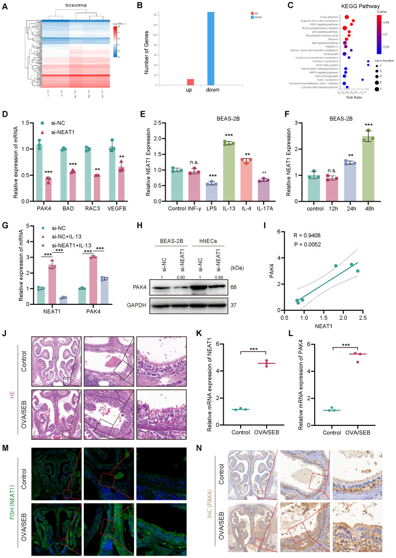
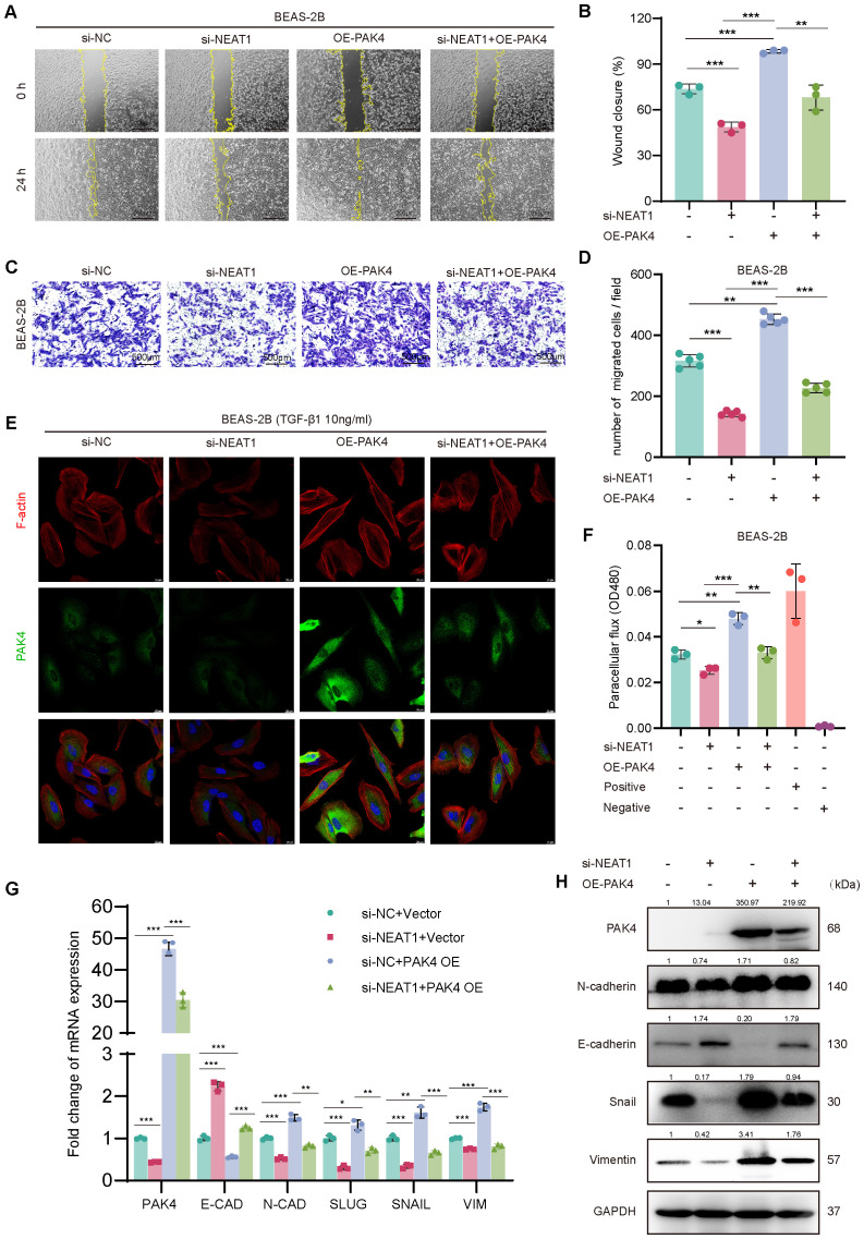
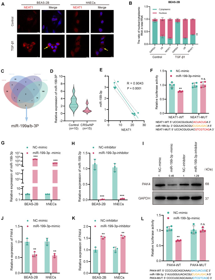
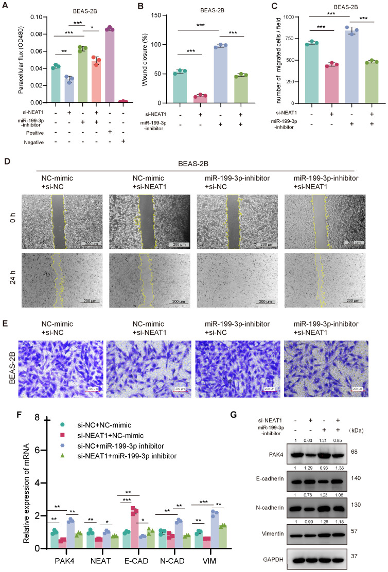
Methods: NEAT1 expression was assessed in nasal epithelial cells from CRSwNP patients using qPCR and FISH. Primary human nasal epithelial cells and BEAS-2B cells were subjected to NEAT1 knockdown via siRNA. Cell migration, barrier function, and cytoskeletal dynamics were evaluated through scratch assays, Transwell migration, FITC-Dextran permeability testing, and phalloidin staining. EMT marker expression was analyzed via Western blotting and immunofluorescence. Transcriptome sequencing identified PAK4 as a downstream effector. In vivo validation was performed using a mouse nasal polyp model, and molecular interactions among NEAT1, miR-199-3p, and PAK4 were confirmed via dual-luciferase reporter assays. Rescue experiments further elucidated mechanistic pathways.

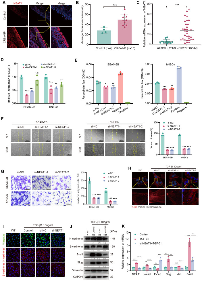
Results: In comparison to controls, NEAT1 expression was significantly elevated in the epithelial tissues of CRSwNP. NEAT1 knockdown inhibited cell migration, enhanced epithelial barrier integrity, and reversed EMT-associated cytoskeletal remodeling. E-cadherin levels increased, while N-cadherin and vimentin decreased. Transcriptomic and functional analyses identified PAK4 as a NEAT1-regulated target. NEAT1 was shown to sponge miR-199-3p, thereby relieving its inhibitory effect on PAK4. Overexpression of miR-199-3p suppressed PAK4 and mitigated EMT-related changes induced by NEAT1.

Conclusion: NEAT1 promotes EMT in nasal polyp epithelial cells by modulating the miR-199-3p/PAK4 axis, highlighting its potential as a diagnostic biomarker and therapeutic target in CRSwNP.

Keywords: CRSwNP; EMT; NEAT1; PAK4; miR-199-3p; non-coding RNA.


Conflict of interest statement

The authors declare that the research was conducted in the absence of any commercial or financial relationships that could be construed as a potential conflict of interest.

Figures

Figure 1
Figure 1
NEAT1 is upregulated in CRSwNP and promotes epithelial–mesenchymal transition (EMT). (A) FISH staining shows elevated NEAT1 expression (red) in CRSwNP tissues, primarily localized in the cytoplasm. Nuclei were counterstained with DAPI (blue). (B) Quantification of average fluorescence intensity in control (n = 4) and CRSwNP (n = 10) tissues. (C) qPCR analysis of NEAT1 expression in nasal tissues from control (n = 12) and CRSwNP patients (n = 32). (D) Validation of NEAT1 knockdown in BEAS-2B and hNECs after siRNA transfection. (E) FITC-dextran assay showing improved epithelial barrier function after NEAT1 knockdown. (F) Wound healing assay demonstrating reduced migration upon NEAT1 knockdown at 48 h (G) Transwell migration assay confirming reduced cell migration in both BEAS-2B and hNECs. (H) Phalloidin staining (Actin-Tracker Red-Rhodamine) showing reduced TGF-β1-induced stress fiber formation after NEAT1 knockdown. (I) Immunofluorescence staining of E-cadherin (red) and vimentin (green) in BEAS-2B cells. (J) Western blot analysis of EMT markers in control, TGF-β1-treated, and NEAT1 knockdown cells. (K) qPCR analysis of EMT-related genes after indicated treatments. Data are shown as mean ± SD; * p < 0.05, **p < 0.01, ***p < 0.001. n.s., not significant.
Figure 2
Figure 2
NEAT1 regulates PAK4 expression via inflammatory stimuli and is upregulated in vivo. (A) Heatmap of differentially expressed genes in BEAS-2B cells after NEAT1 knockdown; si-NEAT1–3 was excluded as an outlier. (B) Bar graph showing the number of upregulated and downregulated genes. (C) KEGG enrichment analysis highlighting significant enrichment in the focal adhesion pathway. (D) qPCR validation of selected downregulated genes after NEAT1 knockdown. (E, F) qPCR analysis of NEAT1 expression in BEAS-2B cells after stimulation with inflammatory cytokines (E) and time-course of IL-13 treatment (F). (G) NEAT1 and PAK4 expression following IL-13 stimulation and/or NEAT1 knockdown. (H) Western blot showing PAK4 protein levels in BEAS-2B and hNECs. (I) Pearson correlation analysis of NEAT1 and PAK4 mRNA levels under IL-13 stimulation. (J) HE staining of nasal mucosa from control and OVA/SEB-induced polyp model mice. (K, L) qPCR analysis of Neat1 and PAK4 mRNA levels in mouse nasal tissue. (M) FISH detection of Neat1 expression in control and model mice. (N) IHC staining of PAK4 in nasal tissues. Data are presented as mean ± SD; **p < 0.01, ***p < 0.001. n.s., not significant.
Figure 3
Figure 3
PAK4 overexpression reverses the inhibitory effects of NEAT1 knockdown on epithelial cell migration and EMT. (A) Wound healing assay of BEAS-2B cells under different treatment conditions (si-NC, si-NEAT1, OE-PAK4, si-NEAT1 + OE-PAK4) at 0 h and 24 h (B) Quantification of wound closure percentage. (C) Transwell assay showing migrated BEAS-2B cells under the same treatments. (D) Quantification of migrated cell numbers per field. (E) Immunofluorescence staining of F-actin (red), PAK4 (green), and nuclei (DAPI, blue) after TGF-β1 (10 ng/mL) stimulation. (F) Paracellular flux (FITC-dextran permeability assay) indicating epithelial barrier integrity. (G) qPCR analysis of EMT-related gene expression under different treatment groups. (H) Western blot analysis of PAK4, E-cadherin, N-cadherin, Snail, and Vimentin protein levels. GAPDH served as a loading control. Data are presented as mean ± SD; *p < 0.05, **p < 0.01, ***p < 0.001.
Figure 4
Figure 4
NEAT1 sponges miR-199-3p to regulate PAK4 via a ceRNA mechanism. (A) RNA-FISH showing NEAT1 localization (red) in BEAS-2B and hNECs, with nuclei counterstained by DAPI (blue); TGF-β1 stimulation increased NEAT1 expression. (B) QPCR analysis of NEAT1 distribution in nuclear and cytoplasmic RNA fractions. (C) Venn diagram integrating ENCORI, TargetScan, and miRDB predicting miR-199-3p as a common target of NEAT1 and PAK4. (D) Violin plot of miR-199-3p expression in CRSwNP (n = 15) vs. control (n = 10) tissues. (E) Pearson correlation analysis showing inverse relationship between NEAT1 and miR-199-3p expression. (F) Dual-luciferase assay validating direct binding of miR-199-3p to NEAT1-WT but not NEAT1-MUT. (G, H) qPCR validation of miR-199-3p overexpression and knockdown in BEAS-2B and hNECs. (J, K) qPCR showing miR-199-3p mimic decreased PAK4 expression, while inhibitor increased it. (I) Western blot confirming miR-199-3p regulation of PAK4 protein levels. (L) Luciferase assay showing miR-199-3p directly targets PAK4 3′UTR; no effect seen in mutant construct. Data are shown as mean ± SD; *p < 0.05, **p < 0.01, ***p < 0.001, n.s., not significant.
Figure 5
Figure 5
miR-199-3p reverses the pro-EMT effects of PAK4 overexpression in BEAS-2B cells. (A) Wound healing assay at 0 h and 24 h showing enhanced cell migration with PAK4 overexpression, which was reversed by co-transfection of miR-199-3p mimic. (B) Quantification of wound closure percentage. (C) Transwell migration assay showing increased migration with PAK4-OE, suppressed by miR-199-3p. (D) Quantification of migrated cells per field. (E) FITC-dextran permeability assay showing PAK4-induced barrier dysfunction and its reversal by miR-199-3p mimic. (F) Phalloidin staining (F-actin in red, PAK4 in green, nuclei in blue) revealing that PAK4 induces cytoskeletal stress fiber formation, which is reduced by miR-199-3p mimic. Data are shown as mean ± SD; *p < 0.05, **p < 0.01, ***p < 0.001.
Figure 6
Figure 6
NEAT1 regulates EMT through the miR-199-3p/PAK4 axis in BEAS-2B cells. (A) FITC-dextran assay confirms that NEAT1 knockdown improves barrier function, which is disrupted by miR-199-3p inhibition. (B–E) Wound healing and Transwell migration assays show that miR-199-3p inhibition reverses NEAT1 knockdown–induced suppression of migration, while subsequent NEAT1 silencing rescues the migratory phenotype. (F, G) qPCR and Western blot analysis show that NEAT1 knockdown upregulates E-cadherin and suppresses PAK4, N-cadherin, Vimentin, Snail, and Slug; miR-199-3p inhibitor reverses this trend. Data are shown as mean ± SD; *p < 0.05, **p < 0.01, ***p < 0.001.
Figure 7
Figure 7
The mechanism diagram of NEAT1 regulating EMT changes in CRSwNP. LncRNA NEAT1 promotes epithelial-mesenchymal transformation of nasal polyp through the up-regulation of PAK4 axis by adsorption of miR-199-3.

Similar articles

References

    1. Mullol J, Bachert C, Amin N, Desrosiers M, Hellings PW, Han JK, et al. Olfactory outcomes with dupilumab in chronic rhinosinusitis with nasal polyps. J Allergy Clin Immunol Pract. (2022) 10:1086–95.e5. doi: 10.1016/j.jaip.2021.09.037 - DOI - PubMed
    1. Papacharalampous GX, Constantinidis J, Fotiadis G, Zhang N, Bachert C, Katotomichelakis M. Chronic rhinosinusitis with nasal polyps (CRSwNP) treated with omalizumab, dupilumab, or mepolizumab: A systematic review of the current knowledge towards an attempt to compare agents’ efficacy. Int Forum Allergy Rhinol. (2024) 14:96–109. doi: 10.1002/alr.23234 - DOI - PubMed
    1. Stevens WW, Schleimer RP, Kern RC. Chronic rhinosinusitis with nasal polyps. J Allergy Clin Immunol Pract. (2016) 4:565–72. doi: 10.1016/j.jaip.2016.04.012 - DOI - PMC - PubMed
    1. Peters AT, Tan BK, Stevens WW. Consultation for chronic rhinosinusitis with nasal polyps and asthma: clinical presentation, diagnostic workup, and treatment options. J Allergy Clin Immunol Pract. (2024) 12:2898–905. doi: 10.1016/j.jaip.2024.07.019 - DOI - PMC - PubMed
    1. Bachert C, Han JK, Wagenmann M, Hosemann W, Lee SE, Backer V, et al. EUFOREA expert board meeting on uncontrolled severe chronic rhinosinusitis with nasal polyps (CRSwNP) and biologics: Definitions and management. J Allergy Clin Immunol. (2021) 147:29–36. doi: 10.1016/j.jaci.2020.11.013 - DOI - PubMed

MeSH terms

LinkOut - more resources