. 2025 Jun 30:16:1585335.
doi: 10.3389/fmicb.2025.1585335. eCollection 2025.

Screening of host proteins interacting with the African swine fever virus outer membrane protein CD2v

Affiliations

Screening of host proteins interacting with the African swine fever virus outer membrane protein CD2v

Xiong-Nan Chen et al. Front Microbiol. .

Abstract

Background: The African swine fever virus (ASFV) is a highly pathogenic double-stranded DNA virus that poses a significant threat to the global swine industry. Although the CD2v protein encoded by ASFV is a key factor in viral immune evasion and pathogenicity, the mechanism underlying its interaction with host proteins remains unclear.

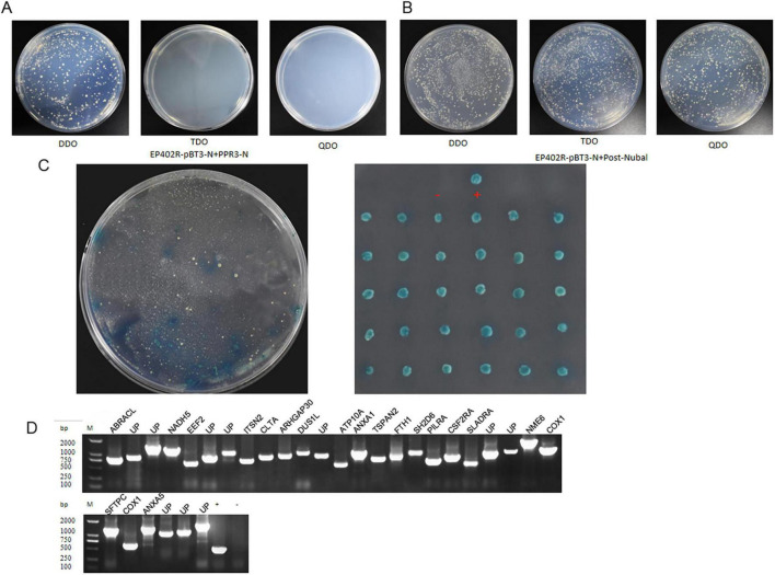
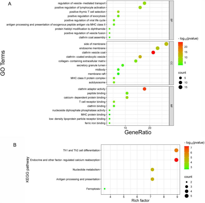
Methods: The aim of this study was to identify a range of potential host proteins that interact with the CD2v protein using the membrane yeast two-hybrid technology.

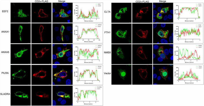
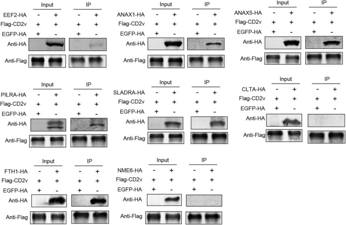
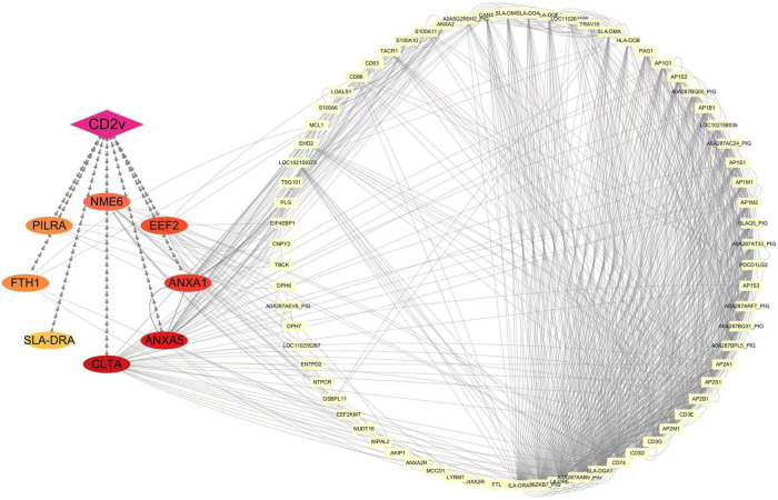
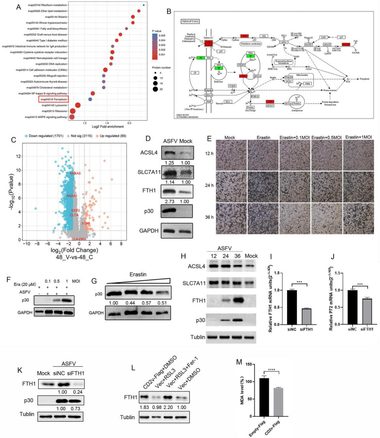
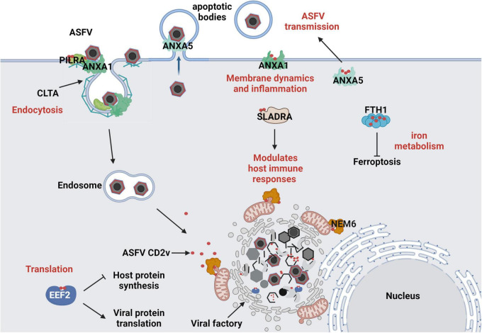
Results: Through subsequent validation experiments and functional analyses, we discovered that these proteins are involved in critical cellular processes such as translational regulation, inflammatory responses, immune signaling, and iron metabolism. Furthermore, interaction network and functional enrichment analyses revealed that ASFV might influence host cell functions through multiple pathways to facilitate viral replication.

Conclusion: This study provides new insights into the pathogenic mechanisms of ASFV and offers valuable clues for identifying antiviral targets.

Keywords: African swine fever virus; CD2v; dual membrane system; interaction network; membrane protein interaction.


Conflict of interest statement

The authors declare that the research was conducted in the absence of any commercial or financial relationships that could be construed as a potential conflict of interest.

Figures

FIGURE 1
FIGURE 1
The DUAL membrane system identified 30 potential host proteins that may interact with the ASFV CD2v protein. (A) Yeast cells co-transformed with the bait plasmid CD2v-pBT3 and the empty prey plasmid pPR3-N were plated on DDO (SD/-Leu/-Trp)/X-α-Gal, TDO (SD/-Leu/-Trp/-His)/X-α-Gal, and QDO (SD/-Leu/-Trp/-His/-Ade)/X-α-Gal selective media. (B) Yeast cells co-transformed with the bait plasmid CD2v-pBT3 and the autoactivation-positive control POST-Nubal were plated on DDO, TDO, and QDO selective media. (C) Yeast cells co-transformed with the bait plasmid CD2v-pBT3 and prey host protein plasmids pPR3-N were plated on TDO selective media, and blue colonies were subsequently plated on QDO selective media. “+” indicates a positive interaction control (co-transformation of PTSU2-APP and PNubG-Fe65), whereas “–” represents a negative interaction control (co-transformation of PTSU2-APP and empty pPR3-N). (D) PCR validation was performed using universal primers for the prey plasmids. Lanes 1–30 represent PCR products from positive clones. Lane “+” is the positive control (PCR using pPR3-N as the template), lane “–” is the negative control, and Lane “UP” is Uncharacterized protein, (PCR using water as the template and all host protein name abbreviations can be found in Supplementary Table S2).
FIGURE 2
FIGURE 2
Co-localization of ASFV CD2v and host proteins. (A) WSL cells were transfected with ASFV CD2v-FLAG (1.25 μg) and host proteins (EEF2, ANXA1, ANXA5, PILRA, SLADRA, CLTA, FTH1, and NME6), HA (1.25 μg), or empty vector control (2.5 μg) plasmids for 24 h, and then the cells were fixed with 4% PFA for immunofluorescence. Immunofluorescence confocal microscopy shows colocalization of the FLAG-tag protein (red) and HA-tag protein (green). Scale bars, 5 μm.
FIGURE 3
FIGURE 3
ASFV CD2v interacts with host proteins. HEK-293T cells were transfected with CD2v-FLAG (2.5 μg) and host proteins (EEF2, ANXA1, ANXA5, PILRA, SLADRA, CLTA, FTH1, and NME6), HA (2.5 μg), or empty vector control (2.5 μg) plasmids for 24 h, followed by co-IP with anti-FLAG magnetic beads and immunoblotting with anti-HA and FLAG antibody.
FIGURE 4
FIGURE 4
Interaction network of ASFV CD2v with host proteins. Protein-protein interactions were analyzed using the STRING database with an interaction score threshold of > 0.4. A total of 48 host proteins were selected to construct the interaction network. Each node represents a protein: on the left are proteins validated in this study interacting with ASFV CD2v, whereas on the right are additional host proteins interacting with the validated ones.
FIGURE 5
FIGURE 5
GO and KEGG pathway enrichment analysis. (A) GO enrichment analysis of biological processes (BP), cellular components (CC), and molecular functions (MF). The x-axis represents the gene ratio, and the y-axis shows the GO terms. (B) KEGG pathway enrichment analysis, where the x-axis indicates the enrichment factor, and the y-axis represents the metabolic signaling pathways.
FIGURE 6
FIGURE 6
ASFV infection regulates cellular sensitivity to ferroptosis. (A) Bubble chart of pathway analysis from 4D proteomics in ASFV-infected cells. Each bubble represents a pathway (top 20 by p-value), with size indicating the number of differential proteins. The x-axis shows pathway impact, and the y-axis shows p-value. (B) Differential proteins in the ferroptosis pathway, with red indicating upregulation and green indicating downregulation. (C) Volcano plot of differentially expressed proteins, highlighting six validated host proteins (EEF2, ANAX1, ANAX5, SLADRA, CLTA, FTH1). (D) ACSL4, SLC7A11, FTH1, and p30 proteins expression in ASFV-infected (MOI = 1) PAM cells at 24 hpi. GAPDH serves as control. (E), WSL cells were inoculated with ASFV (0.1, 0.5, and 1 MOI, 2 h) and incubated in maintenance medium containing erastin for 12, 24, and 36 hpi, cell death was observed under bright-field microscopy (F) ASFV p30 protein expression in ASFV-infected (0.1, 0.5, and 1 MOI) PAM cells at 36 hpi. GAPDH serves as control. (G) PAM cells were inoculated with ASFV (0.1 MOI, 2 h) and incubated in erastin-containing maintenance medium (1, 2.5, 5, and 10 μM), ASFV p30 protein expression levels were determined through Western blot at 36 hpi. (H) WSL cells were inoculated with ASFV MOI = 1, 2 h) for 12, 24, and 36 hpi. Western blotting of ACSL4, SLC7A11, FTH1, and p30 proteins expression levels were determined through Western blot; α-tublin served as control. (I,J) PAM cells were transfected with FTH1 protein-targeting siRNAs or negative control (NC) siRNA (siNC) for 48 h, followed by infection with ASFV (0.1 MOI). mRNA expression of FTH1 and viral P72 protein was determined by qRT-PCR at 24 hpi. (K) Western blotting of FTH1 and p30 proteins from ASFV-infected (1 MOI) siFTH1 and siNC PAM cells at 24 hpi; α-tublin serves as control. (L) WSL cells were transfected with CD2v-Flag plasmids or empty vector control for 24 h and treated with RSL3 (10 μM, a ferroptosis activator) or RSL3 + Ferrostatin-1 (10 μM, a ferroptosis inhibitor) or DMSO for 12 h. (M) WSL cells were transfected with CD2v-Flag plasmids or an empty vector control for 24 h. MDA concentrations in the cell lysates were measured according to the manufacturer’s instructions of the kit.
FIGURE 7
FIGURE 7
Predicted Interaction Model of ASFV CD2v with Host Cells. (1) ASFV hijacks the host translation machinery by interacting with the translation initiation factor EEF2, promoting viral protein synthesis and inhibiting host protein expression. (2) ASFV regulates membrane dynamics and inflammatory responses by interacting with ANXA1, facilitating viral entry and immune evasion. By interacting with ANXA5, it interferes with host membrane-associated functions, aiding ASFV assembly and promoting its propagation within apoptotic bodies. (3) ASFV CD2v interacts with PILRA and CLTA to promote the endocytic entry of ASFV into host cells. (4) ASFV CD2v enhances or suppresses the SLA-DR-mediated antigen presentation process of viral proteins by interacting with SLADRA, thereby modulating the host innate immune response. (5) ASFV regulates intracellular iron metabolism homeostasis through the interaction of CD2v with FTH1, promoting viral replication. (6) ASFV CD2v recruits NME6 to regulate mitochondrial function and maintain cellular homeostasis, thereby providing energy for viral DNA replication.

Similar articles

References

    1. Abkallo H. M., Hemmink J. D., Oduor B., Khazalwa E. M., Svitek N., Assad-Garcia N., et al. (2022). Co-deletion of a238l and ep402r genes from a genotype ix african swine fever virus results in partial attenuation and protection in swine. Viruses 14:2024. 10.3390/v14092024 - DOI - PMC - PubMed
    1. Alejo A., Matamoros T., Guerra M., Andres G. (2018). A proteomic atlas of the african swine fever virus particle. J. Virol. 92:e01293-18. 10.1128/JVI.01293-18 - DOI - PMC - PubMed
    1. Alkafaas S. S., Abdallah A. M., Ghosh S., Loutfy S. A., Elkafas S. S., Abdel F. N., et al. (2023). Insight into the role of clathrin-mediated endocytosis inhibitors in SARS-cov-2 infection. Rev. Med. Virol. 33:e2403. 10.1002/rmv.2403 - DOI - PMC - PubMed
    1. Berri F., Haffar G., Le V. B., Sadewasser A., Paki K., Lina B., et al. (2014). Annexin v incorporated into influenza virus particles inhibits gamma interferon signaling and promotes viral replication. J. Virol. 88 11215–11228. 10.1128/JVI.01405-14 - DOI - PMC - PubMed
    1. Castello A., Quintas A., Sanchez E. G., Sabina P., Nogal M., Carrasco L., et al. (2009). Regulation of host translational machinery by african swine fever virus. PLoS Pathog. 5:e1000562. 10.1371/journal.ppat.1000562 - DOI - PMC - PubMed

LinkOut - more resources