. 2025 Jul 15;135(14):e188492.
doi: 10.1172/JCI188492.

First-generation and preclinical evaluation of an EphA5-targeted antibody-drug conjugate in solid tumors

Affiliations

First-generation and preclinical evaluation of an EphA5-targeted antibody-drug conjugate in solid tumors

Fernanda I Staquicini et al. J Clin Invest. .

Abstract

Contemporary cancer treatment strategies are shifting toward targeted therapies to improve efficacy and minimize toxicity. Here, we report the design and preclinical evaluation of MBRC-101, a first-in-class antibody-drug conjugate (ADC) targeting EphA5, a receptor tyrosine kinase with an established role in embryonic development but not extensively studied in cancer. We show that EphA5 is expressed in multiple solid tumors, including cancers of the aerodigestive (non-small cell lung, head and neck, gastric, colon, and pancreatic) and genitourinary (bladder and ovary) tracts, as well as most breast cancer subsets (including triple-negative tumors), with limited expression in normal tissues. MBRC-101 is a humanized anti-EphA5 antibody conjugated to monomethyl auristatin E (MMAE) through a ThioBridge, thereby ensuring stable drug-to-antibody ratio and reducing off-target effects. MBRC-101 showed potent antitumor activity, achieving complete tumor regression in several patient-derived xenograft models. Preclinical Good Laboratory Practice-compliant toxicology studies in rats and nonhuman primates demonstrated that MBRC-101 is well tolerated, with observed toxicities limited to known MMAE off-target effects. These findings establish EphA5 as a therapeutic target in cancer and support the translational development of MBRC-101 as a promising ADC candidate for clinical evaluation, currently in a first-in-human multicenter investigational trial for patients with advanced solid tumors (ClinicalTrials.gov, NCT06014658).

Keywords: Cancer; Cancer immunotherapy; Oncology; Therapeutics.


Conflict of interest statement

Conflict of interest: FIS, VDO, SYK, YW, JKP, KFB, SCA, and IC are employees of MBrace Therapeutics, Inc (MBrace). WA and RP are cofounders of, consultants for, and equity holders in MBrace. RP and WA, through Rutgers University (Rutgers), have a Sponsored Research Agreement with MBrace, which is managed under the conflict-of-interest policies of Rutgers, The State University of New Jersey. RP, WA, and FIS are listed as inventors on international patent application WO2018/057703 (granted in the United States as patent 11,254,751) filed by STC.UNM and Los Alamos National Security, LLM, on technologies related to this article; and on international patent application WO2023/081766 (pending in the United States as US patent applications 18/052,479, 18/707,250 and 18/360,287) filed by Rutgers and MBrace on technologies related to this article.

Figures

Figure 1
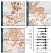
Figure 1. EphA5 expression in multiple cancer indications.
Illustrative images of EphA5 expression in (A) breast, (B) lung, and (C) colon and pancreatic cancers. In all cases, EphA5 expression is observed in both the cytoplasm and on the membrane of cells, with expression restricted to cancer cells and not the surrounding adjacent tissue. (D) Percentage of EphA5-expressing cancer cells detected in archival tumor samples. Scale bar, 200 μm. Data presented as mean ± SEM. ACC, adenocarcinoma; HR, hormone receptor; SCC, squamous cell carcinoma; TNBC, triple-negative breast cancer.
Figure 2
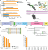
Figure 2. Identification and structural analysis of the binding epitope recognized by the anti-EphA5 antibody.
(A) Antibody-binding epitope of EphA5 mapped by site-directed alanine scanning. (B) AlphaFold-predicted, 3-dimensional, atomic-level structure of full-length human EphA5 containing the identified epitope (green) and excluding low-confidence regions. Consensus protein topology prediction was determined by TOPCONS. The inset visualizes the critical amino acid residues responsible for binding to the anti-EphA5 antibody (green, major binding contributor; orange, binding contributor; yellow, secondary binding contributor). (C) Schematic representation of the identified epitope (residues R306–E330) relative to the established domains of human EphA5. (D) Multiple sequence alignment of the epitope (residues R306–E330) derived from relevant EphA5 orthologs (green, major binding contributor; orange, binding contributor; yellow, secondary binding contributor). (E) Conformational similarity between the extracellular region (residues S58–P561) or epitope (residues R306–E330) of human EphA5 with its corresponding ortholog following best-fit superimposition of AlphaFold-predicted 3-dimensional atomic-level structures. Root mean square deviation was calculated for paired Cα atoms of all corresponding residues. (F) MPA confirmed dose-dependent binding specificity of the anti-EphA5 antibody to EphA5. (G) All other members of the Eph family of receptors were evaluated and did not show cross-reactivity with the anti-EphA5 antibody. SAM, sterile alpha motif.
Figure 3
Figure 3. Binding specificity of the anti-EphA5 antibody and cytotoxicity of MBRC-101 in cells.
(A and B) Flow cytometry analysis shows EphA5 expression on the surface of well-established lung cancer cells. (C) Receptor-mediated antibody internalization in EphA5-positive cells (H460). Internalization was not detected in EphA5-low cells (H226). (D) MBRC-101 consists of a humanized IgG1 mAb linked to MMAE, its cytotoxic payload, through a ThioBridge bis-sulfone conjugation unit. This unit rebridges the interchain disulfides of the antibody and incorporates a protease-cleavable linker, resulting in an ADC with a homogeneous DAR4. (E) MBRC-101 kills EphA5+ cells in a concentration-dependent manner.
Figure 4
Figure 4. MBRC-101 antitumor activity.
(A) IHC of tissue sections from a PDX model of TNBC shows moderate and heterogenous expression of EphA5. Scale bar, 200 μm. Weight of tumors collected at the end of the dose-range-finding study confirmed antitumor activity of MBRC-101 at clinically relevant dose levels. Data are presented as means ± SEM. Statistical tests were ordinary 1-way ANOVA coupled with post hoc Tukey’s multiple comparisons test. (B) Histological analysis of tissue sections of tumors collected at the end of treatment. The pan-tumor marker PAN-CK was used to detect cancer cells, and Ki-67 served to detect proliferating cancer cells (arrows). The Ki-67 proliferation index was assessed by point counting from 500 to 1,000 cells and reported as percent positive cells. Scale bar, 20 μm. (C) MBRC-101 antitumor activity compared with sacituzumab govitecan, an FDA-approved ADC for the treatment of TNBC. Data are presented as means ± SEM. Statistical tests were 2-way ANOVA coupled with post hoc Tukey’s multiple comparisons test among treatment groups.
Figure 5
Figure 5. Antitumor activity of MBRC-101 in various PDX models of human cancers.
(A and B) Head-and-neck squamous carcinoma (ordinary 1-way ANOVA and Tukey’s multiple comparisons test). (C and D) Lung squamous cell carcinoma (unpaired t test). (E and F) Lung adenocarcinoma (unpaired t test). (G and H) TNBC (unpaired t test). Treatments were given weekly for 2 weeks and up to 4 weeks (asterisks). In all cases, complete tumor regression without regrowth was achieved at 5 mg/kg or 10 mg/kg, with no observed weight loss. All data are presented as means ± SEM.
Figure 6
Figure 6. Toxicokinetics of MBRC-101 in rats and monkeys.
(A) Total mAb, (B) total ADC, and (C) unconjugated MMAE concentrations in Sprague-Dawley rats after administration of MBRC-101 via i.v. bolus (days 1 and 22) at doses of 10, 20, and 30 mg/kg. (D) Total mAb, (E) total ADC, and (F) unconjugated MMAE concentrations in cynomolgus monkeys after administration of MBRC-101 via i.v. bolus (days 1 and 22) at doses of 5, 7.5, and 10 mg/kg. All data are presented as means ± SD. BLQ, below the limit of quantification.

References

    1. Colomer R, et al. Herceptin: from the bench to the clinic. Cancer Invest. 2001;19(1):49–56. doi: 10.1081/CNV-100000074. - DOI - PubMed
    1. Lambert JM, Chari RV. Ado-trastuzumab Emtansine (T-DM1): an antibody-drug conjugate (ADC) for HER2-positive breast cancer. J Med Chem. 2014;57(16):6949–6964. doi: 10.1021/jm500766w. - DOI - PubMed
    1. Cardillo TM, et al. Humanized anti-Trop-2 IgG-SN-38 conjugate for effective treatment of diverse epithelial cancers: preclinical studies in human cancer xenograft models and monkeys. Clin Cancer Res. 2011;17(10):3157–3169. doi: 10.1158/1078-0432.CCR-10-2939. - DOI - PMC - PubMed
    1. Starodub AN, et al. First-in-human trial of a novel Anti-Trop-2 antibody-SN-38 conjugate, sacituzumab govitecan, for the treatment of diverse metastatic solid tumors. Clin Cancer Res. 2015;21(17):3870–3878. doi: 10.1158/1078-0432.CCR-14-3321. - DOI - PMC - PubMed
    1. Breij EC, et al. An antibody-drug conjugate that targets tissue factor exhibits potent therapeutic activity against a broad range of solid tumors. Cancer Res. 2014;74(4):1214–1226. doi: 10.1158/0008-5472.CAN-13-2440. - DOI - PubMed

Associated data