. 2025 Jul 15;38(5):130.
doi: 10.1007/s13577-025-01262-3.

Metformin promotes osteogenic differentiation of human periodontal ligament stem cells via KLF2-mediated activation of miR-181a-5p under lipopolysaccharide stimulation

Affiliations

Metformin promotes osteogenic differentiation of human periodontal ligament stem cells via KLF2-mediated activation of miR-181a-5p under lipopolysaccharide stimulation

Xu Zhang et al. Hum Cell. .

Abstract

Periodontal ligament stem cells (PDLSCs) constitute a promising source for successful periodontal regeneration. This study aims to explore roles of metformin, krüppel-like factor 2 (KLF2), and miR-181a-5p in mediating osteogenic differentiation of human PDLSCs (hPDLSCs) following lipopolysaccharide (LPS) stimulation. The osteogenic differentiation potential of hPDLSCs isolated from human premolar root samples were examined by alkaline phosphatase (ALP) staining, ALP activity assay, Alizarin red S staining, and Western blotting of osteogenic markers. Metformin pretreatment at dose of 100 μM significantly resulted in increased ALP activity, elevated protein expressions of osteogenic markers, and more generated mineralized matrix in hPDLSCs with LPS stimulation. KLF2 and miR-181a-5p were found to be increased by metformin pretreatment at dose of 100 μM in hPDLSCs with stimulation but not in hPDLSCs without LPS stimulation. The interaction between the KLF2 and the promoter of miR-181a-5p was noted by the dual-luciferase reporter assay. KLF2 knockdown or miR-181a-5p inhibition notably abrogated the improvements of osteogenic differentiation by metformin pretreatment in LPS-stimulated hPDLSCs. The findings of the study indicate metformin protects hPDLSCs against impaired osteogenic differentiation of hPDLSCs after LPS stimulation by KLF2-mediated activation of miR-181a-5p under inflammation conditions.

Keywords: MicroRNA; Periodontitis; Regeneration; Stem cells; Tissue engineering.


Conflict of interest statement

Declarations. Conflict of interest: No conflict of interest is declared by the authors. Ethical approval and consent to participation: The study protocol complied with the principles of the Declaration of Helsinki and was approved by the Ethics Committee of Changsha Stomatological Hospital. All participants or their guardians had signed the informed consent form prior to recruitment.

Figures

Fig. 1
Fig. 1
Identification of hPDLSCs. A, Representative images (200 ×; 400 ×) of hPDLSC subculture. B, Representative images (200 ×; 400 ×) of osteogenic differentiation of hPDLSCs evaluated by Alizarin red S staining. C, Representative images (200 ×; 400 ×) of adipogenic differentiation of hPDLSCs evaluated by oil red O staining. D, The phenotype analysis of hPDLSCs using flow cytometry
Fig. 2
Fig. 2
Metformin promotes osteogenic differentiation in LPS-stimulated hPDLSCs. A, CCK-8 assays to detect the viability of hPDLSCs after 10-, 100-, 1000- and 2000-μM metformin treatment for 24, 48, and 72 h; results of mean with s.d. were yielded from six biological and technical replicates and analyzed by two-way ANOVA plus Bonferroni post-hoc test; *P < 0.05 compared to other concentrations of metformin. B, Representative images (400 ×) of ALP-positive cells and the ALP activity in LPS-stimulated hPDLSCs with 10-, 100-, and 1000-μM metformin pretreatment after 14 day osteogenic induction. C, Western blotting detections of RUNX2 and OPN in LPS-stimulated hPDLSCs with 10-, 100-, and 1000-μM metformin pretreatment after 7 day osteogenic induction. D, Representative images (400 ×) of Alizarin red-stained nodules and the quantitative analysis (OD value at 562 nm) of mineralized matrix in LPS-stimulated hPDLSCs with 10-, 100-, and 1000-μM metformin pretreatment after 21 day osteogenic induction. For panel B-D, results of mean with s.d. were yielded from six biological and technical replicates and analyzed by one-way ANOVA plus Tukey’s post-hoc test. *P < 0.05 compared to LPS-stimulated hPDLSCs without metformin pretreatment and #P < 0.05 compared to LPS-stimulated hPDLSCs with 100 μM metformin pretreatment
Fig. 3
Fig. 3
Metformin increases KLF2 expression in hPDLSCs under inflammation condition. A, Western blotting detections of KLF2 protein in control hPDLSCs, LPS-stimulated hPDLSCs with or without 10-, 100-, and 1000-μM metformin pretreatment. B, Western blotting detections of KLF2 protein in control hPDLSCs with 10-, 100-, and 1000-μM metformin treatment. C, Representative images (200 ×) showing immunofluorescence staining of the intracellular localization of KLF2 in control hPDLSCs, LPS-stimulated hPDLSCs with or without 10-, 100-, and 1000-μM metformin pretreatment. Results of mean with s.d. were yielded from six biological and technical replicates and analyzed by one-way ANOVA plus Tukey’s post-hoc test; *P < 0.05 compared to control hPDLSCs and #P < 0.05 compared to LPS-stimulated hPDLSCs with 100 μM metformin pretreatment
Fig. 4
Fig. 4
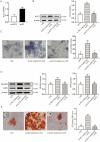
The effects of metformin on LPS-stimulated hPDLSCs are KLF2-dependent. A, The efficiency of KLF2 knockdown by si-KLF2, determined by qRT-PCR; results of mean with s.d. were yielded from six biological and technical replicates and analyzed by unpaired t test; ▲ P < 0.05 compared to si-KLF2. B, Western blotting detections of KLF2 protein in LPS-stimulated hPDLSCs with or without si-KLF2 pre-transfection and metformin pretreatment. C, Representative images (400 ×) of ALP-positive cells and the ALP activity in LPS-stimulated hPDLSCs with or without si-KLF2 pre-transfection and metformin pretreatment after 14 day osteogenic induction. D, Western blotting detections of RUNX2 and OPN in LPS-stimulated hPDLSCs with or without si-KLF2 pre-transfection and metformin pretreatment. E, Representative images (400 ×) of Alizarin red-stained nodules and the quantitative analysis (OD value at 562 nm) of mineralized matrix in LPS-stimulated hPDLSCs with or without si-KLF2 pre-transfection and metformin pretreatment. &P < 0.05 compared to si-NC + metformin + LPS. For panel B-E, results of mean with s.d. were yielded from six biological and technical replicates and analyzed by one-way ANOVA plus Tukey’s post-hoc test
Fig. 5
Fig. 5
KLF2-mediated activation of miR-181a-5p is involved in metformin effects on LPS-stimulated hPDLSCs. A, The qRT-PCR detections of miR-181a-5p in control hPDLSCs with or without 10-, 100-, and 1000-μM metformin treatment, in LPS-stimulated hPDLSCs with or without 10-, 100-, and 1000-μM metformin pretreatment; *P < 0.05 compared to control hPDLSCs and #P < 0.05 compared to LPS-stimulated hPDLSCs with 100 μM metformin pretreatment. B, Western blotting detections of KLF2 protein in control hPDLSCs transfected with oe-KLF2 and miR-181a-5p inhibitor. C, The qRT-PCR detections of miR-181a-5p in control hPDLSCs transfected with oe-KLF2 and miR-181a-5p inhibitor; ▲ P < 0.05 compared to oe-KLF2 + miR-181a-5p inhibitor. D, Three putative binding positions of KLF2 in the miR-181a-5p promoter based on the JASPAR database analysis and named as sites 1–3. E, Dual-luciferase reporter assay was performed to confirm the target area of KLF2 in the promoter of miR-181a-5p; results of mean with s.d. were yielded from six biological and technical replicates and analyzed by unpaired t test; ▼ P < 0.05 compared to si-NC. F, The qRT-PCR detections of miR-181a-5p in LPS-stimulated hPDLSCs with or without oe-KLF2, miR-181a-5p inhibitor pre-transfection, and metformin pretreatment. G, Representative images (400 ×) of ALP-positive cells and the ALP activity in LPS-stimulated hPDLSCs with or without oe-KLF2, miR-181a-5p inhibitor pre-transfection, and metformin pretreatment after 14-day osteogenic induction. H, Western blotting detections of RUNX2 and OPN in LPS-stimulated hPDLSCs with or without oe-KLF2, miR-181a-5p inhibitor pre-transfection, and metformin pretreatment after 7 day osteogenic induction. I, Representative images (400 ×) of Alizarin red-stained nodules and the quantitative analysis (OD value at 562 nm) of mineralized matrix in LPS-stimulated hPDLSCs with or without oe-KLF2, miR-181a-5p inhibitor pre-transfection, and metformin pretreatment after 21 day osteogenic induction. & P < 0.05 compared to oe-NC + miR-181a-5p inhibitor + metformin + LPS. For panel A-C and F-I, results of mean with s.d. were yielded from six biological and technical replicates and analyzed by one-way ANOVA plus Tukey’s post-hoc test

Similar articles

References

    1. Fischer NG, de Souza Araujo IJ, Daghrery A, et al. Guidance on biomaterials for periodontal tissue regeneration: fabrication methods, materials and biological considerations. Dent Mater. 2025;41:283–305. - PubMed
    1. Wang X, Chen J, Tian W. Strategies of cell and cell-free therapies for periodontal regeneration: the state of the art. Stem Cell Res Ther. 2022;13:536. - PMC - PubMed
    1. Wen S, Zheng X, Yin W, et al. Dental stem cell dynamics in periodontal ligament regeneration: from mechanism to application. Stem Cell Res Ther. 2024;15:389. - PMC - PubMed
    1. Trubiani O, Pizzicannella J, Caputi S, et al. Periodontal Ligament stem cells: current knowledge and future perspectives. Stem Cells Dev. 2019;28:995–1003. - PubMed
    1. Liang Q, Du L, Zhang R, Kang W, Ge S. Stromal cell-derived factor-1/Exendin-4 cotherapy facilitates the proliferation, migration and osteogenic differentiation of human periodontal ligament stem cells in vitro and promotes periodontal bone regeneration in vivo. Cell Prolif. 2021;54:e12997. - PMC - PubMed

MeSH terms

LinkOut - more resources