

Placenta segmentation redefined: review of deep learning integration of magnetic resonance imaging and ultrasound imaging
- PMID: 40663247
- PMCID: PMC12263505
- DOI: 10.1186/s42492-025-00197-8
Placenta segmentation redefined: review of deep learning integration of magnetic resonance imaging and ultrasound imaging
Abstract
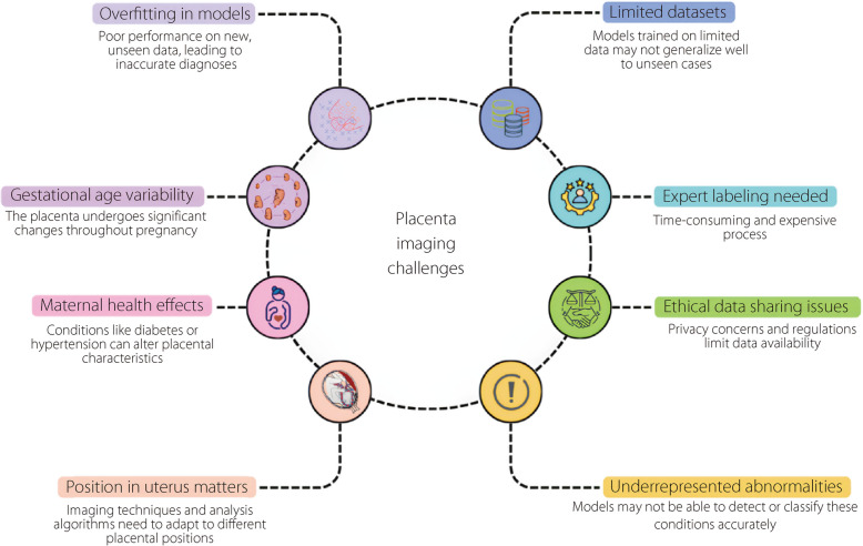
Placental segmentation is critical for the quantitative analysis of prenatal imaging applications. However, segmenting the placenta using magnetic resonance imaging (MRI) and ultrasound is challenging because of variations in fetal position, dynamic placental development, and image quality. Most segmentation methods define regions of interest with different shapes and intensities, encompassing the entire placenta or specific structures. Recently, deep learning has emerged as a key approach that offer high segmentation performance across diverse datasets. This review focuses on the recent advances in deep learning techniques for placental segmentation in medical imaging, specifically MRI and ultrasound modalities, and cover studies from 2019 to 2024. This review synthesizes recent research, expand knowledge in this innovative area, and highlight the potential of deep learning approaches to significantly enhance prenatal diagnostics. These findings emphasize the importance of selecting appropriate imaging modalities and model architectures tailored to specific clinical scenarios. In addition, integrating both MRI and ultrasound can enhance segmentation performance by leveraging complementary information. This review also discusses the challenges associated with the high costs and limited availability of advanced imaging technologies. It provides insights into the current state of placental segmentation techniques and their implications for improving maternal and fetal health outcomes, underscoring the transformative impact of deep learning on prenatal diagnostics.
Keywords: Deep learning; Magnetic resonance imaging; Placenta; Segmentation; Ultrasound.
© 2025. The Author(s).
Conflict of interest statement
Declarations. Competing interests: The authors declare no competing of interest.
Figures


Similar articles
-
Contrast-enhanced ultrasound using SonoVue® (sulphur hexafluoride microbubbles) compared with contrast-enhanced computed tomography and contrast-enhanced magnetic resonance imaging for the characterisation of focal liver lesions and detection of liver metastases: a systematic review and cost-effectiveness analysis.Health Technol Assess. 2013 Apr;17(16):1-243. doi: 10.3310/hta17160. Health Technol Assess. 2013. PMID: 23611316 Free PMC article.
-
Management of urinary stones by experts in stone disease (ESD 2025).Arch Ital Urol Androl. 2025 Jun 30;97(2):14085. doi: 10.4081/aiua.2025.14085. Epub 2025 Jun 30. Arch Ital Urol Androl. 2025. PMID: 40583613 Review.
-
Short-Term Memory Impairment.2024 Jun 8. In: StatPearls [Internet]. Treasure Island (FL): StatPearls Publishing; 2025 Jan–. 2024 Jun 8. In: StatPearls [Internet]. Treasure Island (FL): StatPearls Publishing; 2025 Jan–. PMID: 31424720 Free Books & Documents.
-
Influence of early through late fusion on pancreas segmentation from imperfectly registered multimodal magnetic resonance imaging.J Med Imaging (Bellingham). 2025 Mar;12(2):024008. doi: 10.1117/1.JMI.12.2.024008. Epub 2025 Apr 26. J Med Imaging (Bellingham). 2025. PMID: 40291815
-
A novel UNet-SegNet and vision transformer architectures for efficient segmentation and classification in medical imaging.Phys Eng Sci Med. 2025 Jul 8. doi: 10.1007/s13246-025-01564-8. Online ahead of print. Phys Eng Sci Med. 2025. PMID: 40627277
References
-
- Ramirez Zegarra R, Ghi T (2023) Use of artificial intelligence and deep learning in fetal ultrasound imaging. Ultrasound Obstet Gynecol 62:185–194. 10.1002/uog.26130 - PubMed
-
- Zhang H, Qie Y (2023) Applying deep learning to medical imaging: a review. Appl Sci 13:10521. 10.3390/app131810521
-
- Schuhmann R, Wehler V (1971) Histologische Unterschiede an Placentazotten innerhalb der materno-fetalen Strömungseinheit. Arch Für Gynäkol 210:425–439. 10.1007/BF01628221 - PubMed
Publication types
LinkOut - more resources
Full Text Sources