. 2025 Dec;16(1):2532815.
doi: 10.1080/21505594.2025.2532815. Epub 2025 Aug 3.

Comprehensive analysis of human keratinocyte interactions with Candida albicans and Candida parapsilosis

Affiliations

Comprehensive analysis of human keratinocyte interactions with Candida albicans and Candida parapsilosis

Ádám Novák et al. Virulence. 2025 Dec.

Abstract

In recent years, microbiome studies have revealed that Candida species are common colonizers of the human skin. The distribution of species, however, varies greatly. Although C. parapsilosis is more likely to resemble skin commensals, opinions are divided, and discrepancies are present regarding C. albicans that is also often associated with cutaneous candidiasis. Therefore, we aimed to thoroughly assess the nature of skin epithelial cell - Candida interactions. To study species-specific host responses, we examined internalization, cytokine and metabolic responses in different keratinocytes (HaCaT, HPV-KER) along with host cell damage following fungal stimuli. To rigorously examine yeast-keratinocyte interactions, we applied two distinct isolates of both C. albicans (SC5314, WO-1) and C. parapsilosis (GA1, CLIB214). Comparison of the two fungi's virulence revealed that while C. albicans effectively adheres to human keratinocytes and causes subsequent damage, C. parapsilosis is unable to establish lasting physical contact and causes less harm. In terms of keratinocyte response, both cell lines showed significantly enhanced cellular (internalization), humoral (IL-6, IL-8) and metabolic responses (2-ketoglutaric acid, citric acid, threorine, hypotaurine) to C. albicans strains, while those towards C. parapsilosis remained relatively low or similar to the control condition. Under certain conditions strain preference was also detected. Of the two cell lines, HPV-KER was more sensitive, as besides interspecies differences, intraspecies differences were also measurable. These results suggest that C. albicans triggers an enhanced antifungal response, thus does not closely resemble skin commensals, like C. parapsilosis. Furthermore, HPV-KER might serve as a more applicable tool for studying keratinocyte antifungal responses.

Keywords: Keratinocyte; candida; immune response; interaction; pathogenic fungi.


Conflict of interest statement

No potential conflict of interest was reported by the author(s).

Figures

Figure 1.
Figure 1.
Adhesion of C. parapsilosis and C. albicans to (a) HaCaT and (b) HPV-KER keratinocytes 1 hour post-infection. Adhesion efficiency was determined by CFU count. MOI of 1:1 (Candida: keratinocyte cells). During the experiment the C. parapsilosis GA1 and CLIB214 strains, C. albicans SC5314 and WO-1 strains were used. ***p < 0.001 (unpaired t-test), n = 3.
Figure 2.
Figure 2.
Efficacy of pathogen internalization (% phagocytosis) in the case of HaCaT cells. Uptake efficiency of C. parapsilosis GA1, CLIB214, and C. albicans WO-1 and SC5314 cells by HaCaT keratinocytes 1 hour after co-incubation. The MOI of 5:1 (pathogen:host) was used. **p < 0.01, *p < 0.05 (unpaired t-test), n = 3. Between 9,000–10,000 individual keratinocytes were analysed per experiment using a flow cytometer coupled with image analysis tools. Representative images of yeast uptake are also shown.
Figure 3.
Figure 3.
Efficacy of pathogen internalization (% phagocytosis) in the case of HPV-KER cells. Uptake efficiency of C. parapsilosis GA1, CLIB214, and C. albicans WO-1 and SC5314 cells 1 hour after co-incubation. The MOI of 5:1 (pathogen:host) was used. **p < 0.01, *p < 0.05 (unpaired t-test), n = 2. Between 9,000–10,000 individual keratinocytes were analysed per run using a flow cytometer coupled with image analysis tools. Representative images of yeast uptake are also shown.
Figure 4.
Figure 4.
Host cell damage caused by C. albicans and C. parapsilosis in (a) HaCaT and (b) HPV-KER keratinocytes. The extent of host cell damage was determined 24 hours after infection. LDH activity was measured from the obtained supernatants. LDH activity is determined in % (mean ± standard error) and compared to the positive control (100% cell lysis). During the experiment the C. parapsilosis GA1 and CLIB214 strains, C. albicans SC5314 and WO-1 strains were used. Statistical significance was determined, where **p < 0.01; ****p < 0.0001 (unpaired t-test), compared to uninfected control cells. n = 6.
Figure 5.
Figure 5.
IL-6 production in HaCaT (a) and HPV-KER (b) cells 24 h after C. albicans and C. parapsilosis infection. During the experiment the C. parapsilosis GA1 and CLIB214 strains, C. albicans SC5314 and WO-1 strains were used. Statistical significance was determined, where *p < 0.05; **p < 0.01; ***p < 0.001 (unpaired t-test) compared to uninfected control cells. n = 3.
Figure 6.
Figure 6.
IL-8 production in HaCaT (a) and HPV-KER (b) keratinocytes after 24 h of C. albicans and C. parapsilosis infection. During the experiment the C. parapsilosis GA1 and CLIB214 strains, C. albicans SC5314 and WO-1 strains were used. Statistical significance was determined, where *p < 0.05; **p < 0.01; ***p < 0.001; ****p < 0.0001 (unpaired t-test) compared to uninfected control cells. n = 3.
Figure 7.
Figure 7.
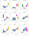
Metabolite secretion by HaCaT cells following 24 h of Candida infection. The MOI of 1:1 was used for C. parapsilosis strains, while the MOI of 1:200 was applied for C. albicans strains. The selection of a given metabolite was based on an f-test for statistical evaluation of the equality of squares of variance (the f-test threshold was 15.0). The X-axis shows the treatments, the Y-axis shows the area under the curve for a given metabolite obtained by GC-MS measurement, which is proportional to the amount of metabolite detected. MC = Medium control (nutrient only group), C = Control (cells with medium).
Figure 8.
Figure 8.
Metabolite secretion by HPV-KER cells following 24 h Candida infection. Metabolite secretion by KPV-KER cells following 24 h of Candida infection. The MOI of 1:1 was used for C. parapsilosis strains, while the MOI of 1:200 was applied for C. albicans strains. The selection of a given metabolite was based on an f-test for statistical evaluation of the equality of squares of variance (the f-test threshold was 15.0). The X-axis shows the treatments, the Y-axis shows the area under the curve for a given metabolite obtained by GC-MS measurement, which is proportional to the amount of metabolite detected. MC = Medium control (nutrient only group), C = Control (cells with medium).
Figure 9.
Figure 9.
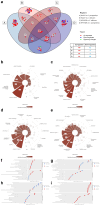
RNA sequencing results from keratinocyte cells. Venn-diagram (a) shows DEGs identified in HaCaT and HPV-KER cells after infection with C. parapsilosis CLIB214 and C. albicans SC5314 strains. Circular bar plots representing overrepresented gene ontology terms in HaCaT vs C. parapsilosis CLIB214 (b), HaCaT vs C. albicans SC5314 (c), HPV-KER vs C. parapsilosis CLIB214 (d) and HPV-KER vs C. albicans SC5314 (e) interactions. The height of the bars indicate the total number of differentially expressed genes contributing to GO terms in the corresponding semantic cluster and the colour depth is proportional to the degree of activation (z-score). Dot plots showing the results of the gene set enrichment analysis (GSEA) of HaCaT vs C. parapsilosis CLIB214 (f), HaCaT vs C. albicans SC5314 (g), HPV-KER vs C. parapsilosis CLIB214 (h) and HPV-KER vs C. albicans SC5314 (i) interactions. The significant results (below 5% FDR) were sorted by their normalized enrichment score and the top 10 terms are visualized in each ontology category. The symbols indicate positive (up-regulated) or negative (down-regulated) concordant enrichment.
Figure 10.
Figure 10.
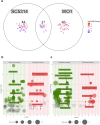
RNA sequencing results from C. albicans strains. Venn diagram of identified DEGs (a). Gene ontology analysis of C. albicans strain SC5314 (b) and WO-1 (c). Significant biological processes and molecular functions (FDR < 5%) are shown, the size of the circles is proportional to the number of genes involved in a given GO term. The z-value on the x-axis represents the theoretical activation or inhibition of the given term, based on the regulation of the involved DEGs. A GO slim set of generic fungal processes was selected and significantly overrepresented GO slim terms are highlighted on the plot.

Similar articles

References

    1. Trofa D, Gácser A, Nosanchuk JD.. Candida parapsilosis, an emerging fungal pathogen. Clin Microbiol Rev. 2008;21(4):606–20. doi: 10.1128/CMR.00013-08 - DOI - PMC - PubMed
    1. Brown GD, Denning DW, Gow NAR, et al. Hidden killers: human fungal infections. Sci Transl Med. 2012;4(165). doi: 10.1126/scitranslmed.3004404 - DOI - PubMed
    1. Gow NAR, Netea MG.. Medical mycology and fungal immunology: new Research perspectives addressing a Major world health challenge. Phil Trans R Soc B: biol Sci. 2016;371(1709):20150462. doi: 10.1098/rstb.2015.0462 - DOI - PMC - PubMed
    1. Guégan S, Lanternier F, Rouzaud C, et al. Fungal skin and soft tissue infections. Curr Opin Infect Dis. 2016;29:124–130. doi: 10.1097/QCO.0000000000000252 - DOI - PubMed
    1. Havlickova B, Czaika VA, Friedrich M. Epidemiological trends in skin mycoses worldwide. Mycoses. 2008;51(s4):2–15. doi: 10.1111/j.1439-0507.2008.01606.x - DOI - PubMed

Publication types

LinkOut - more resources