. 2025 Jul 22;122(29):e2411174122.
doi: 10.1073/pnas.2411174122. Epub 2025 Jul 15.

A genetically defined pontine nucleus essential for ingestion in mice

Affiliations

A genetically defined pontine nucleus essential for ingestion in mice

Selvee Sungeelee et al. Proc Natl Acad Sci U S A. .

Abstract

The first phase of feeding consists in the procurement of solid foods from the environment by biting, and their preparation for swallowing by chewing. These actions require the precise coordination of tens of orofacial muscles for the jaw and tongue. The seat for this motor patterning is known to reside in the reticular formation, a complex and poorly mapped region of the hindbrain, but the neuron groups involved are still elusive. Here, we characterize a group of excitatory reticular interneurons located in the supratrigeminal area that express the homeodomain transcription factor Phox2b. This nucleus-Sup5Phox2b-is premotor to both jaw-closing and jaw-opening motoneurons and receives direct input from cranial sensory afferents, motor cortex, and satiation related nuclei. Its activity differentially tracks lapping, biting, and chewing movements, suggesting its involvement in the elaboration of distinct orofacial motor patterns in vivo. Acute global activation or inhibition of Sup5Phox2b by optogenetics interrupt volitional feeding sequences. Thus, Sup5Phox2b is an obligatory subcortical node, topologically and genetically defined, in the neural circuits that control the oral phase of feeding in mice.

Keywords: brainstem; feeding; motor systems; orofacial movements; systems neuroscience.


Conflict of interest statement

Competing interests statement:The authors declare no competing interest.

Figures

Fig. 1.
Fig. 1.
Genetic markers and ontogeny of Sup5Phox2b (A and B) Coronal sections at P0 through the pons of a Phox2b::Cre;Tau-nlsLacZ (A), and a Vglut2::Cre;Tau-nlsLacZ mouse (B), stained for the indicated markers. (C and D) Sections through the brainstem of a Hoxa2::Cre;Tau-nlsLacZ mouse, parasagittal at embryonic day (E) 13.5 (C, pontine curvature outlined in white) and coronal at P0 (D, principal trigeminal nucleus and Mo5 outlined by stippled line), immunostained for the indicated markers. (E) Coronal section through the brainstem of an Olig3::creERT2;Tau nlsLacZ mouse at P0, labeled with the indicated markers. (F) Flatmount of a hindbrain at E11.5 embryo hybridized with a Phox2b probe [previously analyzed in (25)]. Precursors of Mo7 are still being produced from the pMNv domain of r4 and migrate caudally into r6. Precursors of Mo5 have already migrated dorsally in r2 and settled immediately ventral to the dB2 progeny. Sup5Phox2b neurons are born in the rostral-most pole of dB2, in r1. (G and H) Coronal sections (Inset) through the pontine flexure of E11.5 embryos, heterozygous (G) or homozygous (H), for a null Phox2b allele, labeled with the indicated markers. The dotted outline indicates the recess of the 4th ventricle, above and below which, r1 and r3 are transversely sectioned, providing mirror images of the progeny of dB2 (Phox2b+) and of dB3 (unlabeled and giving rise to Lmx1b + postmitotic neurons (green arrowheads). The yellow arrowhead indicates the onset of Lmx1b expression in the dB2 progeny (red arrowheads) in r1 (i.e. the prospective Sup5Phox2b), as well as in r3. In the absence of Phox2b (H) the dB2 domain does not generate postmitotic neurons (white asterisk in H) but postmitotic Lmx1b neurons are still observed in r1. IV, fourth ventricle; C, caudal; Mo5, trigeminal motor nucleus; Mo7, facial motor nucleus; MnV, branchiovisceral motoneuronal precursors; R, rostral; r, rhombomere; Sup5, supratrigeminal nucleus; V, ventral. [Scale bars, (A), 200 µm; (B): 500 µm; (CE) 500 µm; (F), 400 µm; (G and H), 200 µm.]
Fig. 2.
Fig. 2.
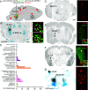
Synaptic targets of Sup5Phox2b. (AC) Monosynaptically restricted labeling of premotor neurons from the masseter in P8 wild-type pups. (A) Schematic of the strategy: G-deficient rabies virus (RV) expressing mCherry is coinjected into the masseter along with a YFP-expressing helper virus HSV-YFP-G, providing G-complementation. (B) Coronal section through the trigeminal region at P8 (higher magnification as Inset), showing mCherry and YFP coinfected “seed cells” in Mo5 (yellow). (C) Coronal section through the pons showing premotor neurons (red) that express Phox2b (green) in ipsilateral (right-hand side of the image) and contralateral Sup5 (higher magnification as Insets). Red arrowhead: commissural fibers. (DO) Anterograde tracing from Sup5Phox2b neurons. (D) Schematic of the injection strategy with summary of the main projection sites. (E) Infected neurons in Sup5Phox2b express mGFP (green) and syp-Ruby (red). Inset: high magnification of an infected neuron in Sup5Phox2b itself covered with mGFP+/syp-Ruby+ boutons. (F) Dense syp-Ruby puncta from infected Phox2b+ cells in the Sup5 region itself. (GO) Coronal sections through the brainstem at low (G, J, and M), and higher (H, K, N) magnifications showing anterograde labeling (gray) of cranial motor nuclei (blue) from Sup5Phox2b. (I, L, and O) Close-ups of motoneurons in the respective motor nuclei, showing axons (mGFP, green) and terminal boutons (syp-Ruby, red puncta) on the cell bodies. [Scale bar, G, J, and M, 500 µm; B, C, E, H, K, and N, 200 µm.]
Fig. 3.
Fig. 3.
Inputs to Sup5Phox2b. (A) Strategy for monosynaptic retrograde tracing from Sup5Phox2b and summary of the major inputs. (B) Low magnification (Left panel) and higher magnifications (Right panels) of coronal sections in the Sup5 region. The infected cells of Sup5Phox2b (black on the Left panel) include GFP+/mCherry+ seed cells (green arrows in Right panels) and GPF/mCherry+ input cells (white arrows), among which many are Phox2b+ (red nuclei). (C) Brain-wide proportions of inputs to Sup5Phox2b. Regions constituting less than 0.3% of total input have been omitted. Note that the contributions of Sup5Phox2b and parabrachial nuclei are pooled, on account that the “parabrachial” of the Allen Brain Atlas includes a large portion of Sup5Phox2b as defined in this study. A small fraction of these cells likely do belong to the parabrachial (medial and lateral) as shown in SI Appendix, Fig. S3. (DG) Other sites of inputs shown on coronal sections showing direct inputs to Sup5Phox2b (mCherry+ cells, black in wide views, red in close ups). (D) Ipsilateral first-order sensory neurons of the mesencephalic trigeminal nucleus (Me5). (E and F) The ipsilateral extended amygdala [i.e., the bed nucleus of the stria terminalis (BNST) (E) and the central amygdaloid nucleus (CeA) (F)]. Most of the inputs from CeA are PKC-∂ (red arrowhead and Right Inset) with occasional PKC-∂+ inputs (white arrowhead and Left Inset); (G) the lateral dentate cerebellar nucleus (Lat); the same plane of section captures input from the reticular formation (IRt and PCRt), bilaterally; Other sites of input are in SI Appendix, Fig. S3. [Scale bar (applies to all panels): 500 µm (B and G), 1 mm (DF).]
Fig. 4.
Fig. 4.
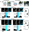
Global activation and inhibition of Sup5Phox2b. (A and B) Schematic representation of viral injection in Sup5Phox2b and fiber-optic implantation, and coronal section of the hindbrain showing infected Sup5Phox2b neurons and the optic fiber track (asterisk). (A′–B″) Photoactivation (A′ and A″) and photoinhibition (B′ and B″) of Sup5Phox2b during lapping (A′ and B′) and chewing (A″ and B″). Black tick marks indicate jaw movement onsets during chewing or lapping, while histograms show the frequency of licks or chews (n = 15 to 25 trials, n = 3 mice). Blue bars indicate the laser stimulation period and the corresponding photograph is framed in blue. (C and D) Box-and-whisker plots of jaw displacement caused by activation and inhibition of Sup5Phox2b during lapping (C) and chewing (D), relative to the maximal open state during each behavior.
Fig. 5.
Fig. 5.
Activity of Sup5Phox2b during biting and chewing. (A) Schematic of viral injection and fiber-optic implantation above Sup5Phox2b, and transverse section through the hindbrain showing transduced Sup5Phox2b neurons and the optic fiber track (asterisk). (B) Example frames of a mouse during different phases of biting and chewing almond: preparing (Left), biting (Middle), chewing (Right). (C) (Top) Representative photometry traces of Sup5Phox2b activity (ΔF/F) over 20 s of chewing almond. Red- and gray-shaded areas indicate biting and chewing epochs, respectively (see SI Appendix, Fig. S5 for quantification). (Bottom) Continuous wavelet transform of the corresponding photometry signals. Signals around 5 Hz are prominent during almond chewing but diminish during biting. (D) Line plot of bite-evoked calcium transients (32 episodes, n = 3 mice): calcium transients were isolated within ±100 ms of 70% of bite onsets (gray shading = SD). (E) Event-triggered averages of Sup5Phox2b activity aligned to bite onset (vertical dashed line) (average of 32 episodes in n = 3 mice, each color represents a different mouse). Shaded area = SEM. (F) Example frames of a mouse during different phases of biting and chewing pasta. (G) Representative photometry traces of Sup5Phox2b activity (Top) and Continuous wavelet transform (Bottom) as in (C), but for chewing pasta instead of almond. (H) Example kernel density estimate of dominant frequency distribution of the continuous wavelet transform during almond (purple) and pasta (red) consumption. (Inset) Dominant frequency variability across conditions (8 sessions, n = 3 mice) (SD of frequency distributions). [Scale bar: (A) 500 µm.]
Fig. 6.
Fig. 6.
Schematic of the “extended enteric nervous system” of vertebrates. Schematic of a mouse with Phox2b-expressing neurons relevant for feeding (represented in red) including two orofacial premotor centers (highlighted in blue). AP: area postrema (involved in vomiting and conditioned food aversion); ENS: enteric nervous system; gVII, gIX, gX: geniculate, glossopharyngeal, and vagal ganglia for all visceral sensations, including taste; IRtPhox2b: Phox2b+ neuron group in the intermediate reticular nucleus (that harbors a lapping center). MoA: nucleus ambiguus (involved in esophageal mobility); Mo7: facial motor nucleus (that mobilizes orofacial muscles); Mo10: dorsal motor nucleus of the vagus nerve (preganglionic to the enteric nervous system); nTS: nucleus of the solitary tract (site of projection of all visceral afferents, including taste); ParaS: parasympathetic ganglia for salivary glands; PG: pelvic ganglion (that innervates pelvic organs including rectum and bladder, the latter shown in gray); SG: sympathetic ganglia (that input to the enteric nervous system); Sup5Phox2b, supratrigeminal nucleus (essential promotor center for food ingestion, this study).

Comment in

Similar articles

References

    1. Yoshida Y., Miyazaki T., Hirano M., Shin T., Kanaseki T., Topographic arrangement of motoneurons innervating the suprahyoid and infrahyoid muscles. A horseradish peroxidase study in cats. Ann. Otol. Rhinol. Laryngol. 92, 259–266 (1983). - PubMed
    1. Dempsey B., et al. , A medullary centre for lapping in mice. Nat. Commun. 12, 6307 (2021). - PMC - PubMed
    1. Dellow P. G., Lund J. P., Evidence for central timing of rhythmical mastication. J. Physiol. 215, 1–13 (1971). - PMC - PubMed
    1. Grill H. J., Norgren R., The taste reactivity test. II. Mimetic responses to gustatory stimuli in chronic thalamic and chronic decerebrate rats. Brain Res. 143, 281–297 (1978). - PubMed
    1. Lund J. P., Dellow P. G., The influence of interactive stimuli on rhythmical masticatory movements in rabbits. Arch. Oral Biol. 16, 215–223 (1971). - PubMed

LinkOut - more resources