. 2025 Jul 16;10(1):221.
doi: 10.1038/s41392-025-02305-9.

DDX39B K63-linked ubiquitination mediated by TRIM28 promotes NSCLC metastasis by enhancing ECAD lysosomal degradation

Affiliations

DDX39B K63-linked ubiquitination mediated by TRIM28 promotes NSCLC metastasis by enhancing ECAD lysosomal degradation

Hang Yuan et al. Signal Transduct Target Ther. .

Abstract

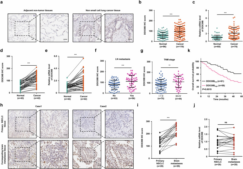
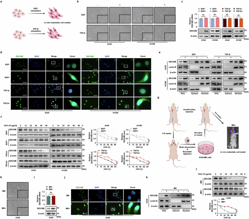
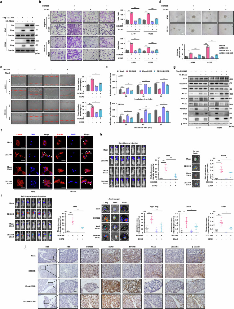
Metastasis is a leading cause of treatment failure and high mortality in non-small cell lung cancer (NSCLC). Recently, we demonstrated that DEAD box helicase 39B (DDX39B) was upregulated and activated metabolic reprogramming in colorectal cancer and hepatocellular carcinoma. However, the function of DDX39B and the therapeutic potential for targeting DDX39B in NSCLC remain unclear. Herein, we discovered that DDX39B was an independent marker for poor survival in NSCLC patients. Strikingly, DDX39B protein, but not its mRNA, was elevated in clinical metastatic brain lesions and metastatic cell models (in vitro EMT-metastatic and in vivo carotid artery injection-induced brain-metastatic cell model). Mechanistically, DDX39B interacted with E3 ubiquitin ligase TRIM28 via Pro322 residue and underwent TRIM28-mediated K63-linked ubiquitination at Lys241, Lys384, and Lys398, leading to DDX39B protein stabilization and upregulation. Subsequently, DDX39B directly bound to ECAD and promoted ECAD lysosomal degradation by recruiting Src and Hakai, which was independent of its RNA helicase activity, followed by activating β-catenin oncogenic signaling and facilitating NSCLC aggressive phenotype. According to structure-based virtual screening, we discovered a clinical antimalarial drug, artesunate, that disrupted the association of DDX39B-TRIM28 complex, resulting in DDX39B degradation and blocking the pro-metastatic effects of DDX39B. Overall, our findings uncover that TRIM28/DDX39B/ECAD axis contributes to NSCLC metastasis and targeting DDX39B degradation by artesunate is an effective and promising therapeutic approach for the treatment of NSCLC.


Conflict of interest statement

Competing interests: The authors declare no competing interests.

Figures

Fig. 1
Fig. 1
Upregulated DDX39B correlates with metastasis and predicts poor prognosis in NSCLC patients. The protein expression of DDX39B in NSCLC (n = 119) and adjacent non-tumor tissues (n = 96) was determined by immunohistochemistry analysis, (a) representative images and (b) relative intensities of DDX39B were shown. c The mRNA expression of DDX39B in NSCLC (n = 76) and adjacent non-tumor tissues (n = 60) was measured by RT‒qPCR assay. d The relative intensities of DDX39B protein staining or (e) mRNA level were analyzed in primary NSCLC and patient-matched normal tissues (n = 42). f DDX39B protein expression in primary NSCLC tissues with lymph node (LN) metastasis (n = 56) or without LN metastasis (n = 63). g DDX39B protein expression in primary NSCLC tissues at early (I + II) (n = 75) or advanced (III + IV) (n = 44) TNM stages. DDX39B expression in metastatic brain tissues and matched primary NSCLC tissues (n = 20) was determined by (h, i) immunohistochemistry analysis or (j) RT‒qPCR. h Representative images of DDX39B expression in primary tissue and metastatic brain lesions were shown. i The relative intensities of DDX39B protein staining or (j) mRNA level were analyzed. k Kaplan‒Meier estimates of overall survival probability based on DDX39B protein expression in NSCLC patients (n = 119). Scale bars, 50 µm. P values were determined by using the two-tailed unpaired t test (b, c, f, g), two-tailed paired t test (d, e, i, j), or log-rank test (k). ns, not significant, **P < 0.01, ***P < 0.001
Fig. 2
Fig. 2
DDX39B protein is highly expressed during metastasis in multiple metastasis models. a Scheme for the in vitro establishment of EGF- or TGF-β-induced EMT cell models (by Figdraw). be NSCLC cell lines (A549 and H1299) were starved overnight and then treated with or without 30 ng/ml EGF or 10 ng/ml TGF-β for 72 h. b Cell morphology was analyzed using microscopy. c The expression of DDX39B was determined by RT‒qPCR (n = 3) and Western blotting. d The expression and distribution of DDX39B protein were examined by immunofluorescence staining. e DDX39B expression in the indicated cell fractions was detected by Western blot analysis. WCL: whole cell lysates. f NSCLC cells were starved overnight and treated with 10 µg/ml cycloheximide (CHX) for the indicated times (0, 12, 24, 36, 48 and 60 h) in the absence or presence of EGF (30 ng/ml) or TGF-β (10 ng/ml). Cell lysates were immunoblotted with antibodies against DDX39B and β-actin, and the ratio of DDX39B to β-actin was calculated. The line graph represents the rate of DDX39B degradation (n = 3). g Scheme for the in vivo selection process of the isolation of brain-metastasis (BM) cells from A549-luc cells (by Figdraw). h Cell morphology of A549-parental and BM cell lines. i The expression of DDX39B in A549-parental and BM cell lines was determined by RT‒qPCR (n = 3) and Western blotting. j The expression and distribution of DDX39B protein in A549-parental and BM cell lines were examined by immunofluorescence staining. k DDX39B expression in the indicated cell fractions of A549-parental and BM cells was detected by Western blot analysis. WCL: whole cell lysates. l A549-parental and BM cells were treated with 10 µg/ml CHX for the indicated times (0, 12, 24, 36, 48 and 60 h). Cell lysates were immunoblotted with antibodies against DDX39B and β-actin, and the ratio of DDX39B to β-actin was calculated. The line graph represents the rate of DDX39B degradation (n = 3). Scale bars, 50 µm. Graphs represent data as the mean±s.d. P values were determined by a two-tailed unpaired t test (c, i) or two-way ANOVA (f, l). ns, not significant, **P < 0.01, ***P < 0.001
Fig. 3
Fig. 3
DDX39B interacts with TRIM28 via P322 residue. a Schematic workflow for exploring DDX39B interactors by co-IP coupled with LC-MS/MS (by Figdraw). b Protein‒protein interaction network of DDX39B from the Biogrid database (https://thebiogrid.org/, minimum evidence: 10). c Venn diagram shows the overlap of potential E3 ligase (TRIM28) identified from our DDX39B interactors data, Biogrid database, and iUUCD 2.0 database. d, e Exogenous and (f, g) endogenous associations between DDX39B and TRIM28 were determined by immunoprecipitation analysis in NSCLC cell lines (A549 and H1299). h GST pull-down assay of purified GST-DDX39B and His-TRIM28 proteins. i Indicated BiFC plasmids were transfected into NSCLC cells, in vivo interaction between DDX39B and TRIM28 was detected by BiFC analysis. j NSCLC cell lines were treated with EGF (30 ng/ml) or TGF-β (10 ng/ml) for 72 h, the interaction between DDX39B and TRIM28 was examined. k The interaction between DDX39B and TRIM28 was examined in A549-parental and BM cell lines. l The binding interface between DDX39B and TRIM28 protein was shown. m GST pull-down assay of purified GST-TRIM28 and the WT or the indicated mutant forms of His-DDX39B. Scale bars, 50 µm
Fig. 4
Fig. 4
TRIM28 enhances the stabilization of DDX39B protein by inducing K63-linked ubiquitination at Lys241, Lys284, and Lys398 residues of DDX39B. NSCLC cells were transfected with Flag-TRIM28, and the (a) mRNA (n = 3) and (b) protein expression of DDX39B were determined by RT‒qPCR and Western blotting, respectively. c The expression of TRIM28 on DDX39B protein degradation was detected by CHX chase analysis (n = 3). d Cells were co-transfected with Flag-TRIM28 and HA-ub, and the effect of TRIM28 on DDX39B total ubiquitination was examined by immunoprecipitation assay. e 293T cells were co-transfected with the indicated ubiquitin plasmids and Flag-TRIM28, and DDX39B ubiquitination were analyzed by immunoprecipitation assay. f 293T cells were co-transfected with Flag-TRIM28, HA-ub-K63 and Myc-DDX39B wild-type (WT) or indicated lysine to arginine (K to R) mutants, and DDX39B ubiquitination were analyzed by immunoprecipitation assay. g 293T cells were co-transfected with Flag-TRIM28, HA-ub-K63 and Myc-DDX39B WT or indicated lysine to arginine (K to R) mutants (K241R, K384R, K398R or 3KR (K241R, K384R and K398R)), and DDX39B ubiquitination were analyzed by immunoprecipitation assay. h 293T cells were co-transfected with Flag-TRIM28, HA-ub-K63 and Myc-DDX39B WT or indicated mutants (P322A, 3KR), and DDX39B ubiquitination were analyzed by immunoprecipitation assay. i The impact of TRIM28 WT or ΔRING (RING domain deletion) on the K63-linked ubiquitination of purified DDX39B WT, P322A or 3KR was examined by in vitro ubiquitination assay. j Mass spectrometry analysis reveals the K63-linked ubiquitination sites of DDX39B at K241, K384, K398 residues. k Sequence alignment around K241, K384, K398 residues of DDX39B in different species was shown. l, m Indicated NSCLC cells were transfected with DDX39B WT, P322A or 3KR mutants, and then treated with CHX (10 μg/ml) for the indicated times (0, 12, 36 and 60 h). The ratio of DDX39B to β-actin was calculated. The line graph represents the rate of DDX39B degradation (n = 3). n, o IHC assay was performed to examine the relationship between DDX39B and TRIM28 expression in primary NSCLC tissues (n = 119). n Representative images of DDX39B and TRIM28 expression were shown. o The correlation between DDX39B and TRIM28 expression was evaluated. Scale bars, 50 µm. Graphs represent data as the mean ± s.d. P values were determined by one-way ANOVA (a), two-way ANOVA (c, l, m) or Pearson correlation coefficient analysis (o). ns, not significant, **P < 0.01, ***P < 0.001
Fig. 5
Fig. 5
DDX39B promotes EMT reprogramming and metastasis in NSCLC cells. a Reconstituted expression of the wild-type (WT), P322A and 3KR (K241R, K384R and K398R) of Flag-DDX39B in NSCLC cell lines (A549 and H1299) was verified by Western blotting. The effects of DDX39B WT, P322A and 3KR mutant on (b) cell migration and invasion, (c) wound closure, (d) 3D sphere formation and (e) adhesion in NSCLC cell lines were analyzed (n = 3). f F-actin and nuclei were stained using rhodamine-phalloidin and DAPI, respectively. Representative images were shown, and the white triangles represented cilia and pseudopodia. g Protein expression of mesenchymal markers (NCAD, Vimentin and Snail) and epithelial markers (ZO-1, Occludin, KRT18, ECAD and EPCAM) in indicated NSCLC cells were measured by Western blotting. h Indicated A549 cells were injected into carotid artery. Six weeks later, the representative bioluminescence images and intensities (p/s/cm2/sr) of mice and isolated brain were presented and quantified (n = 7/group). i Indicated A549 cells were orthotopically injected into the left pulmonary tissue. Eight weeks later, the representative bioluminescence images and intensities (p/s/cm2/sr) of mice and isolated organs (including the right lung, brain, and liver) were shown and quantified (n = 7/group). Scale bars, 50 µm. Graphs represent data as the mean ± s.d. P values were determined by one-way ANOVA (b–e, h, i). **P < 0.01, ***P < 0.001
Fig. 6
Fig. 6
DDX39B interacts with and accelerates ECAD ubiquitination and lysosomal degradation by recruiting Src and Hakai. a KEGG enrichment analysis of the DDX39B interactome. b, cThe endogenous association between DDX39B and ECAD was determined by immunoprecipitation analysis. d GST pull down assay of purified GST-ECD (GST-tagged ECAD cytoplasmic domain) and His-DDX39B protein. e Indicated BiFC plasmids were transfected into NSCLC cells, and in vivo interaction between DDX39B and ECAD was detected by BiFC analysis. f The effect of DDX39B on ECAD protein stability was detected by CHX analysis (n = 3). g DDX39B-overexpressed NSCLC cells were treated with chloroquine (50 μM, 8 h) or MG132 (10 μM, 8 h), and ECAD expression was detected by Western blot analysis. h The colocalization of ECAD and LAMP1 (a lysosomal marker) in DDX39B-overexpressed cells was determined by immunofluorescence staining. i Cells were co-transfected with Myc-DDX39B and Flag-ub, and the effect of DDX39B on ECAD ubiquitination was examined by immunoprecipitation assay. j The effect of DDX39B on the interaction between ECAD and Hakai was determined by immunoprecipitation analysis. k DDX39B-deficient NSCLC cells were transfected with or without HA-Hakai plasmid. The expression and ubiquitination of ECAD protein were detected. l The pan-pTyr and (m) Tyr754-phosphorylation of ECAD were examined in DDX39B-overexpressed cells. n The effect of DDX39B on the interaction of ECAD with Src was evaluated by immunoprecipitation assay. o DDX39B-silenced cells were transfected with or without HA-Src plasmid, and pECADY754 was analyzed by Western blot analysis. p 293T cells were co-transfected with Flag-DDX39B and HA-ECAD plasmids, and the lysates were processed for tandem affinity purification by using anti-Flag and anti-HA magnetic beads. The effects of DDX39B WT, P322A or 3KR (K241R, K384R and K398R) mutant on (q) the interaction between ECAD and Hakai or Src, (r) the expression of ECAD and ECAD ubiquitination, and (s) the expression of pECADY754. t, u IHC assay was performed to examine the relationship between DDX39B and ECAD expression in primary NSCLC tissues (n = 119). t Representative images of DDX39B and ECAD expression were shown. u The correlation between DDX39B and ECAD expression was evaluated. v Kaplan‒Meier estimates of overall survival probability based on ECAD protein expression in NSCLC patients (n = 119). w Prognostic value of DDX39B combined with ECAD expression in NSCLC patients (n = 119). Scale bars, 50 µm. Graphs represent data as the mean ± s.d. P values were determined by two-way ANOVA (f). Pearson correlation coefficient analysis (u), and log-rank test (v, w). ***P < 0.001
Fig. 7
Fig. 7
The overexpression of ECAD is sufficient to abolish DDX39B-mediated EMT reprogramming and metastasis in NSCLC in vitro and in vivo. a Reintroduced ECAD expression was performed in DDX39B-overexpressed NSCLC cell lines (A549 and H1299) and verified by Western blotting. b Cell migration and invasion, (c) wound closure, (d) sphere formation and (e) adhesion in indicated NSCLC cell lines were determined (n = 3). f F-actin and nuclei were stained using rhodamine-phalloidin and DAPI, respectively. Representative images were shown, and the white triangles represented cilia and pseudopodia. g The expression of EMT markers was detected by Western blot analysis. h Indicated A549 cells were injected into carotid artery. Six weeks later, the representative bioluminescence images and intensities (p/s/cm2/sr) of mice and isolated brain were presented and quantified (n = 7/group). i Indicated A549 cells were orthotopically injected into the left pulmonary tissue. Eight weeks later, the representative bioluminescence images and intensities (p/s/cm2/sr) of mice and isolated organs (including the right lung, brain, and liver) were shown and quantified (n = 7/group). j Immunohistochemistry analysis was performed to examine the relationship between DDX39B expression and EMT markers (ECAD, EPCAM, NCAD, Vimentin, β-catenin) in indicated orthotopic lung cancer tissues. Representative images were shown. Scale bars, 50 µm. Graphs represent data as the mean ± s.d. P values were determined by one-way ANOVA (be, h, i). *P < 0.05, **P < 0.01, ***P < 0.001
Fig. 8
Fig. 8
Artesunate inhibits DDX39B protein stability and tumor metastasis by blocking the DDX39B-TRIM28 interplay in NSCLC. a The overall structure of the complex and two-dimensional ligand-interaction maps of the binding of artesunate to DDX39B were revealed by a computational model. DDX39B (green), artesunate (orange) (left). The interacting amino acids of DDX39B to artesunate were shown (right). b The binding affinity of artesunate to DDX39B was determined by bio-layer interferometry (BLI) assay. c The equilibrium dissociation constant (KD) of artesunate interacting with DDX39B was calculated using surface plasmon resonance (SPR) assay. d Cellular thermal shift assay (CETSA) was performed in NSCLC cell lines treated with DMSO or 100 μM artesunate. A loading control was established using β-actin. e The indicated cells were treated with or without 20 µM artesunate for 24 h. The interaction between DDX39B and TRIM28 was analyzed by immunoprecipitation assay. f NSCLC cells with or without stable expression of TRIM28 were treated with artesunate (20 μM, 24 h), and the effect of artesunate on DDX39B expression was analyzed. g TRIM28-overexpressing NSCLC cells were co-treated with or without artesunate (20 μM) and CHX (10 μg/ml) for the indicated times (0, 12, 36 and 60 h). The ratio of DDX39B to β-actin was calculated. The line graph represents the rate of DDX39B degradation (n = 3). NSCLC cells were treated with artesunate at the indicated (h) doses and (i) times, and the expression of DDX39B and ECAD was detected by Western blotting. j NSCLC cells with or without stable expression of DDX39B were treated with artesunate (20 μM, 24 h), and the effect of artesunate on DDX39B-induced ECAD degradation was analyzed. The indicated NSCLC cells were treated with or without artesunate (20 μM) (n = 3). The abilities of (k) cell migration, invasion and (l) sphere formation were investigated. m Indicated A549 cells were injected into carotid artery. Two weeks later, mice were intraperitoneally injected with artesunate (100 mg/kg per two day and per mice) for four weeks. Representative bioluminescence images and intensities (p/s/cm2/sr) of mice and isolated brain were presented and quantified (n = 7/group). n Indicated A549 cells were orthotopically inoculated into the left pulmonary tissue. Three weeks later, the mice were intraperitoneally injected with artesunate (100 mg/kg per two day and per mice) for five weeks, the representative bioluminescence images and intensities (p/s/cm2/sr) of mice and isolated organs (including the right lung, brain, and liver) were shown and quantified (n = 7/group). o Schematic illustration of TRIM28/DDX39B/ECAD axis-mediated metastasis in NSCLC (by Figdraw). Scale bars, 50 µm. Graphs represent data as the mean ± s.d. P values were determined by two-way ANOVA (g) or one-way ANOVA (kn). **P < 0.01, ***P < 0.001

References

    1. Siegel, R. L., Miller, K. D., Fuchs, H. E. & Jemal, A. Cancer statistics, 2022. CA-Cancer J. Clin.72, 7–33 (2022). - PubMed
    1. Chen, Z., Fillmore, C. M., Hammerman, P. S., Kim, C. F. & Wong, K. K. Non-small-cell lung cancers: a heterogeneous set of diseases. Nat. Rev. Cancer14, 535–546 (2014). - PMC - PubMed
    1. Liu, W., Powell, C. A. & Wang, Q. Tumor microenvironment in lung cancer-derived brain metastasis. Chin. Med. J.135, 1781–1791 (2022). - PMC - PubMed
    1. Yousefi, M. et al. Lung cancer-associated brain metastasis: Molecular mechanisms and therapeutic options. Cell. Oncol.40, 419–441 (2017). - PubMed
    1. Li, K. et al. ZNF32 induces anoikis resistance through maintaining redox homeostasis and activating Src/FAK signaling in hepatocellular carcinoma. Cancer Lett.442, 271–278 (2019). - PubMed

MeSH terms

Substances