. 2025 Jul 15;16(1):6503.
doi: 10.1038/s41467-025-61187-1.

Variable lymphocyte receptor F is generated via somatic diversification and expressed by lamprey T-like cells

Affiliations

Variable lymphocyte receptor F is generated via somatic diversification and expressed by lamprey T-like cells

Sabyasachi Das et al. Nat Commun. .

Abstract

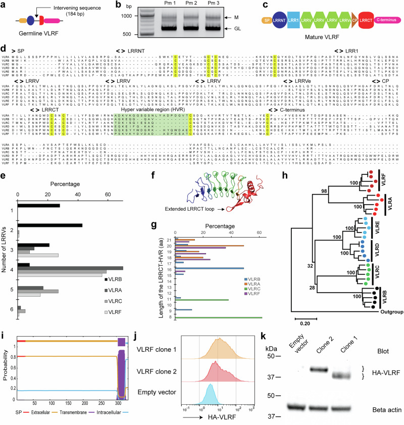
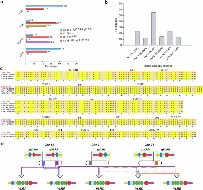
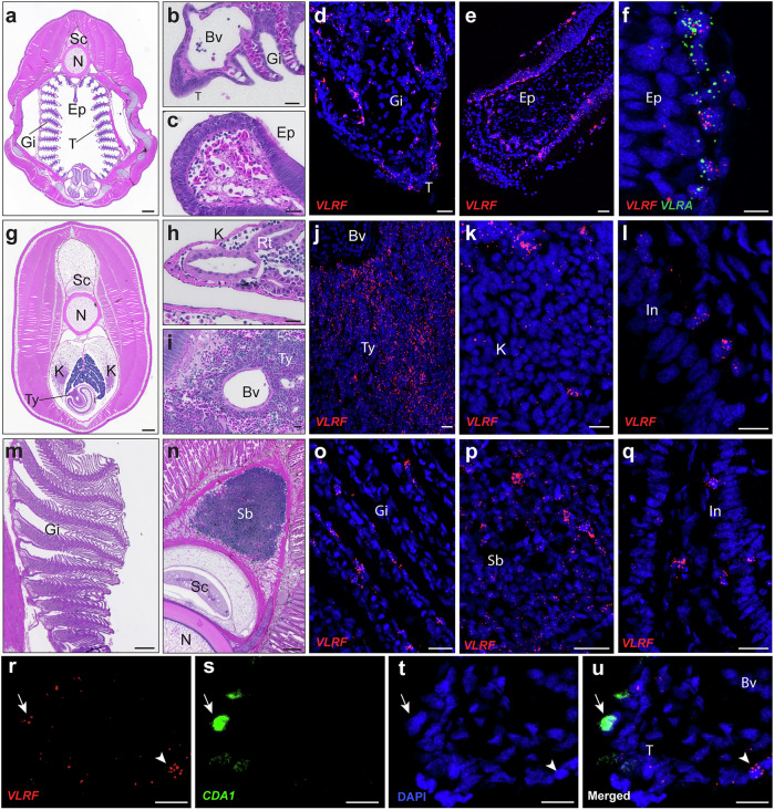
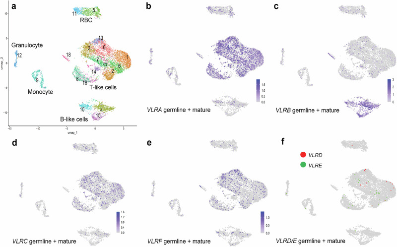
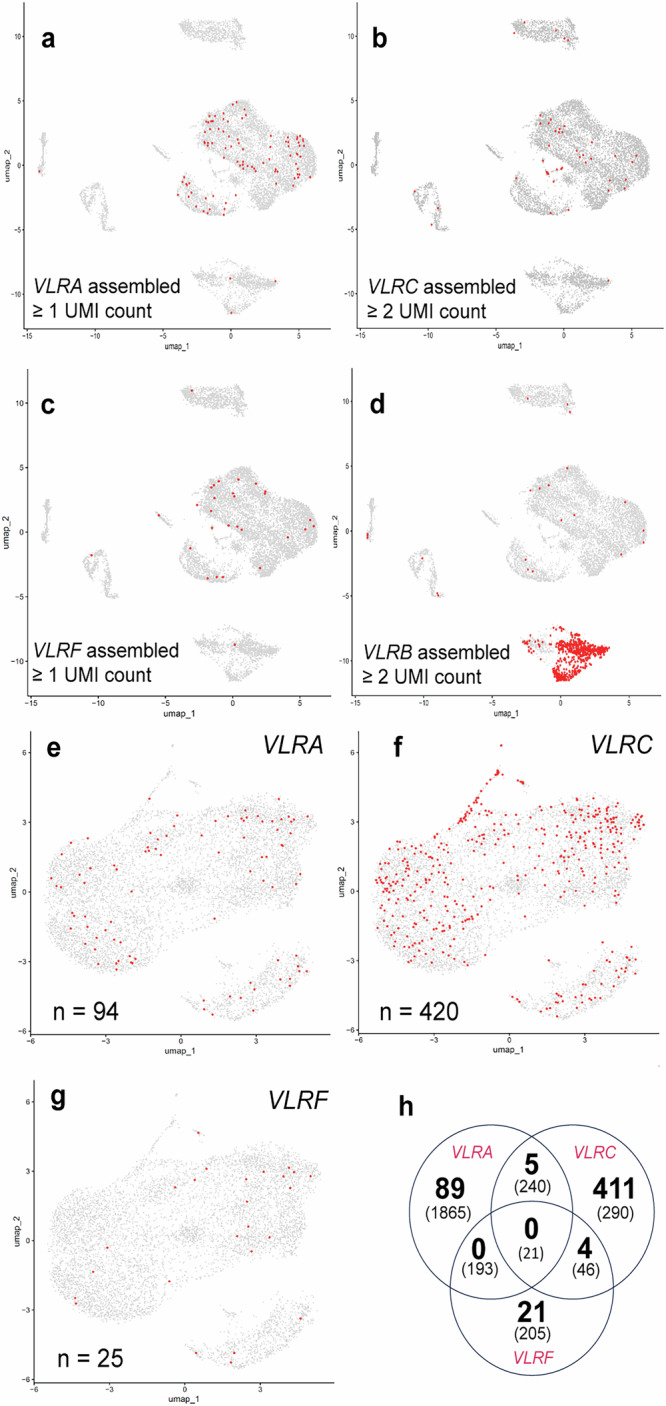
All extant jawless vertebrates (lampreys and hagfishes) possess a unique adaptive immune system characterized by highly variable lymphocyte receptors (VLR) that are assembled in developing lymphocytes using leucine-rich-repeat donor cassettes. Five VLR types have been identified in lampreys: VLRA, VLRB, VLRC, VLRD, and VLRE. VLRB-expressing lymphocytes are functional analogs to B cells, whereas VLRA, VLRC, VLRD, and VLRE-expressing lymphocytes are more akin to T cells of jawed vertebrates. Here we define an additional VLR, designated VLRF. VLRF is phylogenetically closest to VLRA, with which it likely shares a common ancestral gene of at least 250 million years in the past. VLR assembly analyses show that VLRA, VLRC, VLRD, VLRE, and VLRF share donor cassettes through long-range intra- and inter-chromosomal interactions, whereas VLRB utilizes a distinct, dedicated cassette set. The pattern of gene expression, donor cassette usage, and distinctive amino acid composition in the C-terminal stalk suggest that VLRF⁺ lymphocytes may represent an additional T-like sub-lineage, adding further complexity to the VLR-based adaptive immune system.


Conflict of interest statement

Competing interests: The authors declare no competing interests.

Figures

Fig. 1
Fig. 1. VLRF in the lamprey is a member of the VLR family.
a Germline VLRF gene in the sea lamprey. b Isolation of mature (M) and germline (GL) VLRF sequences from electrophoretic bands (n = 3). c Schematic of the mature VLRF sequence. d Alignment of representative mature VLRA, VLRB, VLRC, VLRD, VLRE, and VLRF sequences in the sea lamprey. Conserved cysteine residues in LRRNT and LRRCT are highlighted in yellow. e Number of LRRV modules in VLRA, VLRB, VLRC, and VLRF repertoires in the sea lamprey. The ending-LRRVe module is included in the number of LRRVs of a mature VLR sequence. 100 mature sequences of each VLR isotype are analyzed. f Predicted 3D structure of VLRF, highlighting the protruding loop in the LRRCT region of this assembly (GenBank accession no. PQ159758). The LRRNT and LRR1 regions are shown in blue, LRRVs are shown in green, while the CP and LRRCT regions are represented in red. g Analysis of the length of the HVR in the LRRCT domain. The trends in the HVR length distribution of VLRB are distinct from those of VLRA, VLRC and VLRF. h A phylogenetic tree constructed using five representative mature sequences for each VLR isotype in the sea lamprey. Bootstrap support values are provided for interior branches. The scale bar represents sequence substitution per site. i Prediction of the transmembrane domain (TM) in VLRF. j Histograms showing anti-HA staining on transfected cells detected by flow cytometry. HEK 293 cells were transfected with an Empty vector, HA epitope-tagged VLRF clone 1 or VLRF clone 2. k Whole cell lysates from transfected HEK 293 cells were analyzed by immunoblotting for the presence of HA epitope (VLRF) and beta actin loading control. Brackets indicate the expected bands for VLRFs with three (clone 1) and four (clone 2) LRRV domains. Each blot is a representative of two independent experiments.
Fig. 2
Fig. 2. VLRA, VLRC and VLRF locus organization in sea lamprey.
a Germline VLRA and VLRF genes are located on chromosome 58, with donor cassettes distributed in two genomic clusters (Cluster I and Cluster II) on chromosome 58. b Genomic organization of the sea lamprey VLRA and VLRF loci on chromosome 58. Cluster I and Cluster II on chromosome 58 contain 129 and 144 donor cassettes, respectively. c Germline VLRC and adjacent donor cassettes are located on chromosome 7. Chromosome 7 is much larger than chromosome 58, as indicated by the gap. d Germline VLRC and the donor cassette cluster on chromosome 7. This cluster contains 18 donor cassettes primarily used for VLRC assembly. For figures (b and d), the cassettes are presented according to their genomic spacing, although the icons are not to scale. Filled triangles above each donor cassette and the germline VLR gene indicate the transcriptional orientation.
Fig. 3
Fig. 3. Genomic donor cassette usage and sharing among assembled VLRA, VLRC, VLRD, VLRE, and VLRF in Sea Lampreys.
a Donor cassette usage from different genomic locations. Donor cassettes from cluster I (Ch.58-cI) and cluster II (Ch.58-cII) of chromosome 58 are mainly used in VLRA and VLRF assemblies. In contrast, VLRC assemblies predominantly use cassettes from Ch.58-cII, followed by chromosome 7 and Ch.58-cI, respectively. For VLRD/VLRE assemblies, donor cassettes from chromosome 75 and Ch.58-cII, along with a small number of cassettes from Ch.58-cI, are used. VLRD and VLRE are considered together due to the limited number of assembled sequences available and their high similarity. b Patterns of donor cassette sharing among VLRs analyzed in relation to the total cassette usage in a dataset comprising 100 VLRA, 100 VLRC, 60 VLRD/VLRE, and 100 VLRF mature sequences. Notably, the highest percentage of cassette sharing is observed between VLRF and VLRA. c Examples of donor cassette sharing with representative sequences. Cassettes incorporated into mature VLR sequences are depicted in red, with the accession numbers of the mature sequences provided in parentheses. Only regions with 100% sequence identity between the donor cassettes and mature sequences are shown, and the corresponding encoded LRR modules are indicated. Alternative codons are highlighted in yellow and orange. d Schematic representation of donor cassette usage among mature VLRA, VLRC, VLRD, VLRE, and VLRF sequences. Genomic donor cassette clusters on chromosomes 58, 7, and 75 are highlighted in color-coded rectangles. Solid lines represent major contributions, while dotted lines denote minor contributions of donor cassettes from each genomic cluster to the mature sequences. Line colors correspond to their respective donor clusters. The graphic was created using BioRender.
Fig. 4
Fig. 4. The relationship among VLR sequences across lamprey species.
aVLRF and VLRA are phylogenetically close in all lamprey species. The phylogenetic tree is constructed by using the protein-coding segments of germline VLR genes in six different lamprey species. Bootstrap values are provided for all branches. Mm, Mordacia mordax; Ga, Geotria australis; Pm, Petromyzon marinus; Et, Entosphenus tridentatus; Lp, Lampetra planeri; Lr, Lethenteron reissneri. Note that the current version of the Mordacia mordax genome lacks detectable orthologs for VLRD. b Genomic map of germline VLRA and VLRF in short-headed lamprey (Mordacia mordax), Far Eastern brook lamprey (Lethenteron reissneri) and Pacific lamprey (Entosphenus tridentatus). The distance between germline VLRA and VLRF is much larger in L. reissneri and E. tridentatus than in M mordax, as indicated by the gap. The map is not drawn to scale. Filled triangles above the germline genes indicate transcription orientation.
Fig. 5
Fig. 5. Cellular and tissue expression of the VLRF gene in sea lampreys.
a Expression of the VLRF gene in different lymphocyte populations in lamprey larvae. TN represents the triple-negative (VLRA/VLRB/VLRC) lymphocyte population. Bars indicate the standard error of the mean in each experiment (n = 6). b Assembly status of VLRF in total RNA extracted from purified lymphocyte populations. bp, base pairs; GL, germline transcripts; M, transcripts of assembled VLRF gene. A+, VLRA+; B+, VLRB+; C+, VLRC+. Each experiment was repeated three times independently with similar results. c, d Tissue expression profiles for VLRF in larvae (c) and adult (d) lampreys. Transcripts are analyzed by real-time RT-PCR with beta-actin as a normalization control for both cellular and tissue distribution analyses. Bars indicate the standard error of the mean for three lamprey larvae (c) and three adult lampreys (d) in each experiment. Bars indicate the standard error of the mean for four lamprey larvae (c) and four adult lampreys (d) in each experiment. The corresponding data points were shown as dot plots.
Fig. 6
Fig. 6. Photomicrographs of hematoxylin and eosin (H&E)-stained or in situ hybridized cross-sections from larvae and adult sea lampreys using VLRF, VLRA, and CDA1 probes.
a H&E-stained cross-section of the anterior region of lamprey larvae, including the epipharyngeal ridge (Ep), thymoids (T), and gills (Gi). b, c Higher magnification of H&E-stained gill filaments and thymoids (b) and the epipharyngeal ridge (c). df In situ hybridization for VLRF (red) in larval immune tissues. VLRF + cells are dispersed throughout the gill filaments and thymoids (d). In the epipharyngeal ridge, VLRF expression is observed in cells lining the epithelial layer (e). Co-localization of VLRF (red) and VLRA (green) in the epipharyngeal ridge (f). g H&E-stained cross-section of the posterior body, depicting the kidney (K) and typhlosole (Ty) in larvae. h, i Higher magnification views of the H&E-stained kidney (h) and typhlosole regions (i) showing lymphoid cells located adjacent to the renal tubules (Rt) in the kidney and surrounding the main blood vessel (Bv) in the typhlosole. jl In situ hybridization for VLRF (red) in posterior immune tissues. VLRF + cells localize around blood vessels within the typhlosole (j) and appear scattered in the kidney (k). Panel (l) shows VLRF+ cells beneath the intestine (ln). m, n H&E-stained sections of the gills (m) and the supraneural body (Sb) (n) of adults. oq in situ hybridization for VLRF (red) in adult immune tissues. VLRF+ cells line the gill filament epithelium (o). Supraneural body contains numerous VLRF+ cells in the lymphoid area (p). VLRF+ cells are observed beneath the intestinal epithelium in adults (q). ru In situ hybridization showing expression of VLRF (red) and CDA1 (green) in the thymoid. A VLRF⁺CDA1⁺ cell is indicated by an arrow, and an arrowhead marks a VLRF⁺CDA1⁻ cell. Nuclei are counterstained with DAPI. Abbreviations: Sc, spinal cord; N, notochord; Ep, epipharyngeal ridge; T, thymoids; Gi, gills; K, kidney; In, intestine; Ty, typhlosole; Bv, blood vessel; Rt, renal tubules; Sb, supraneural body. Scale bars: (a, g, m) 400 µm; (b, c, d, h, i, j, o, p) 20 µm; (e) 25 µm; (f) 5 µm; (n) 100 µm; (k, l, qu) 10 µm. Representative images shown; histological patterns were consistent across all specimens analyzed (n = 4).
Fig. 7
Fig. 7. Expression of VLR invariant sequence in a single-cell RNA sequencing analysis of sea lamprey white blood cells.
a Cluster designations were assigned to known cell type by VLR (B- and T-like cells) and marker gene expression. bf Expression of VLR invariant sequence segments of (b) VLRA, (c) VLRB, (d) VLRC, (e) VLRF, and (f) VLRD (red dots) and VLRE (green dots). Note that in figures (bf), detection of the VLR invariant regions cannot distinguish between germline or assembled VLR transcription. For (f), positive cells are designated without reference to expression level due to very low expression of VLRD and VLRE.
Fig. 8
Fig. 8. T-like cells in sea lamprey tend to express only one type of VLR.
ad Cells expressing assembled VLR genes in an analysis of all white blood cells: VLRA (a), VLRC (b), VLRF (c), and VLRB, (d). eh Overlap among cells that are positive for assembled VLRA, VLRC, and VLRF sequence in a reanalysis of only cells from T-like cell clusters (see Fig. 7) showing cells positive for at least one assembled sequence for VLRA (e) VLRC (f) and VLRF (g). h A Venn diagram showing the numbers of overlapping cells expressing at least one sequence from an assembled T-like cell VLR gene (bold numbers) or VLR invariant regions (numbers in parentheses). Note that because the distribution of 10 x sequence along the VLR transcripts in this analysis is highly biased toward the 5′ end of most transcripts, assembled sequences are significantly underrepresented, but are nonetheless localized to T-like (VLRA, VLRC, VLRF) and B-like (VLRB) cell clusters. VLRB and VLRC assemblies are more prevalent than VLRA and VLRF assemblies, so a higher cut-off of ≥ 2 UMI counts was set for assembled VLRC (b) and VLRB (d) to reduce background from ambient (non-cell-associated) RNA. For much rarer VLRA and VLRF assemblies, all detectable cells with assembled sequences are highlighted.

Similar articles

References

    1. Holland, N. D. & Chen, J. Origin and early evolution of the vertebrates: new insights from advances in molecular biology, anatomy, and palaeontology. Bioessays23, 142–151 (2001). - PubMed
    1. Janvier, P. Early jawless vertebrates and cyclostome origins. Zool. Sci.25, 1045–1056 (2008). - PubMed
    1. Kuratani, S. Evo-devo studies of cyclostomes and the origin and evolution of jawed vertebrates. Curr. Top. Dev. Biol.141, 207–239 (2021). - PubMed
    1. Marletaz, F. et al. The hagfish genome and the evolution of vertebrates. Nature627, 811–820 (2024). - PMC - PubMed
    1. Timoshevskaya, N. et al. An improved germline genome assembly for the sea lamprey Petromyzon marinus illuminates the evolution of germline-specific chromosomes. Cell Rep.42, 112263 (2023). - PMC - PubMed

LinkOut - more resources