. 2025 Jul 15;15(1):121.
doi: 10.1038/s41408-025-01309-6.

Age-dependent phenotypic and molecular evolution of pediatric MDS arising from GATA2 deficiency

Affiliations

Age-dependent phenotypic and molecular evolution of pediatric MDS arising from GATA2 deficiency

Lili Kotmayer et al. Blood Cancer J. .

Abstract

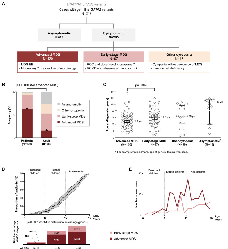
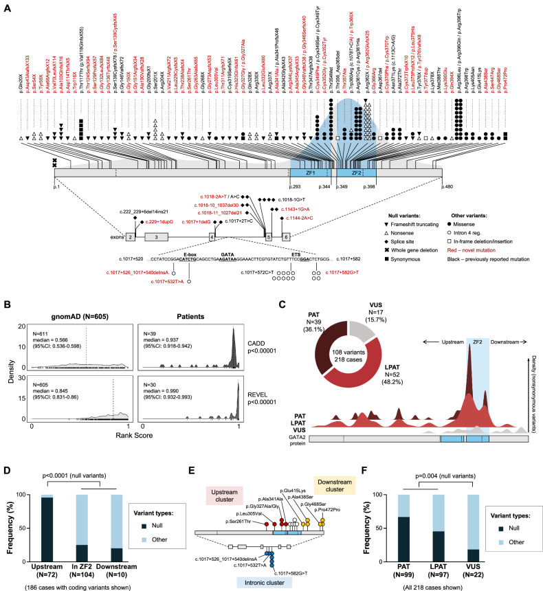
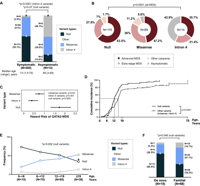
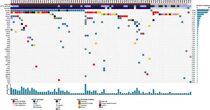
GATA2 deficiency is an autosomal dominant transcriptopathy disorder with high risk for myelodysplastic syndrome (MDS). To elucidate genotype-phenotype associations and identify new genetic risk factors for MDS, we analyzed 218 individuals with germline heterozygous GATA2 variants. We observed striking age-dependent incidence patterns in GATA2-related MDS (GATA2-MDS), with MDS being absent in infants, rare before age 6 years, and steeply increasing in older children. Among 108 distinct GATA2 variants (67 novel), null mutations conferred a 1.7-fold increased risk for MDS, had earlier MDS onset compared to other variants (12.2 vs. 14.6 years, p = 0.009) and were associated with lymphedema and deafness. In contrast, intron 4 variants exhibited reduced penetrance and lower risk for MDS development. Analysis of the somatic landscape revealed unique patterns of clonal hematopoiesis. SETBP1 mutations occurred exclusively in patients with monosomy 7 and their frequency decreased with age. Conversely, the frequency of STAG2 mutations and trisomy 8 increased with age and appeared protective against early development of advanced MDS. Overall, the majority (73.9%) of mutation-positive cases harbored monosomy 7, suggesting it serves as a major driver in malignant progression. Our findings provide evidence for age-appropriate surveillance, and a foundation for genotype-driven risk stratification in GATA2 deficiency.


Conflict of interest statement

Competing interests: Claudia Wehr: honoraria from Takeda, Grifols, Jazz Pharmaceuticals and MSD (not related to the work in this manuscript). Marcin Wlodarski: Retro bio, Guidepoint and OLG Research & Consulting (consultancy) not related to the work in this manuscript.

Figures

Fig. 1
Fig. 1. Distribution of germline GATA2-related hematological phenotypes in the study cohort.
A Hematological phenotypes in 218 individuals (205 symptomatic and 13 asymptomatic) with confirmed germline GATA2 variants. In symptomatic cohort, 3 clinically relevant phenotypic categories were established. Early-stage MDS encompasses cases with refractory cytopenia of childhood and refractory cytopenia with multilineage dysplasia in adults. Details are shown in Table S1. B Frequency of phenotypes compared between pediatric and adult patients (N = 218, Fisher’s exact test). C Age at diagnosis across different phenotypic categories (N = 218, Non-parametric Mann-Whitney U test). D Time to disease in pediatric GATA2-MDS cohort (N = 167). By age 6 years, 7.2% of the cohort developed MDS, increasing to 45.5% by age 12 years (gray area: 95% confidence intervals). E Number of new diagnoses across age in pediatric GATA2-MDS cohort (advanced MDS and early-stage MDS) (N = 167, Chi-square test). LPAT likely pathogenic; MDS myelodysplastic syndrome, PAT pathogenic, RCC refractory cytopenia of childhood, RCMD refractory cytopenia with multilineage dysplasia, VUS variant of unknown significance.
Fig. 2
Fig. 2. Germline mutational landscape of GATA2.
A Landscape of 108 distinct GATA2 variants across 218 individuals. 65 variants were not previously reported (red). Most nonsynonymous variants (variants predicted to alter protein structure/function) are localized within the ZF2 domain. Details in Table S2. B In silico prediction of GATA2 variants in our cohort using CADD (N = 39) and REVEL (N = 30). Distribution of rank scores from computational tools for GATA2 substitutions shows a distinct clustering of variants in patients compared to control cases from gnomAD. Only substitution variants were included because their effect can be predicted with CADD and REVEL. (ANOVA test used for discrimination between cohorts). Details in Tables S3 and S4. C Distribution of variants according to predicted pathogenicity (based on ACMG-AMP evidence codes) and spatial distribution of nonsynonymous variants across GATA2 protein. Details in Tables S2 and S5. D Clustering analysis showing differences of variant type localization (N = 186 cases with coding variants, Fisher’s exact test). E Map of 17 VUSs identified in 22 cases (18 pedigrees). All are novel variants and cluster primarily upstream and downstream of ZF2, and within intron 4. F Differences between variant pathogenicity (PAT, LPAT and VUS) and variant types (null and other). The VUS group had significantly more non-null (“other”) variants compared to PAT/LPAT variants (N = 218 cases, p = 0.004, Fisher’s exact test). LPAT likely pathogenic, PAT pathogenic, VUS variant of unknown significance, ZF2 zinc finger 2.
Fig. 3
Fig. 3. Association of GATA2 genotypes with clinical phenotypes.
A Relationship between GATA2 variant types and disease status. Intron 4 variants were predominantly found in asymptomatic carriers, demonstrating reduced penetrance, while null variants trended toward enrichment in symptomatic individuals (Fisher’s exact test). In-frame variants (2 deletions and 1 insertion) and missense variants (N = 86) were analyzed as a combined group. B Distribution of hematologic manifestations across GATA2 variant types. Cox’s regression model was used to compare the proportion of MDS cases (advanced MDS and early-stage MDS) across variant types. C Comparative risk assessment for MDS development by variant type (Hazard risk calculation). D Cumulative incidence of MDS according to variant type in the total cohort (N = 218) (Cox’s regression model). Cases with no MDS diagnosis (other cytopenia and asymptomatic group) were censored at diagnosis time point (corresponding to last follow up). E Age-specific prevalence of GATA2 variants (Fisher’s exact test). F Distribution of variant types in familial versus de novo disease (Fisher’s exact test). MDS myelodysplastic syndromes.
Fig. 4
Fig. 4. Somatic patterns in GATA2 deficiency.
A Frequency of karyotype abnormalities across pediatric and adult patients. B Somatic mutation burden and its relationship with karyotypes. Horizontal axis represents the number of mutations identified. Within the box, proportion of patients carrying somatic mutations across 3 predominant karyotype groups are shown, as indicated in the legend. C Co-occurrence matrix of somatic mutations and karyotypes. For each somatic event (columns) rates of co-occurrence with other cytogenetic changes and somatic mutations (rows) are shown. D Age at diagnosis in patients with common somatic alterations (Non-parametric Mann-Whitney U test used to compare median age at diagnosis for each somatic alteration, bolded p values are significant). E Prevalence of somatic alterations across age groups (Linear regression model, bolded p values are significant). Youngest patients with abnormal karyotype (monosomy 7): 2.8 and 3.9 years old (RCC). Youngest patient with somatic mutations: 4.4 years old (MDS-EB). Details in Table S6. -7 monosomy 7, +8 trisomy 8, NA not available, NK normal karyotype.
Fig. 5
Fig. 5. Somatic landscape of patients with GATA2 deficiency.
Molecular characterization identified somatic mutations in 88/143 patients. Complete dataset available in Table S6. -7 monosomy 7, +8 trisomy 8, NA not available, NK normal karyotype.

References

    1. Hahn CN, Chong CE, Carmichael CL, Wilkins EJ, Brautigan PJ, Li XC et al. Heritable GATA2 mutations associated with familial myelodysplastic syndrome and acute myeloid leukemia. Nat Genet. 2011;43:1012–7. - PMC - PubMed
    1. Hsu AP, Sampaio EP, Khan J, Calvo KR, Lemieux JE, Patel SY et al. Mutations in GATA2 are associated with the autosomal dominant and sporadic monocytopenia and mycobacterial infection (MonoMAC) syndrome. Blood. 2011;118:2653–5. - PMC - PubMed
    1. Ostergaard P, Simpson MA, Connell FC, Steward CG, Brice G, Woollard WJ et al. Mutations in GATA2 cause primary lymphedema associated with a predisposition to acute myeloid leukemia (Emberger syndrome). Nat Genet. 2011;43:929–31. - PubMed
    1. Pasquet M, Bellanne-Chantelot C, Tavitian S, Prade N, Beaupain B, Larochelle O et al. High frequency of GATA2 mutations in patients with mild chronic neutropenia evolving to MonoMac syndrome, myelodysplasia, and acute myeloid leukemia. Blood. 2013;121:822–9. - PMC - PubMed
    1. Hirabayashi S, Wlodarski MW, Kozyra E, Niemeyer CM. Heterogeneity of GATA2-related myeloid neoplasms. Int J Hematol. 2017;106:175–82. - PubMed

Substances