. 2025 Jul 15;8(1):1054.
doi: 10.1038/s42003-025-08441-w.

Exo-miR-1911-5p regulates ferroptosis to promote macrophages M2 polarization-mediated gastric cancer cisplatin resistance via MYB/AKR1B10/ACC

Affiliations

Exo-miR-1911-5p regulates ferroptosis to promote macrophages M2 polarization-mediated gastric cancer cisplatin resistance via MYB/AKR1B10/ACC

Zihao Kong et al. Commun Biol. .

Abstract

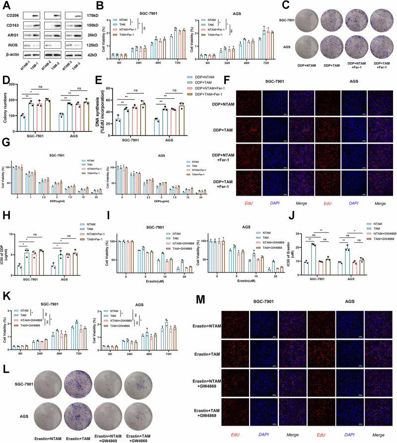
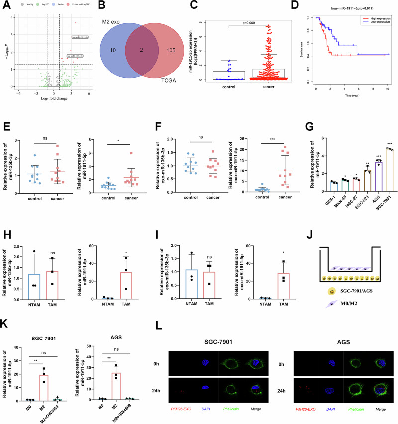
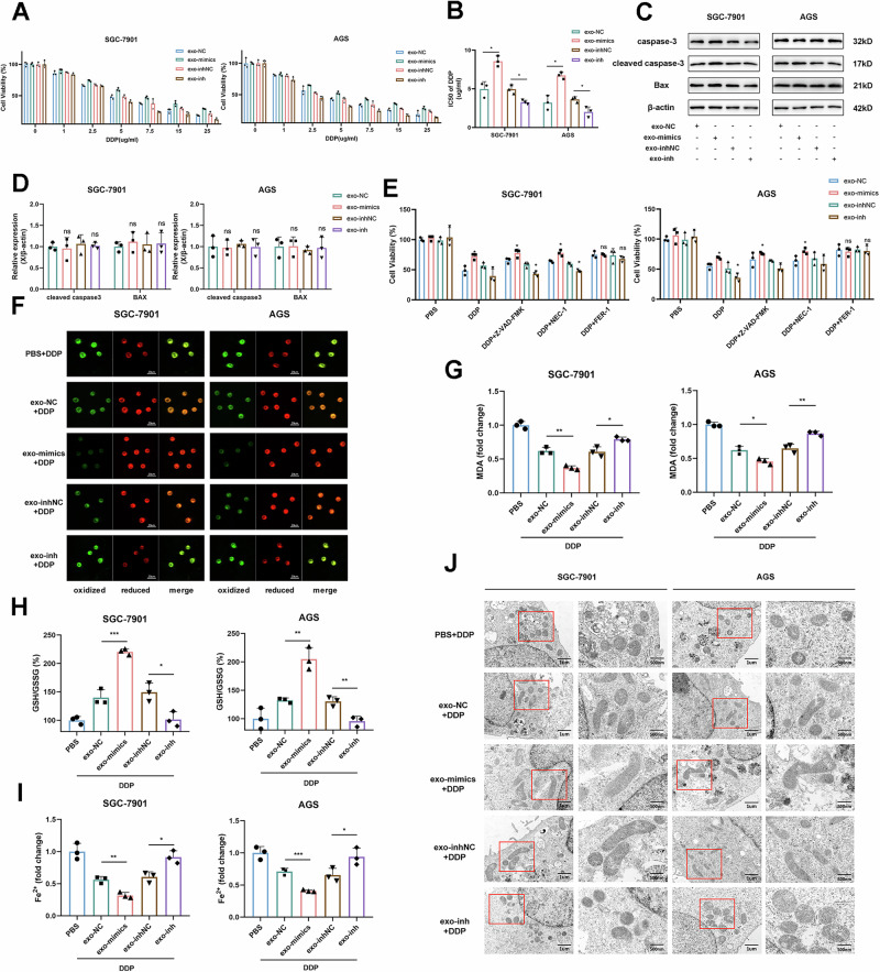
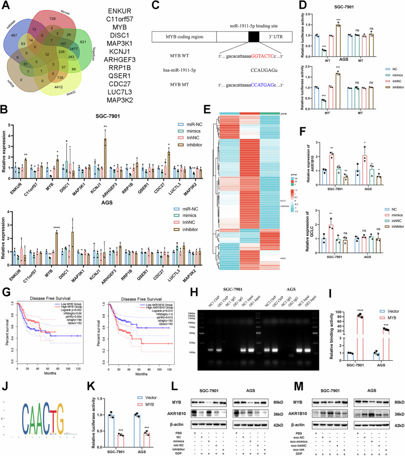
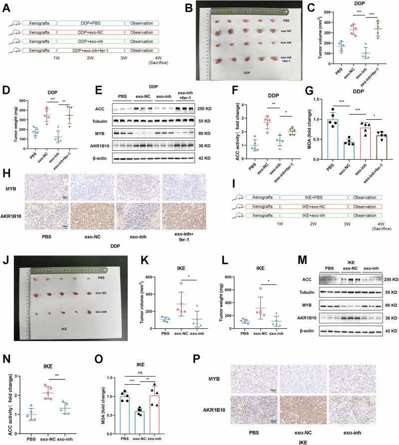
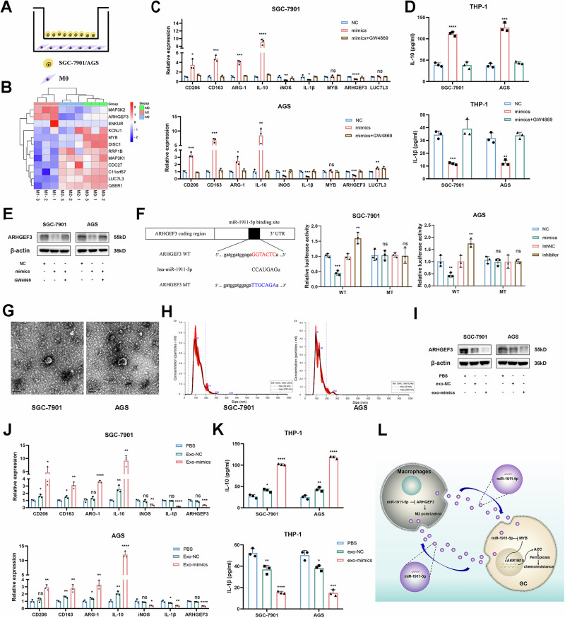
Tumor-associated macrophages (TAMs) have been implicated in fostering various hallmarks of cancer progression in gastric cancer (GC). However, the intricate molecular mechanisms underlying TAM-induced chemoresistance remain incompletely understood. Exosomes emerge as key players, mediating TAM-induced resistance to cisplatin (DDP) by regulating ferroptosis. Our investigation reveals that exo-miR-1911-5p, delivered to GC cells from TAMs, significantly contributes to cisplatin resistance. Specifically, direct modulation of MYB by MiR-1911-5p leads to decreased expression of AKR1B10, a crucial factor in preventing ferroptosis. Further exploration confirms the regulation of ACC by AKR1B10. Through targeting the MYB/AKR1B10/ACC axis, exo-miR-1911-5p inhibits ferroptosis to enhances cisplatin resistance. Additionally, exo-miR-1911-5p promotes M2 polarization of TAMs by targeting ARHGEF3. Collectively, our findings highlight the critical role of exo-miR-1911-5p in mediating cisplatin resistance through modulating the cross-talk between TAMs and GC. Targeting exo-miR-1911-5p could represent a promising strategy for overcoming DDP resistance in GC.


Conflict of interest statement

Competing interests: The authors declare no competing interests.

Figures

Fig. 1
Fig. 1. TAM inhibits ferroptosis to induce cisplatin resistance in GC.
A Validation of macrophage polarization: Protein levels of CD206, CD163, ARG1, iNOS in TAMs from human GC tissues vs. NTAMs from normal tissues by Western blot. β-actin served as a loading control (n  =  3); BF Co-culture with TAMs/NTAMs in the presence or absence of Fer-1 to investigate DDP-induced malignant transformation via (B: CCK-8, C, D: Colony formation, E, F: EdU) (n  =  3); G Relative viability of GC cells co-cultured with TAMs/NTAMs exposed to cisplatin at the indicated concentrations (n  =  3); H The IC50 of GC cells treated with cisplatin (n  =  3); I Relative viability of GC cells co-cultured with TAMs/NTAMs exposed to Erastin at the indicated concentrations (n  =  3). J The IC50 of GC cells treated with Erastin; KM Co-culture with TAMs/NTAMs in the presence or absence of Fer-1 to investigate Erastin-induced malignant transformation via (K: CCK-8, L: Colony formation, M: EdU) (n  =  3). Representative of at least 3 experiments, data displayed as mean ± SD. ****p  <  0.0001, ***p  <  0.001, **p  <  0.01, *p  <  0.05.
Fig. 2
Fig. 2. High expression of exo-miR-1911-5p is transferred from TAMs to GC.
A Volcano map of different miRNA expression levels in M2-exo VS M0-exo in GSE97467; B Venn diagram of the intersection of differential miRNAs; C Expression level of miR-1911-5p in TCGA-STAD; D Survival curve of miR-1911-5p in patients treated with chemotherapy from TCGA-STAD; E, F qRT-PCR of miR-1911-5p and miR-135b-3p expression in GC tissues (E) (n  =  10), exosomes from GC tissues (F) (n  =  10); G Comparison of miR-1911-5p expression in GC cell lines and GES-1 (n  =  3); H, I qRT-PCR of miR-1911-5p and miR-135b-3p expression in TAMs (H) (n  =  3), exosomes from TAMs (I) (n  =  3); J Co-culture pattern of GC cells and M2-like macrophages; K qRT-PCR analysis of miR-1911-5p expression in GC cells after co-culture and addition of GW4869 (n  =  3); L Internalization of PKH26-labelled exosomes (red) in GC cells were observed through confocal microscopy. Representative of at least 3 experiments, data displayed as mean ± SD. ****p  <  0.0001, ***p  <  0.001, **p  <  0.01, *p  <  0.05.
Fig. 3
Fig. 3. Exosomal miR-1911-5p enhances cisplatin resistance in GC by inhibiting ferroptosis.
A Relative viability of GC cells co-cultured with exo-mimics/inhibitor exposed to cisplatin at the indicated concentrations (n  =  3); B The IC50 of GC cells treated with cisplatin; C, D Expression of apoptosis-related proteins detected by Western blot (n  =  3); E Cell viability of GC cells regulated by cell death inhibitors (Z-VAD-FMK, Nec-1, Fer-1) in response to DDP treatment (n  =  3); FI The phenotype of ferroptosis modulated by exo-miR-1911-5p (F Lipid peroxidation product MDA; G Lipid ROS; H GSH/GSSG ratio; I Fe²⁺) (n  =  3); J Electron microscopy used to view mitochondria in GC cells. Representative of at least 3 experiments, data displayed as mean ± SD. ****p  <  0.0001, ***p  <  0.001, **p  <  0.01, *p  <  0.05.
Fig. 4
Fig. 4. miR-1911-5p regulates ferroptosis via MYB/AKR1B10 signaling pathway.
A The prediction of the miR-1911-5p target genes from miRDB, RNA22, TargetScan, Microt4, and miRWalk; B The expression of screened target genes transfected with miR-1911-5p (n  =  3); C The mutant and putative binding site of miR-1911-5p with MYB; D Dual luciferase reporter genes to verify the regulatory relationship between miR-1911-5p and MYB (n  =  3); E PCR array screening for ferroptosis-related genes regulated by miR-1911-5p (n  =  3); F qRT-PCR for validation of miR-1911-5p-regulated ferroptosis-related genes (n  =  3); G Disease-free survival curves for MYB and AKR1B10 in TCGA-STAD; H, I Chip-qpcr to verify the binding of MYB to AKR1B10; J Motif of MYB from JASPAR; K Dual luciferase reporter gene to verify that MYB negatively regulates AKR1B10 (n  =  3); L, M The protein level of MYB and AKR1B10 after transfection with miR-1911-5p or exo-miR-1911-5p. Representative of at least 3 experiments, data displayed as mean ± SD. ****p  <  0.0001, ***p  <  0.001, **p  <  0.01, *p  <  0.05.
Fig. 5
Fig. 5. Exo-miR-1911-5p/MYB/AKR1B10 regulates ferroptosis through ACC pathway.
A Cell viability after DDP stimulation detected by CCK-8 after MYB knockdown (n  =  3); B, C Lipid peroxidation levels were detected after MYB knockdown (B Lipid ROS; C MDA) (n  =  3); D Western blot was performed to detect protein levels of MYB and AKR1B10 after MYB knockdown (n  =  3). E Detection of the GSH/GSSG ratio after knocking down MYB (n  =  3); F Subcellular localization of AKR1B10 and ACC in GC cell lines; G, H The binding of AKR1B10 to ACC was verified by COIP experiments. I The expression of ACC protein were detected after exosomes addition (n  =  3). J ACC activities in GC cells exposed to DDP treated with exo-mimics/inhibitor (n  =  3); K ACC activities in GC cells exposed to DDP treated with exo-inhibitor/si-MYB (n  =  3); LN Phenotypic changes in ferroptosis after the addition of ACC inhibitors (L lipid ROS; M. MDA; N:GSH/GSSG) (n  =  3). Representative of at least 3 experiments, data displayed as mean ± SD. ****p  <  0.0001, ***p  <  0.001, **p  <  0.01, *p  <  0.05.
Fig. 6
Fig. 6. Exo-miR-1911-5p is a therapeutic target for cisplatin resistance in GC.
A Groups of C57BL6 mice injected with DDP; B Xenograft tumors of sacrificed mice treated with cisplatin at the experimental endpoint (n  =  5); C Tumor volume in each group after DDP treatment (n  =  5); D Tumor weight after treatment with DDP in each group (n  =  5); E Western blotting indicates expression levels of MYB, AKR1B10, and ACC (n  =  5). F ACC activity in each group stimulated by DDP (n  =  5); G Exo-miR-1911-5p suppresses MDA levels (n  =  5); H IHC revealed the expression levels of MYB and AKR1B10 (n  =  5). I Pattern of three groups of mice injected with IKE; J Xenograft tumors of sacrificed mice treated with IKE at the experimental endpoint (n  =  5); K, L Tumor volume (K) and weight (L) of nude mice in each group after IKE treatment (n  =  5); M The expression levels of MYB, AKR1B10, and ACC were determined by Western blot (n  =  5); N ACC activity was stimulated by IKE in each group (n  =  5); O IHC revealed expression levels of MYB and AKR1B10 (n  =  5); PExo-miR-1911-5p inhibits MDA levels in the IKE group (n  =  5). Representative of at least 3 experiments, data displayed as mean ± SD. ****p  <  0.0001, ***p  <  0.001, **p  <  0.01, *p  <  0.05.
Fig. 7
Fig. 7. Exo-miR-1911-5p promotes M2 polarization in macrophages via ARHGEF3.
A Co-culture pattern diagrams of M0 type macrophages and GC cell lines; B Heat map of miR-1911-5p target gene expression in M0, M1, M2-like macrophages (n  =  3); C The expression of the M1/M2-like macrophages marker and miR-1911-5p differential target genes were analyzed using qRT-PCR (n  =  3); D Detection of IL-10 and IL-1β in macrophage culture medium by Elisa (n  =  3); E Western blot revealed protein expression of ARHGEF3 after transfection and treatment with GW4869 (n  =  3); F Dual luciferase reporter gene reveals that miR-1911-5p negatively regulates ARHGEF3 (n  =  3); G Morphology of exosomes under TEM; H The size of exosomes detected by NTA; I Western blot demonstrates protein expression of ARHGEF3 after exosome therapy (n  =  3); J The expression of the M1/M2-like macrophahes marker and miR-1911-5p differential target genes after exosome stimulation analyzed by qRT-PCR (n  =  3); K Detection of IL-10 and IL-1β in macrophage culture medium by Elisa (n  =  3); L Mechanism diagram of this article. Representative of at least 3 experiments, data displayed as mean ± SD. ****p  <  0.0001, ***p  <  0.001, **p  <  0.01, *p  <  0.05.

References

    1. Sung, H. et al. Global cancer statistics 2020: GLOBOCAN estimates of incidence and mortality worldwide for 36 cancers in 185 countries. CA Cancer J. Clin.71, 209–249 (2021). - PubMed
    1. Van Cutsem, E., Sagaert, X., Topal, B., Haustermans, K. & Prenen, H. Gastric cancer. Lancet388, 2654–2664 (2016). - PubMed
    1. Blanco-Fernandez, B., Gaspar, V. M., Engel, E. & Mano, J. F. Proteinaceous hydrogels for bioengineering advanced 3D tumor models. Adv. Sci. (Weinh.)8, 2003129 (2021). - PMC - PubMed
    1. Binnewies, M. et al. Understanding the tumor immune microenvironment (TIME) for effective therapy. Nat. Med.24, 541–550 (2018). - PMC - PubMed
    1. Christofides, A. et al. The complex role of tumor-infiltrating macrophages. Nat. Immunol.23, 1148–1156 (2022). - PMC - PubMed

MeSH terms