. 2025 Jul 15;145(1):375.
doi: 10.1007/s00402-025-05977-8.

Glenoid morphology in patients undergoing reverse total shoulder arthroplasty due to fracture

Affiliations

Glenoid morphology in patients undergoing reverse total shoulder arthroplasty due to fracture

Jackson S Hamersly et al. Arch Orthop Trauma Surg. .

Abstract

Introduction: Glenoid morphology in patients undergoing reverse total shoulder arthroplasty (rTSA) due to arthritis has been previously studied; however, it has not been as thoroughly evaluated in fracture populations. The purpose of this study is to utilize pre-operative computed tomography (CT) scans to better understand the glenoid anatomy of those patients undergoing rTSA due to fracture.

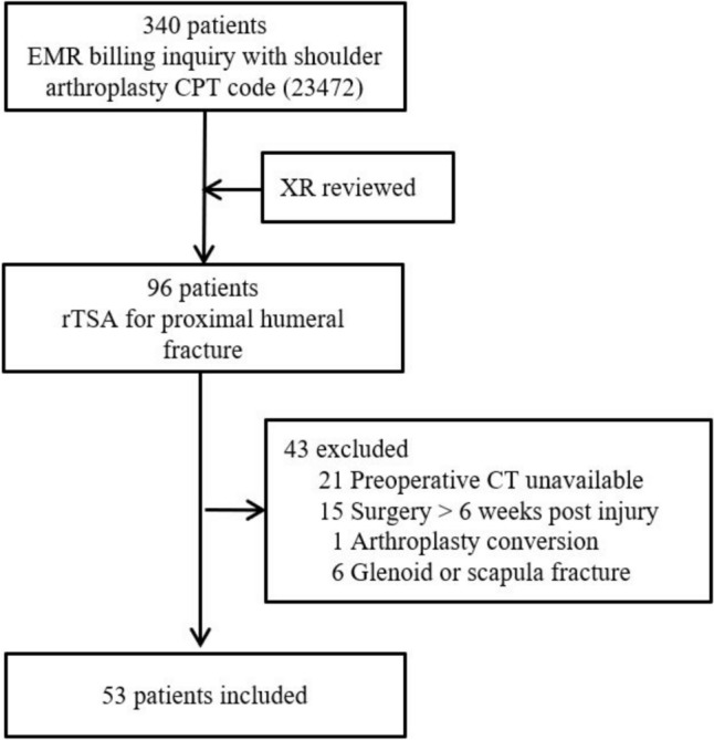
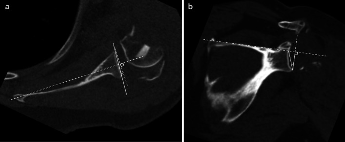
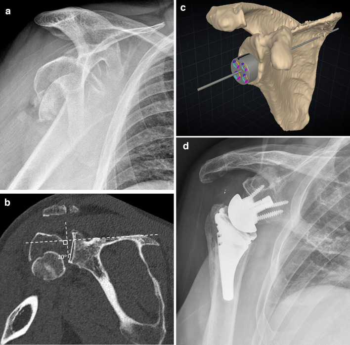
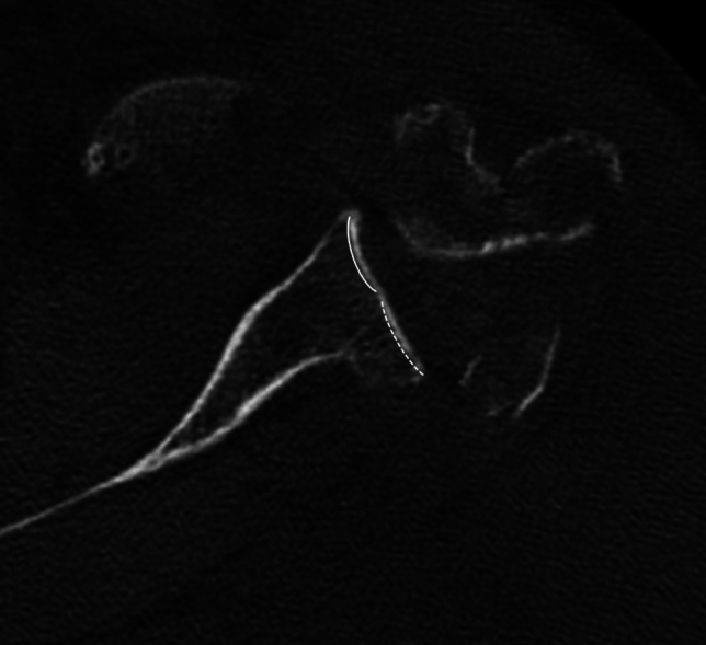
Materials and methods: Patients over the age of 18 who underwent rTSA for proximal humerus fractures from January 1, 2015 to October 31, 2023 at two university health system affiliated hospitals were included if they had a CT scan available for review and image reconstruction. Patients were excluded if a pathologic fracture was identified, surgery was performed greater than 6 weeks after the initial injury, surgery was a conversion or revision surgery, or if a glenoid fracture was present. Glenoid version and reverse shoulder arthroplasty (RSA) angles were measured by a musculoskeletal fellowship-trained radiologist and a shoulder and elbow fellowship-trained orthopaedic surgeon and averaged for final values. Glenoid morphologies were determined using the Walch and Favard classifications.

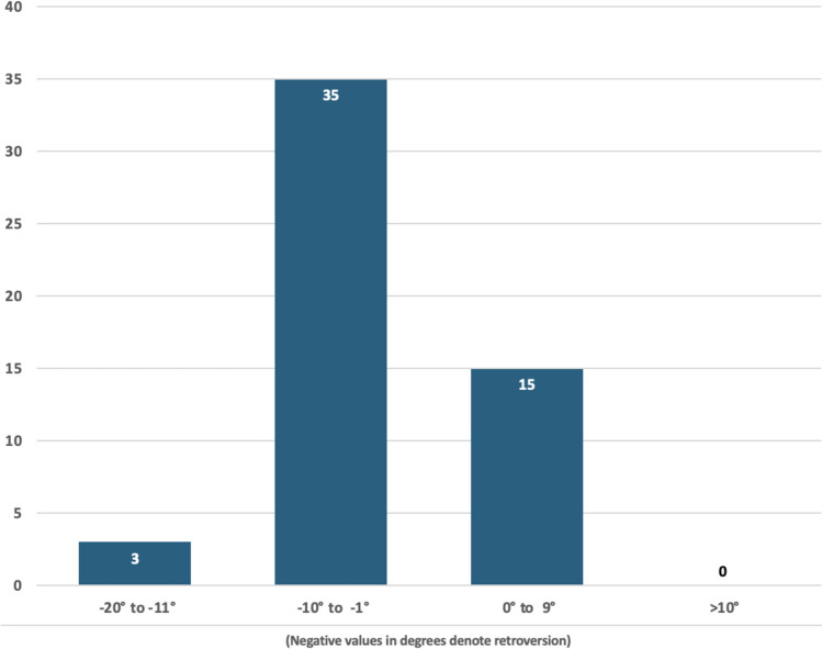
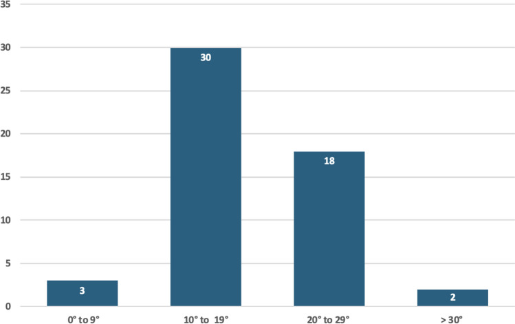
Results: A total of 53 patients with a mean age of 70.4 years (range 36.6-91.2) were included in this study, 84.9% of which were female. Walch A1 glenoid morphology was noted in 92.5% of patients, and Favard E0 morphology was present in 98.1% of patients. Median glenoid version was 3° of retroversion. Median RSA angle was 19°. Of note, 37.7% of patients had a RSA angle of ≥ 20°.

Conclusions: Patients undergoing rTSA for fracture may not have significant glenoid deformity from arthritic wear. However, surgeons should be aware of variations in glenoid version and RSA angle. In this study population, over one-third of patients had a RSA angle of ≥ 20°. Thus, surgeons should take these findings into account when performing rTSA for fracture.

Keywords: Computed tomography; Glenoid anatomy; RSA angle; Reverse total shoulder arthroplasty; Trauma.


Conflict of interest statement

Declarations. Conflict of interest: Jackson Hamersly: None Mason Tippy: None James Slaven: None Yohan Jang: Payments (AO Trauma North America, Orthopaedic Trauma Association) Lauren Ladd: Committee/Panel Member (Skeletal Society of Radiology, Radiologic Society of North America, American Association for Women Radiologists, Indiana Radiologic Society) Mark Dillon: Educational Grants (Depuy Synthes), Paid Consultant (Arthrex), Committee Member (American Academy of Orthopaedic Surgeons and American Shoulder and Elbow Surgeons), Stock (ActivArmor).

Figures

Fig. 1
Fig. 1
Flowchart of selection of patients for inclusion in present study. EMR electronic medical record, CPT current procedural terminology, rTSA reverse total shoulder arthroplasty
Fig. 2
Fig. 2
a Axial CT image demonstrating Glenoid Version measurement by the Friedman line technique. b Coronal CT image showing the reverse shoulder arthroplasty (RSA) angle of glenoid inclination by Boileau technique
Fig. 3
Fig. 3
Graph demonstrating the number of patients in each group of Glenoid Version, grouped by severity in both anteversion (positive angle) and retroversion (negative angle)
Fig. 4
Fig. 4
Graph demonstrating the number of patients in each group of RSA angles, grouped by severity
Fig. 5
Fig. 5
Placement of anatomic glenoid components typically utilize the entire glenoid face (A). However, in rTSA only the inferior portion of the glenoid is used, which can lead to inadvertent superior tilt of the baseplate unless accounted for when planning (B). Reprinted with permission from Boileau et al. [15]
Fig. 6
Fig. 6
A Scapular Y view radiograph of the shoulder showing a comminuted proximal humeral surgical neck fracture with impaction, displacement, and extension through the greater tuberosity. B Coronal CT image through the scapula and glenohumeral joint in the same patient, again showing the comminuted, displaced proximal humeral fracture, as well as measurement of the RSA angle (20 degrees in this patient). C Reconstructed 3D image of the scapula from prior CT scan on the same patient with preoperative templating software used to determine glenoid baseplate position and size. Due to the patient’s anatomy and specifically the RSA angle, an augmented component was utilized. D Grashey radiograph of the right shoulder in the same patient status post rTSA placement demonstrates appropriate position and alignment of the prosthesis, specifically with neutral to slightly inferior alignment of the baseplate. Note is made of incidental metal debris in the subacromial area
Fig. 7
Fig. 7
Axial CT image through the mid glenoid showing Walch class B2 morphology with a biconcave articular surface, including the expected concavity anteriorly (solid line) and secondary concavity posteriorly from osseous wear (dashed line)

Similar articles

References

    1. Baron JA, Karagas M, Barrett J, Kniffin W, Malenka D, Mayor M et al (1996) Basic epidemiology of fractures of the upper and lower limb among Americans over 65 years of age. Epidemiology 7:612–618. 10.1097/00001648-199611000-00008 - PubMed
    1. Hanson B, Neidenbach P, de Boer P, Stengel D (2009) Functional outcomes after nonoperative management of fractures of the proximal humerus. J Shoulder Elbow Surg 18:612–621. 10.1016/j.jse.2009.03.024 - PubMed
    1. Dillon MT, Prentice HA, Burfeind WE, Chan PH, Navarro RA (2019) The increasing role of reverse total shoulder arthroplasty in the treatment of proximal humerus fractures. Injury 50:676–680. 10.1016/j.injury.2019.01.034 - PubMed
    1. Critchley O, McLean A, Page R, Taylor F, Graves S, Lorimer M et al (2020) Reverse total shoulder arthroplasty compared to stemmed hemiarthroplasty for proximal humeral fractures: a registry analysis of 5946 patients. J Shoulder Elbow Surg 29:2538–2547. 10.1016/j.jse.2020.04.005 - PubMed
    1. Cuff DJ, Pupello DR (2013) Comparison of hemiarthroplasty and reverse shoulder arthroplasty for the treatment of proximal humeral fractures in elderly patients. J Bone Joint Surg 95:2050–2055. 10.2106/JBJS.L.01637 - PubMed

LinkOut - more resources