

Intraneural nodular fasciitis in peripheral nerves: report of two cases and literature review
- PMID: 40665257
- PMCID: PMC12261800
- DOI: 10.1186/s12891-025-08926-z
Intraneural nodular fasciitis in peripheral nerves: report of two cases and literature review
Abstract
Background: Although nodular fasciimmon and can occur in various anatomical locations, its occurrence within a nerve is extremely rare. Nodular fasciitis usually resolves spontaneously after partial resection. However, it often presents diagnostic challenges due to its resemblance to malignant diseases, resulting in excessive treatments such as extended nerve excision and nerve transplantation.
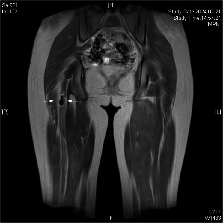
Case presentation: We report two cases of intraneural nodular fasciitis. A 37-year-old woman presented with left upper limb numbness and pain, without trauma history. Preoperative ultrasound was performed. Subtotal resection of the mass in the superficial branch of the radial nerve was conducted. Postoperative pathology and immunohistochemistry confirmed intraneural nodular fasciitis. At 9-month follow-up, symptoms resolved with no recurrence of the mass. A 15-year-old female presented with progressive right lower limb numbness, later accompanied by pain, distal muscle weakness, and difficulty in lifting the foot. Preoperative ultrasound and magnetic resonance imaging were performed. The mass within the sciatic nerve was completely removed. Postoperative pathology and immunohistochemistry confirmed intraneural nodular fasciitis. At 3-month follow-up, symptoms resolved with no recurrence of the mass.
Conclusions: Accurate diagnosis of intraneural nodular fasciitis is crucial to prevent unnecessary treatment. Its ultrasound and magnetic resonance imaging features lack specificity. Preoperative biopsy using ultrasound or computed tomography guidance may be considered if necessary and safe. The histopathological features for intraneural nodular fasciitis exhibits spindle cells in a tissue-culture-like pattern within a richly myxoid matrix, abundant capillaries, inflammatory cell infiltration, frequent mitotic figures without atypia, and infiltrative margins. Immunohistochemically, intraneural nodular fasciitis is characterized by SMA(+) and S100(-). Surgical excision of the lesion is necessary to prevent neurological deficits. And the vast majority of intraneural nodular fasciitis cases spontaneously regress after subtotal resection. A comprehensive diagnostic approach is recommended when intraneural nodular fasciitis is suspected. This article analyzes the diagnostic workup and pathogenesis of all 13 reported intraneural nodular fasciitis cases (including our two), aiming to aid clinicians in achieving precise diagnosis and avoiding overtreatment.
Keywords: Diagnosis; Intraneural nodular fasciitis; Nodular fasciitis; Peripheral nerve tumors; Radial nerve; Sciatic nerve.
© 2025. The Author(s).
Conflict of interest statement
Declarations. Ethics approval and consent to participate: The study was approved by the institutional review board of Shandong Provincial Hospital Affiliated to Shandong First Medical University. This study was approved by Shandong Provincial Hospital Ethics Committee. Approval number (SWYX: NO.2023 − 348). Written informed consent was obtained from all patients/legally authorized representatives of under 16 age participants. Consent for publication: Written informed consent for publication of their clinical details and clinical images was obtained from all the patients/parents. Competing interests: The authors declare no competing interests.
Figures


Similar articles
-
Intraneural Nodular Fasciitis of the Dorsal Scapular Nerve: Case Report and Review of the Literature.J Neurol Surg A Cent Eur Neurosurg. 2023 Jul;84(4):404-407. doi: 10.1055/s-0041-1739218. Epub 2021 Dec 12. J Neurol Surg A Cent Eur Neurosurg. 2023. PMID: 34897609 Review.
-
123I-MIBG scintigraphy and 18F-FDG-PET imaging for diagnosing neuroblastoma.Cochrane Database Syst Rev. 2015 Sep 29;2015(9):CD009263. doi: 10.1002/14651858.CD009263.pub2. Cochrane Database Syst Rev. 2015. PMID: 26417712 Free PMC article.
-
Nodular fasciitis: a case series unveiling novel and rare gene fusions, including two cases with aggressive clinical behavior.Virchows Arch. 2025 Jun;486(6):1235-1245. doi: 10.1007/s00428-025-04040-6. Epub 2025 Feb 6. Virchows Arch. 2025. PMID: 39912885
-
Signs and symptoms to determine if a patient presenting in primary care or hospital outpatient settings has COVID-19.Cochrane Database Syst Rev. 2022 May 20;5(5):CD013665. doi: 10.1002/14651858.CD013665.pub3. Cochrane Database Syst Rev. 2022. PMID: 35593186 Free PMC article.
-
Bioengineered nerve conduits and wraps for peripheral nerve repair of the upper limb.Cochrane Database Syst Rev. 2022 Dec 7;12(12):CD012574. doi: 10.1002/14651858.CD012574.pub2. Cochrane Database Syst Rev. 2022. PMID: 36477774 Free PMC article.
References
-
- <subcutaneous pseudosarcomatous fibro source am j clin pathol so 1955 mar 25 3 241 52.pdf>. - PubMed
-
- Luna A, Molinari L, Bollea Garlatti LA, Ferrario D, Volonteri V, Roitman P, Galimberti G, Mazzuoccolo L. Nodular fasciitis, a forgotten entity. Int J Dermatol. 2019;58(2):190–3. - PubMed
-
- Wang X, De Schepper A, Vanhoenacker F, De Raeve H, Gielen J, Aparisi F, Rausin L, Somville J. Nodular fasciitis: correlation of MRI findings and histopathology. Skeletal Radiol. 2002;31(3):155–61. - PubMed
-
- Lu L, Lao IW, Liu X, Yu L, Wang J. Nodular fasciitis: a retrospective study of 272 cases from China with clinicopathologic and radiologic correlation. Ann Diagn Pathol. 2015;19(3):180–5. - PubMed
-
- Kleinstiver BJ, Rodriguez HA. Nodular fasciitis. A study of forty-five cases and review of the literature. J Bone Joint Surg Am. 1968;50(6):1204–12. - PubMed
Publication types
MeSH terms
Grants and funding
LinkOut - more resources
Full Text Sources
