Alzheimer's disease and its co-pathologies: Implications for hippocampal degeneration, cognitive decline, and the role of APOE ε4
- PMID: 40665478
- PMCID: PMC12263346
- DOI: 10.1002/alz.70483
Alzheimer's disease and its co-pathologies: Implications for hippocampal degeneration, cognitive decline, and the role of APOE ε4
Abstract
Introduction: In neurodegenerative dementias, the co-occurrence and interaction of amyloid β peptide (Aβ), tau pathology, and other pathological lesions confound their individual contributions to neurodegeneration and their modulation by risk factors.
Methods: We analyzed 480 post mortem human brains (ages 50-99) using regression and structural equation models to assess the relationships among Aβ, tau, limbic-predominant age-related TDP-43 encephalopathy neuropathological changes (LATE-NC), α-synuclein, other age-related lesions, and apolipoprotein E (APOE) ε4, as well as their effects on CA1 neuronal density, brain weight, and cognitive status.
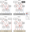
Results: Aβ, tau, LATE-NC, and amygdala-predominant α-synuclein pathology were mutually interdependent. Tau was the strongest predictor of global neurodegeneration, while LATE-NC primarily, but not exclusively, affected hippocampal neuron loss. Small vessel disease correlated with both LATE-NC and α-synuclein, while APOE ε4 was mainly associated with extracellular parenchymal and capillary Aβ pathology.
Discussion: Although Alzheimer's disease pathology plays a central role in brain degeneration, coexisting pathologies can both exacerbate and independently contribute to it. These factors should be considered in patient stratification.
Highlights: In aging individuals, amyloid β peptide (Aβ), tau pathology, limbic-predominant age-related TDP-43 encephalopathy neuropathological changes (LATE-NC), and amygdala-predominant α-synuclein pathology were interrelated but contributed independently to neurodegeneration. LATE-NC was the strongest driver of CA1 neuronal loss, while tau burden was the strongest predictor of global brain degeneration. Apolipoprotein E ε4 was associated with both extracellular and capillary Aβ deposits, but not with tau burden. Temporal lobe small vessel disease was associated with both LATE-NC and amygdala-predominant α-synuclein pathology. Neural network models can reliably identify hippocampal pyramidal neurons on hematoxylin-stained histological slides.
Keywords: amyloid β; apolipoprotein E ε4; cerebral amyloid angiopathy; digital pathology; hippocampal degeneration; limbic‐predominant age‐related TDP‐43 encephalopathy neuropathological changes; medial temporal lobe; mixed dementia; small vessels disease; tau; α synuclein.
© 2025 The Author(s). Alzheimer's & Dementia published by Wiley Periodicals LLC on behalf of Alzheimer's Association.
Conflict of interest statement
DRT and SOT received consultant honoraria from Muna Therapeutics (Belgium). DRT collaborated with Novartis Pharma AG (Switzerland), and GE Healthcare (UK). CAFvA has received honoraria for serving on the scientific advisory board of Biogen, Roche, Novo Nordisk, BioNTech, Lilly, Dr Willmar Schwabe GmbH & Co.KG and MindAhead UG. Additionally, CAFvA has received travel funding and speaker honoraria from Biogen, Lilly, Novo Nordisk, Roche Diagnostics AG, Novartis, Medical Tribune Verlagsgesellschaft mbH, Landesvereinigung für Gesundheit und Akademie für Sozialmedizin Niedersachsen e. V., FomF GmbH | Forum für medizinische Fortbildung, and Dr Willmar Schwabe GmbH & Co.KG. Research support was received from Roche Diagnostics AG, and funding was provided by the Innovationsfond (Fund of the Federal Joint Committee, Gemeinsamer Bundesausschuss, G‐BA; Grants No. VF1_2016‐201; 01NVF21010; 01VSF21019). MO has provided scientific advice to Fujirebio, Roche, Biogen, Lilly, and Axon. RV's institution has clinical trial agreements (RV as PI) with Alector, AviadoBio, Biogen, Denali, J&J, Eli Lilly, and UCB. RV's institution has consultancy agreements (RV as member of DSMB) with AC Immune. All other authors had nothing to disclose. Author disclosures are available in the supporting information.
Figures


Similar articles
-
APOE4 impact on soluble and insoluble tau pathology is mostly influenced by amyloid-β.Brain. 2025 Jul 7;148(7):2373-2383. doi: 10.1093/brain/awaf016. Brain. 2025. PMID: 39817469 Free PMC article.
-
Neuropathological hallmarks in the post-mortem retina of neurodegenerative diseases.Acta Neuropathol. 2024 Aug 19;148(1):24. doi: 10.1007/s00401-024-02769-z. Acta Neuropathol. 2024. PMID: 39160362 Free PMC article.
-
Limbic-predominant age-related TDP-43 encephalopathy (LATE): consensus working group report.Brain. 2019 Jun 1;142(6):1503-1527. doi: 10.1093/brain/awz099. Brain. 2019. PMID: 31039256 Free PMC article.
-
CSF tau and the CSF tau/ABeta ratio for the diagnosis of Alzheimer's disease dementia and other dementias in people with mild cognitive impairment (MCI).Cochrane Database Syst Rev. 2017 Mar 22;3(3):CD010803. doi: 10.1002/14651858.CD010803.pub2. Cochrane Database Syst Rev. 2017. PMID: 28328043 Free PMC article.
-
Dysfunction of the blood-brain barrier in Alzheimer's disease: Evidence from human studies.Neuropathol Appl Neurobiol. 2022 Apr;48(3):e12782. doi: 10.1111/nan.12782. Epub 2022 Feb 2. Neuropathol Appl Neurobiol. 2022. PMID: 34823269
References
-
- Nichols E, Merrick R, Hay SI, et al. The prevalence, correlation, and co‐occurrence of neuropathology in old age: harmonisation of 12 measures across six community‐based autopsy studies of dementia. The Lancet Healthy Longevity. 2023;4(3):e115‐e125. doi: 10.1016/S2666-7568(23)00019-3 - DOI - PMC - PubMed
MeSH terms
Substances
Grants and funding
- G0F8516N/Fonds Wetenschappelijk Onderzoek
- G065721N/Fonds Wetenschappelijk Onderzoek
- G024925N [DRT]/Fonds Wetenschappelijk Onderzoek
- 1225725N [SOT]/Fonds Wetenschappelijk Onderzoek
- C14/17/107/KU Leuven Onderzoeksraad
- C14/22/132/KU Leuven Onderzoeksraad
- C3/20/057 [DRT]/KU Leuven Onderzoeksraad
- PDMT2/21/069 [SOT]/KU Leuven Onderzoeksraad
- 22-AAIIA-963171 [DRT]/ALZ/Alzheimer's Association/United States
- AARF-24-1300693 [SOT]/ALZ/Alzheimer's Association/United States
- TH-624-4-1/Deutsche Forschungsgemeinschaft
- TH-624-4-2/Deutsche Forschungsgemeinschaft
- TH-624-6-1 [DRT]/Deutsche Forschungsgemeinschaft
- SFB1279 [MO]/Deutsche Forschungsgemeinschaft
- #10810/Alzheimer Forschung Initiative
- #13803 [DRT]/Alzheimer Forschung Initiative
- A2022019F[SOT]/BrightFocus Foundation
- FTLDc01GI1007A [MO]/German Federal Ministry of Education and Research
- D.5009 [MO]/Boehringer Ingelheim Ulm University BioCenter
- 01EW2008 [MO]/EU Moodmarker program
- D.2468/Thierry Latran Foundation
- D.2468 [MO]/Thierry Latran Foundation
- D.3830 [MO]/Foundation of the State of Baden-Württemberg
- 01GI1007A [MO]/BonaRes
- [MO]/Roux Program of the Martin Luther University Halle-Wittenberg
- 01ED2008A [MO]/EU Joint Programme-Neurodegenerative Diseases network Genfi-Prox
LinkOut - more resources
Full Text Sources
Medical
Miscellaneous

