

Morphological and Histological Variations of the Reproductive Organs During the Annual Cycle in a Neotropical Bat: Peters' Ghost-Faced Bat Mormoops megalophylla (Chiroptera: Mormoopidae)
- PMID: 40665677
- PMCID: PMC12264469
- DOI: 10.1002/jmor.70066
Morphological and Histological Variations of the Reproductive Organs During the Annual Cycle in a Neotropical Bat: Peters' Ghost-Faced Bat Mormoops megalophylla (Chiroptera: Mormoopidae)
Abstract
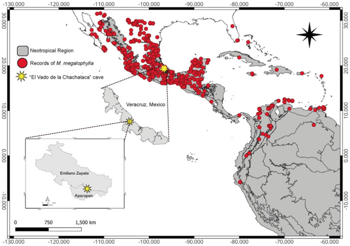
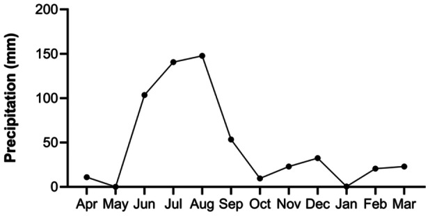
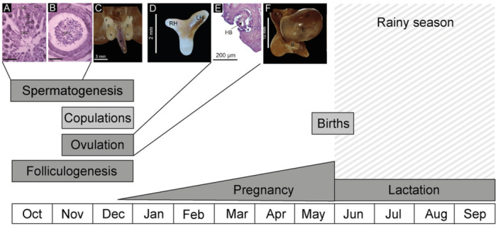
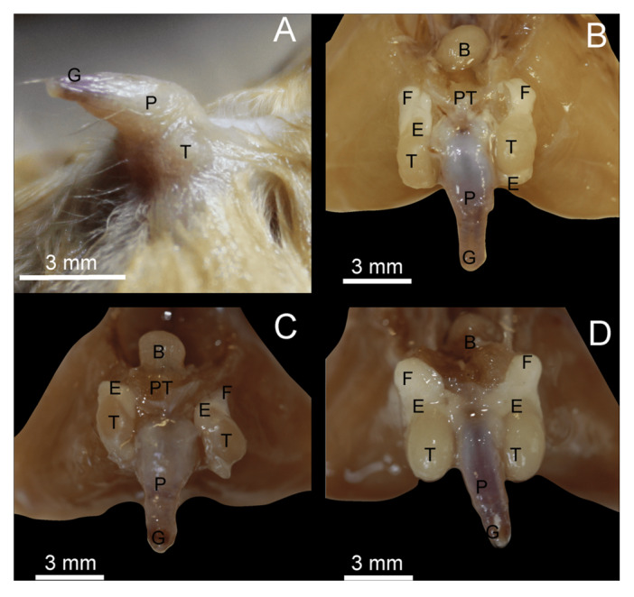
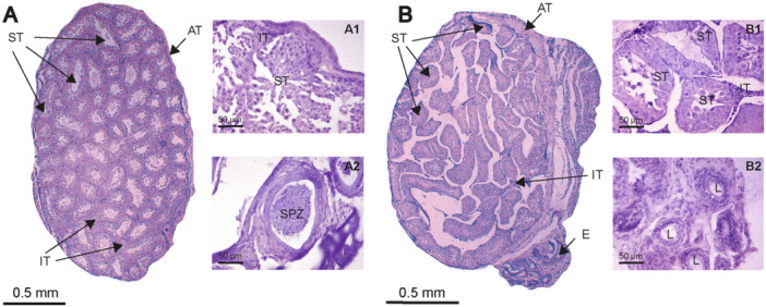
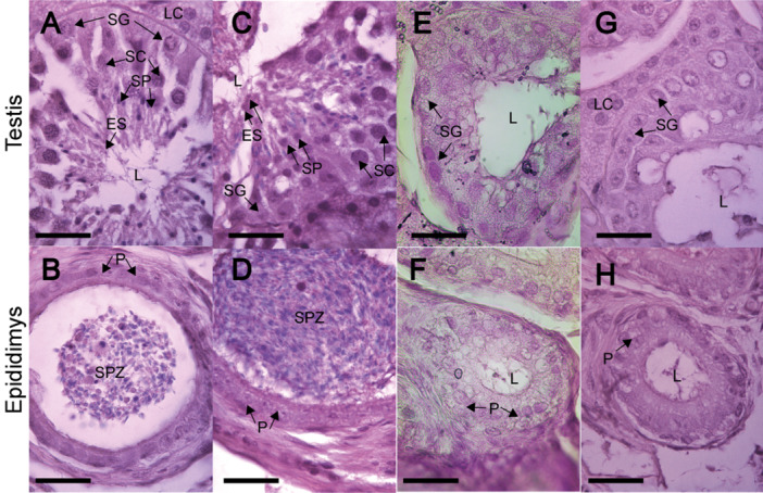
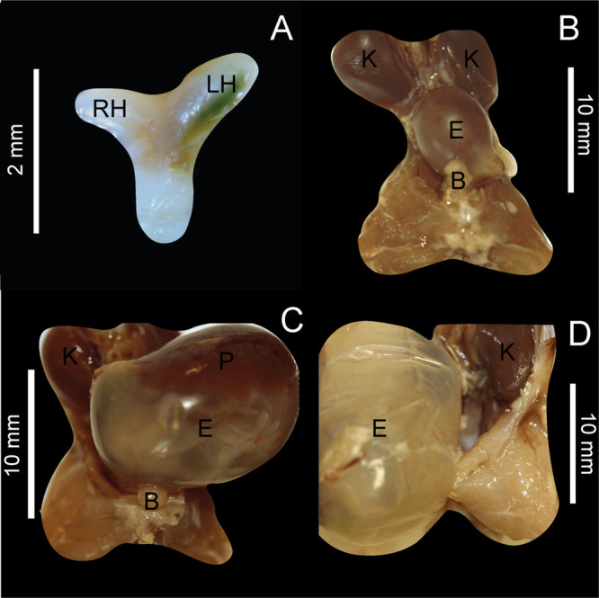
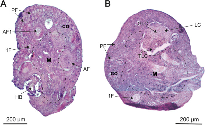
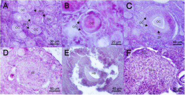
Recent studies have emphasized the ecological significance of bats as insect regulators. This recognition has prompted an increased scientific interest in Mormoops megalophylla, a notable neotropical insectivorous bat species. The extant literature on its biology remains limited and substantial knowledge gaps persist, particularly regarding its reproductive cycle. This study sought to examine morphological and histological variations in the reproductive organs of male and female M. megalophylla over the annual cycle to elucidate the stages of its reproductive process. Over the course of the year, five sexually mature individuals of each sex were sampled monthly, culminating in a total of 120 specimens, to document variations in external sexual characters, the testes, epididymides, uterus, and ovaries; all sampled individuals underwent morphological, morphometric, and histological analyses. The findings indicate that M. megalophylla has migratory testes with seasonal spermatogenesis occurring from October to December, a bicornuate uterus, and a single folliculogenic period that is synchronized with spermatogenesis. This suggests a monoestrous, seasonal, and synchronous reproductive pattern. At the population level, copulation occurs between November and December, gestation occurs between December and May, and parturition occurs between late May and early June. The lactation period extended from June to September.
Keywords: biology; folliculogenesis; reproduction; sinistral dominance; spermatogenesis.
© 2025 The Author(s). Journal of Morphology published by Wiley Periodicals LLC.
Figures
Similar articles
-
Sexual Harassment and Prevention Training.2024 Mar 29. In: StatPearls [Internet]. Treasure Island (FL): StatPearls Publishing; 2025 Jan–. 2024 Mar 29. In: StatPearls [Internet]. Treasure Island (FL): StatPearls Publishing; 2025 Jan–. PMID: 36508513 Free Books & Documents.
-
Seasonal Analysis of the Male Reproductive Tract and Germ Cell Proliferation in Leptodactylus podicipinus (Anura).J Morphol. 2025 Aug;286(8):e70072. doi: 10.1002/jmor.70072. J Morphol. 2025. PMID: 40778485 Free PMC article.
-
The Black Book of Psychotropic Dosing and Monitoring.Psychopharmacol Bull. 2024 Jul 8;54(3):8-59. Psychopharmacol Bull. 2024. PMID: 38993656 Free PMC article. Review.
-
Morphological, functional and neurological outcomes of craniectomy versus cranial vault remodeling for isolated nonsyndromic synostosis of the sagittal suture: a systematic review.JBI Database System Rev Implement Rep. 2015 Sep;13(9):309-68. doi: 10.11124/jbisrir-2015-2470. JBI Database System Rev Implement Rep. 2015. PMID: 26470674
-
FSH replaced by low-dose hCG in the late follicular phase versus continued FSH for assisted reproductive techniques.Cochrane Database Syst Rev. 2013 Mar 28;2013(3):CD010042. doi: 10.1002/14651858.CD010042.pub2. Cochrane Database Syst Rev. 2013. PMID: 23543584 Free PMC article.
References
-
- Albuja, L. 1999. Murciélagos del Ecuador. Cicetronic Cía. Ltda. Offset. 2nd ed.
-
- Álvarez‐Castañeda, S. T. , Álvarez T., and González‐Ruiz N.. 2017. Keys for Identifying Mexican Mammals. JHU Press. 10.1353/book.50028. - DOI
-
- Ammerman, L. K. , Hice C. L., and Schmidly D. J.. 2012. Bats of Texas (Vol. 43). Texas A&M University Press.
-
- Anónimo . 2010. Lineamientos para la conducción ética de la investigación, docencia y difusión. División de Ciencias Biológicas y de la Salud, Universidad Autónoma Metropolitana, Iztapalapa, Ciudad de México, México.
-
- Anthony, E. L. P. 1988. “Age Determination in Bats.” In Ecological and Behavioral Methods for the Study of Bats, edited by Kunz T. H., 47–58. Smithsonian Institution Press.
MeSH terms
LinkOut - more resources
Full Text Sources
