. 2025 Jul 11:17:299-312.
doi: 10.2147/ORR.S525292. eCollection 2025.

Combination Hyaluronic Acid and Multipotent Stromal Cells Fails to Improve Rat Knee OA Outcomes Compared to Cells Alone

Affiliations

Combination Hyaluronic Acid and Multipotent Stromal Cells Fails to Improve Rat Knee OA Outcomes Compared to Cells Alone

Kennedy Michele Davis et al. Orthop Res Rev. .

Abstract

Introduction: Multipotent Stromal Cells (MSCs) are utilized as therapeutic agents for addressing musculoskeletal conditions, including knee osteoarthritis (OA). However, major challenges in the clinical application include maintenance of the cells in the joint capsule. Hyaluronic acid (HA) is endogenous in synovial joints and commercially available as a joint lubricant. We tested the hypothesis that delivery of MSCs in HA into an OA rat knee model could improve outcomes.

Methods: Rat bone marrow MSCs were suspended in a commercially available HA paste, and cell viability measured with live/dead stains. Biomarkers for MSC chondrogenesis and osteogenesis were monitored with PCR. MSCs with or without HA were injected into the knees of OA rats and histology conducted 6 weeks later.

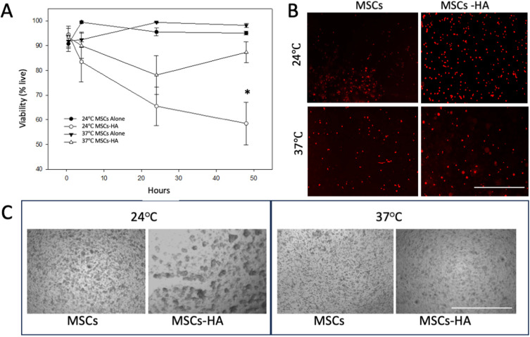
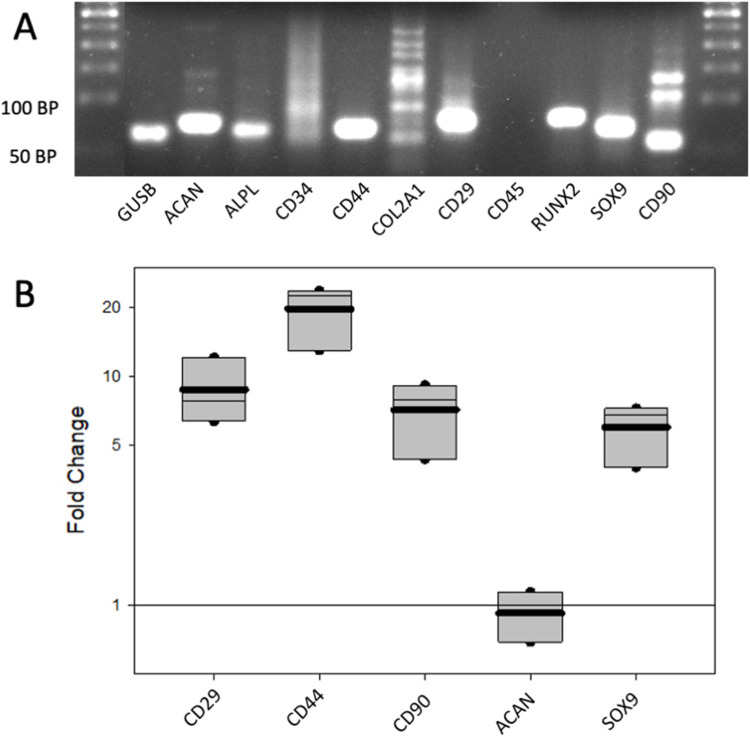
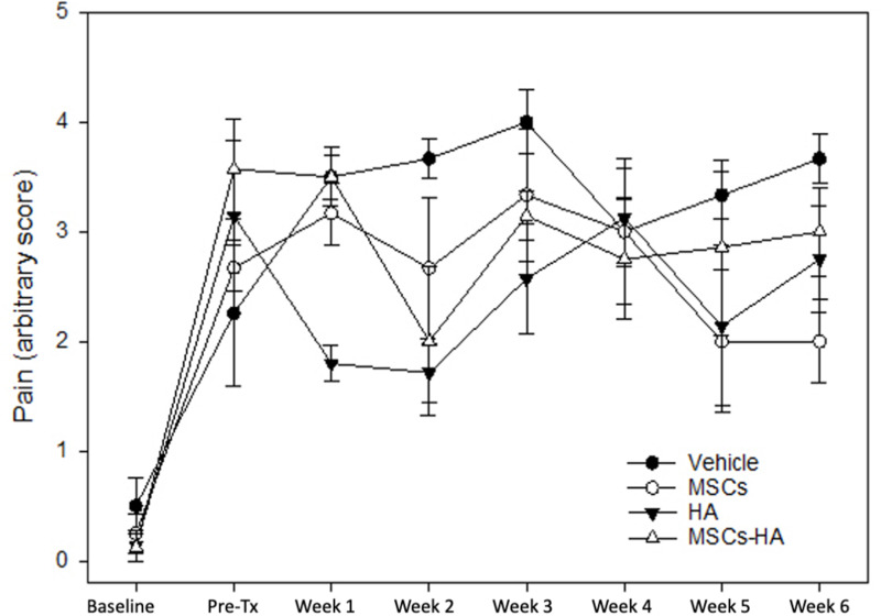
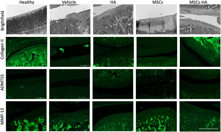
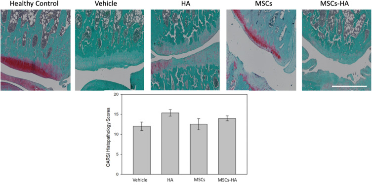
Results: Suspending MSC in HA resulted in a slight reduction in viability. The gene expression profile showed an increase in MSC biomarkers for cells in HA with a decrease in osteogenic markers. Four groups of treatment (vehicle, MSCs alone, HA alone, MSCs + HA) were injected into the knees of osteoarthritic rats. Pain scores, collected weekly, showed no difference between the groups. Immunohistochemistry for inflammatory markers illustrated no obvious differences between groups. Proteoglycans, indicative of cartilage, showed a loss in the vehicle group and modest signs of cartilage with MSCs alone, but when mixed with the HA, any benefit was lost. OARSI Histological Scoring completed by 2 independent technicians concluded no improvement in joint integrity with the addition of HA.

Conclusion: A commercially available HA failed to enhance joint regeneration compared to MSCs alone.

Keywords: Hyaluronic acid; multipotent stem cell; osteoarthritis.


Conflict of interest statement

MH, DM, AW, SH and LSB are employees of Likarda, Inc, which gains no benefit from the results of this study. LSB is also the founder and has equity in Likarda, Inc. The authors report no other conflicts of interest in this work.

Figures

Figure 1
Figure 1
Live/Dead Staining of HA-Entrapped MSCs. (A) Cells were cultured in standard conditions (37°C and 5%CO2) or at room temperature (24°C) in either standard cell culture media or suspended in a media-HA paste (MSCs-HA). The viability of the cells in standard culture media was higher than cells in HA, especially when held at room temperature (p < 0.05 at 48 hours). (B) The images provide examples of the dead cells (red) in each condition at the first time point at 48 hours. Scale bar = 500 μm. (C) Changes in cell morphology were observed under brightfield microscopy of MSCs alone and MSC-HA suspension over time. At the final reading of 48 hours, cells suspended in HA displayed greater amounts of cell clustering than MSCs alone when stored at 4°C and 24°C. When cultured in an incubator at 37°C, both groups had similar cell morphology. Scale bar = 1000 μm.
Figure 2
Figure 2
Gene Expression Profile of MSCs in HA. (A) The gel electrophoresis of the amplified probes shows clear bands of the anticipated size, demonstrating the efficiency of the extraction method. CD34 and CD45 are negative markers for MSCs. (B) The fold change in the dCT levels after 7 days of suspension in HA are shown. The panel of biomarkers used to identify MSCs showed clear positive amplification of CD29, CD44, and CD90. Biomarkers of chondrogenic differentiation were either the same between groups (ACAN) or higher with HA (SOX9). Probes that averaged a Ct value of 35 or greater were defined as not detected and not included in analysis.
Figure 3
Figure 3
Pain Scores of Rats with Knee OA. OA was induced in rat knees by destabilizing the medial meniscus. Prior to the destabilization procedure, each animal was tested for pain in the knees using a standard rodent pain assessment, providing a baseline measurement of non-painful joints. After the knee destabilization but before injection of the treatments (Pre-Tx) the pain levels went up in all 4 groups. A week after the intra-articular injections of the treatments, only animals receiving HA alone had a transient reduction in pain for 2 weeks. By week 3, there were no differences between scores for the 4 groups.
Figure 4
Figure 4
Immunohistochemistry of Rat Knee Joints. Knee joints were stained for markers of early and advanced OA. The knees of healthy (non-injury model) animals were included as a comparison. The brightfield images illustrated the abnormal surface of the joints with crevices and fibrillations in the knees of the OA rats regardless of the treatment group. Collagen II was clearly identified in the healthy animal but not in other groups. Staining for the metalloproteinase, ADAMTS5, illustrated a low level of punctate staining in joints from all groups. MMP-13 staining was similar across groups. The scale bar = 1mm.
Figure 5
Figure 5
Blinded Scoring of Knee Joint Integrity. Joint sections were stained with Fast Green and Safranin O (red) to visualize the bone morphology and proteoglycan level (red), indicative of cartilage. An image of a healthy knee shows red staining (proteoglycan) along the surface of the bone. However, animals that underwent OA induction and received the different treatments all showed lower levels of proteoglycans and a rougher bony surface. Multiple images from each animal were scored by blinded scientists using the OARSI Histopathology Scoring System. Lower scores indicate better joint integrity. The results showed no statistical difference between the treatment groups.

Similar articles

References

    1. Cui A, Li H, Wang D, Zhong J, Chen Y, Lu H. Global, regional prevalence, incidence and risk factors of knee osteoarthritis in population-based studies. EClin Med. 2020;2020:29–30. - PMC - PubMed
    1. Arslan I, Damen J, deWilde M, et al. Incidence and prevalence of knee osteoarthritis using codified and narrative data from electronic health records: a population-based study. Arthritis Care Res. 2022;74(6):937–944. doi: 10.1002/acr.24861 - DOI - PMC - PubMed
    1. Verges J, Vitaloni M, Bibas M, et al. Global management of patients with knee osteoarthritis begins with quality of life assessment: a systematic review. BMC Musculoskelet Disord. 2019;20(1):493. doi: 10.1186/s12891-019-2895-3 - DOI - PMC - PubMed
    1. Kawano M, Araujo I, Castro M, Matos M. Assessment of quality of life in patients with knee osteoarthritis. Acta Ortop Bras. 2015;23(6):307–310. doi: 10.1590/1413-785220152306150596 - DOI - PMC - PubMed
    1. Fuggle N, Cooper C, Oreffo R, et al. Alternative and complementary therapies in osteoarthritis and cartilage repair. Aging Clin Exp Res. 2020;32(4):547–560. doi: 10.1007/s40520-020-01515-1 - DOI - PMC - PubMed

LinkOut - more resources