. 2025 Jul 1:13:1551010.
doi: 10.3389/fcell.2025.1551010. eCollection 2025.

Repurposing piroxicam enhances the antineoplastic effects of docetaxel and enzalutamide in prostate cancer cells using 2D and 3D in vitro culture models

Affiliations

Repurposing piroxicam enhances the antineoplastic effects of docetaxel and enzalutamide in prostate cancer cells using 2D and 3D in vitro culture models

Amani Yehya et al. Front Cell Dev Biol. .

Abstract

Introduction: Drug repurposing is gaining consideration in cancer due to the challenges of poor outcomes and resistance associated with the current conventional modalities. Non-steroidal anti-inflammatory drugs (NSAIDs), widely used for treating inflammation, are being explored for their potential efficacy in cancer treatment, including prostate cancer (PCa). This study aims to evaluate the efficacy of Piroxicam (PXM), an NSAID, in enhancing the sensitivity of PCa cells to chemotherapy and hormonal drugs.

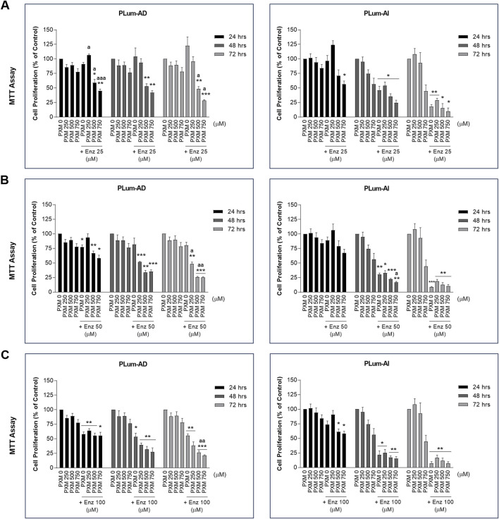
Methods: Computational analysis was conducted to identify differentially expressed genes between our established murine PCa cell models, PLum-AD (androgen-dependent) and PLum-AI (androgen-independent), to uncover potential therapeutic targets. In two-dimensional (2D) cell culture, cell proliferation, viability, and migration assays were performed on PLum-AD and PLum-AI cells treated with PXM alone or in combination with docetaxel (Doc) or enzalutamide (Enz). Additionally, the impact of these treatments on stem-like progenitor cells was assessed using three-dimensional (3D)-Matrigel™-based sphere-forming and organoid formation assays.

Results: Transcriptomic analysis revealed that inflammatory pathways are enriched during PCa progression, making them viable targets for NSAID-based interventions. Single treatment of PXM demonstrated significant anti-cancer effects on PLum-AD and PLum-AI cells, evidenced by reduced cell proliferation, viability, migration, sphere growth, and organoid growth.

Discussion: Importantly, PXM treatment in combination with Doc or Enz resulted in more pronounced antineoplastic effects compared to single-drug exposure. Our work suggests PXM as a potential adjunctive therapy to enhance the efficacy of conventional treatments in PCa patients.

Keywords: docetaxel; drug repurposing; enzalutamide; organoids; piroxicam; prostate cancer; tumor spheres.


Conflict of interest statement

The authors declare that the research was conducted in the absence of any commercial or financial relationships that could be construed as a potential conflict of interest. The author(s) declared that they were an editorial board member of Frontiers, at the time of submission. This had no impact on the peer review process and the final decision.

Figures

FIGURE 1
FIGURE 1
Inflammatory and epithelial-mesenchymal transition pathways are implicated in murine PCa progression. (A) Venn diagram representing the significantly differentially expressed genes (DEGs) between murine PLum-AI and PLum-AD PCa cells obtained through analysis of GSE151187 microarray data using GEO2R–GEO tool (Adjusted p-value and Log 2-fold change threshold set at 0.01 and 2, respectively; (B) Volcano plot representing the downregulated (blue dots) and upregulated (red dots) genes between PLum-AI and PLum-AD cells obtained through analysis of GSE151187 microarray data using GEO2R–GEO tool (Adjusted p-value and Log 2-fold change threshold set at 0.01 and 2, respectively; (C) Clustergram representing the enriched terms obtained by pathway analysis of the DEGs in the murine PCa cells using the Enrichr tool MSigDB Hallmark 2020 database; (D) Protein-protein interaction network for DEGs derived from the selected pathways of relevance to PCa and inflammation that included epithelial-mesenchymal transition, interferon gamma response, TNF-alpha signaling via NF-kB, IL-6/JAK/STAT3 signaling, and inflammatory response. Functional protein association network was established using STRING tool (version 12.0). DEGs: differentially expressed genes; EMT: epithelial-mesenchymal transition; TNFα: tumor necrosis factor-alpha; NF-kB: nuclear factor kappa-light-chain-enhancer of activated B cells; IFNγ: interferon-gamma; IR: inflammatory response.
FIGURE 2
FIGURE 2
Increasing concentrations of piroxicam, alone or in combination with docetaxel, reduced the proliferation of murine prostate cancer cells. The antiproliferative effect of 250, 500, and 750 μM of piroxicam on PLum-AD and PLum-AI cells alone or in combination with 1 (A), 5 (B), and 10 (C) nM of docetaxel was assessed in triplicates using thiazolyl blue tetrazolium bromide (MTT) assay at 24, 48, and 72 h post-treatment. Results are expressed as a percentage of the treated groups compared to the control at each time point and data are reported as mean ± SEM of at least three independent experiments (*P < 0.05; **P < 0.01; ***P < 0.001 indicate significance when comparing to the untreated group and a P < 0.05; aa P < 0.01 indicate significance when comparing the piroxicam/docetaxel combination treated groups to docetaxel treated group). PXM: piroxicam; Doc: docetaxel.
FIGURE 3
FIGURE 3
Increasing concentrations of piroxicam, alone or in combination with enzalutamide, reduced the proliferation of murine prostate cancer cells. The antiproliferative effect of 250, 500, and 750 μM of piroxicam on PLum-AD and PLum-AI cells alone or in combination with 25 (A), 50 (B), and 100 (C) μM of enzalutamide was assessed in triplicates using thiazolyl blue tetrazolium bromide (MTT) assay at 24, 48, and 72 h post-treatment. Results are expressed as a percentage of the treated groups compared to the control at each time point and data are reported as mean ± SEM of at least three independent experiments (*P < 0.05; **P < 0.01; ***P < 0.001 indicate significance when comparing to the untreated group and a P < 0.05; aa P < 0.01; aaa P < 0.001 indicate significance when comparing the piroxicam/enzalutamide combination treated groups to enzalutamide treated group). PXM: piroxicam; Enz: enzalutamide.
FIGURE 4
FIGURE 4
Increasing concentrations of piroxicam, alone or in combination with docetaxel or enzalutamide, reduced the viability of murine prostate cancer cells in a dose-dependent manner. The effect of 250 and 500 μM of PXM on the viability of PLum-AD and PLum-AI cells alone or in combination with 1 and 5 nM of docetaxel (A) or 25 and 50 μM of enzalutamide (B) was determined in duplicates using the trypan blue exclusion assay at 24 and 48 h post-treatment. Results are expressed as a percentage of the treated groups compared to the control at each time point and data are reported as mean ± SEM of three independent experiments (***P < 0.001 indicates significance when comparing to the untreated group, a P < 0.05; aa P < 0.01; aaa P < 0.001 indicate significance when comparing the piroxicam/1 nM of docetaxel and piroxicam/25 μM of enzalutamide combination treated groups to 1 nM of docetaxel and 25 μM of enzalutamide treated group, respectively, **P < 0.01; ***P < 0.001 indicate significance when comparing the piroxicam/5 nM of docetaxel and piroxicam/50 μM of enzalutamide combination treated groups to 5 nM of docetaxel and 50 μM of enzalutamide treated group, respectively). PXM: piroxicam; Doc: docetaxel; Enz: enzalutamide.
FIGURE 5
FIGURE 5
Piroxicam, alone or in combination with docetaxel or enzalutamide, reduced the migration indices of murine prostate cancer cells. Representative bright-field images of the wounds in PLum-AD at 0 h and 16 h and in PLum-AI cells at 0 h and 24 h post-treatment with PXM alone or in combination with docetaxel (A) or enzalutamide (B). Images were acquired using Axiovert microscope from Zeiss at ×5 magnification. PXM: piroxicam; Doc: docetaxel; Enz: enzalutamide; T: hours post-treatment.
FIGURE 6
FIGURE 6
Increasing concentrations of Piroxicam, alone or in combination with docetaxel or enzalutamide, reduced the migration indices of murine prostate cancer cells. The effect of 250 and 500 μM of PXM on the migration of PLum-AD and PLum-AI cells alone or in combination with 1 and 5 nM of docetaxel (A) or 25 and 50 μM of enzalutamide (B) was determined using the wound healing assay. A uniform scratch was made in PLum-AD and PLum-AI confluent monolayer of cells using 200 μL micropipette tips. The wound area was monitored over time and serial images were captured at ×5 magnification at 0 h and up to 16 and 24 h post-treatment for PLum-AD and PLum-AI, respectively. Quantification of the open wound area was assessed over time using ImageJ tool. For each group, results are presented as the percentage of wound area at 16 h for PLum-AD cells and 24 h for PLum-AI cells over the percentage of wound area at time zero. Data are reported as mean ± SEM of four independent experiments (*P < 0.05; **P < 0.01; ***P < 0.001 indicate significance when comparing to the untreated group, a P < 0.05 indicates significance when comparing the piroxicam/1 nM of docetaxel and piroxicam/25 μM of enzalutamide combination treated groups to 1 nM of docetaxel and 25 μM of enzalutamide treated group, respectively, bb P < 0.01 indicates significance when comparing the piroxicam/5 nM of docetaxel and piroxicam/50 μM of enzalutamide combination treated groups to 5 nM of docetaxel and 50 μM of enzalutamide treated group, respectively). PXM: piroxicam; Doc: docetaxel; Enz: enzalutamide.
FIGURE 7
FIGURE 7
Piroxicam, alone or in combination with docetaxel or enzalutamide, reduced the growth of murine prostatospheres. Representative images of PLum-AD (A) and PLum-AI (B) cells-derived spheres treated with piroxicam and docetaxel or enzalutamide after 7 to 8 and 4–5 days post-seeding, respectively, are shown (scale bar = 100 μm). Images were acquired using Axiovert inverted microscope equipped with Carl Zeiss Zen 2012 image software. PXM: piroxicam; Doc: docetaxel; Enz: enzalutamide.
FIGURE 8
FIGURE 8
Increasing concentrations of piroxicam, alone or in combination with docetaxel or enzalutamide, reduced the growth of murine prostatospheres in a dose-dependent manner. The effect of 150, 250, and 350 μM of piroxicam on the sphere-forming ability of PLum-AD and PLum-AI cells alone or in combination with the indicated concentrations of docetaxel (A and B) or enzalutamide (C and D) was assessed using the sphere formation assay. Cells were seeded in MatrigelTM with or without treatment(s) which was replenished every other day. The number of formed spheres was counted 7 to 8 and 4–5 days post-seeding of PLum-AD and PLum-AI cells, respectively. Results are expressed as percentage of sphere-formation unit (SFU) determined based on: SFU= (number of counted spheres/number of input cells) × 100. The average diameter (μm) of at least 30 spheres per condition was determined. Data are reported as mean ± SEM of at least three independent experiments (*P < 0.05; **P < 0.01; ***P < 0.001 indicate significance when comparing to the untreated group, a P < 0.05; aa P < 0.01; aaa P < 0.001 indicate significance when comparing the piroxicam/0.1 nM of docetaxel and piroxicam/1 μM of enzalutamide combination treated groups to 0.1 nM of docetaxel and 1 μM of enzalutamide treated group, respectively, bb P < 0.01, bbb P < 0.001 indicate significance when comparing the piroxicam/0.5 nM of docetaxel and piroxicam/5 μM of enzalutamide combination treated groups to 0.5 nM of docetaxel and 5 μM of enzalutamide treated group, respectively, c P < 0.05; ccc P < 0.001 indicate significance when comparing the piroxicam/1 nM of docetaxel and piroxicam/10 μM of enzalutamide combination treated groups to 1 nM of docetaxel and 10 μM of enzalutamide treated group, respectively). PXM: piroxicam; Doc: docetaxel; Enz: enzalutamide.
FIGURE 9
FIGURE 9
Piroxicam, alone or in combination with docetaxel or enzalutamide, reduced the growth and size of PLum-AD cell-derived organoids. Representative images of PLum-AD (A) and PLum-AI (B) cell-derived organoids treated with piroxicam and docetaxel or enzalutamide after 7 to 8 and 4–5 days post-seeding, respectively, are shown (scale bar = 100 μm). Images were acquired using Axiovert inverted microscope equipped with Carl Zeiss Zen 2012 image software. PXM: piroxicam; Doc: docetaxel; Enz: enzalutamide.
FIGURE 10
FIGURE 10
Piroxicam, in combination with docetaxel or enzalutamide, reduced the growth and size of Plum-AD cell-derived organoids. The effect of 150 and 250 μM of piroxicam on the organoid-forming ability of PLum-AD and PLum-AI cells alone or in combination with the indicated concentrations of docetaxel (A and B) or enzalutamide (C and D) was assessed using the cell-derived organoid formation assay. Cells were seeded in MatrigelTM with or without treatment(s) which was replenished every other day. The number of formed organoids was counted 7 to 8 and 4–5 days post-seeding of PLum-AD and PLum-AI cells, respectively. The OFC was calculated as the number of formed organoids starting with the same number of seeded cells for the different conditions. The average area (μm2) of at least 30 organoids per condition was determined. Data are reported as mean ± SEM of at least three independent experiments (*P < 0.05; **P < 0.01; ***P < 0.001 indicate significance when comparing to the untreated group, a P < 0.05; aa P < 0.01; aaa P < 0.001 indicate significance when comparing the piroxicam/0.5 nM of docetaxel and piroxicam/2.5 μM of enzalutamide combination treated groups to 0.5 nM of docetaxel and 2.5 μM of enzalutamide single treated group, respectively). PXM, piroxicam; Doc, docetaxel; Enz, enzalutamide.

References

    1. Abou-Kheir W., Hynes P. G., Martin P., Yin J. J., Liu Y.-N., Seng V., et al. (2011). Self-renewing Pten-/-TP53-/-protospheres produce metastatic adenocarcinoma cell lines with multipotent progenitor activity. PloS one 6 (10), e26112. 10.1371/journal.pone.0026112 - DOI - PMC - PubMed
    1. Aurilio G., Cimadamore A., Mazzucchelli R., Lopez-Beltran A., Verri E., Scarpelli M., et al. (2020). Androgen receptor signaling pathway in prostate cancer: from genetics to clinical applications. Cells 9 (12), 2653. 10.3390/cells9122653 - DOI - PMC - PubMed
    1. Bahmad H. F., Cheaito K., Chalhoub R. M., Hadadeh O., Monzer A., Ballout F., et al. (2018). Sphere-formation assay: three-dimensional in vitro culturing of prostate cancer stem/progenitor sphere-forming cells. Front. Oncol. 8, 347. 10.3389/fonc.2018.00347 - DOI - PMC - PubMed
    1. Bahmad H. F., Daher D., Aljamal A. A., Elajami M. K., Oh K. S., Alvarez Moreno J. C., et al. (2021b). Repurposing of anticancer stem cell drugs in brain tumors. J. Histochem Cytochem 69 (12), 749–773. 10.1369/00221554211025482 - DOI - PMC - PubMed
    1. Bahmad H. F., Demus T., Moubarak M. M., Daher D., Alvarez Moreno J. C., Polit F., et al. (2022). Overcoming drug resistance in advanced prostate cancer by drug repurposing. Med. Sci. 10 (1), 15. 10.3390/medsci10010015 - DOI - PMC - PubMed

LinkOut - more resources