[Preprint]. 2025 Jun 28:2025.06.26.25330266.
doi: 10.1101/2025.06.26.25330266.

Missense variants in TUBA4A cause myo-tubulinopathies

Affiliations

Missense variants in TUBA4A cause myo-tubulinopathies

Mridul Johari et al. medRxiv. .

Abstract

Tubulinopathies encompass a wide spectrum of disorders resulting from variants in genes encoding α- and β-tubulins, the key components of microtubules. While previous studies have linked de novo or dominantly inherited TUBA4A missense variants to neurodegenerative phenotypes, including amyotrophic lateral sclerosis, frontotemporal dementia, hereditary spastic ataxia, and more recently, an isolated report of congenital myopathy, the full phenotypic and genotypic spectrum of TUBA4A-related disorders remains incompletely characterised. In this multi-centre study, we identified 13 novel TUBA4A missense variants in 31 individuals from 19 unrelated families. Remarkably, affected individuals in 17 families presented with a primary axial myopathy without any identified CNS involvement or history of such disease. In the remaining two families, we observed probands with cerebellar ataxia and epilepsy accompanying proximal and axial muscle weakness, establishing the first documented association between TUBA4A variants and multisystem proteinopathy. Our cohort exhibited diverse genotypes and associated inheritance patterns: four families demonstrated autosomal dominant transmission through heterozygous variants in TUBA4A, three probands had homozygous TUBA4A variants, where the biallelic genotype was found to be associated with the disease, and the heterozygous carriers were asymptomatic; five probands carried de novo variants, and nine probands with heterozygous TUBA4A variants were classified as "isolated-sporadic cases" where parental samples were unavailable. Clinical phenotypes ranged from mild to severe myopathy, predominantly affecting the axial and paraspinal muscles. We observed a range of disease onset, from congenital to late adulthood. Creatine kinase levels were also variable, ranging from normal to highly elevated. Cardiac function remained preserved across the cohort. Muscle biopsies revealed a range of pathologies, including myofibre size variation, myofibre atrophy, nemaline bodies, core-like regions, internal nuclei, and endomysial fibrosis. Immunohistochemical staining showed evidence of proteinopathy, with autophagic features and TUBA4A accumulation in patient myofibres. Complementary in silico and in vitro investigations suggested that the identified TUBA4A substitutions cause significant protein abnormalities and may differentially impact microtubule dynamics. Our findings establish myo-tubulinopathies as distinct clinical entities, encompassing both primary myopathies and multisystem proteinopathies with muscle involvement. This study broadens the phenotypic and genotypic spectrum of TUBA4A-related disorders beyond autosomal dominant or de novo mechanisms and neurodegenerative presentations. These results underscore the importance of considering TUBA4A variants in the differential diagnosis of axial myopathies and multisystem proteinopathies, regardless of central nervous system (CNS) involvement.

Keywords: autophagy; genotype-phenotype correlation; protein aggregate myopathy; tubulin; tubulinopathy.


Conflict of interest statement

Competing interests The authors report no competing interests.

Figures

Fig.1:
Fig.1:. TUBA4A-related myopathy presents with variable patterns of muscle weakness, most consistently affecting axial muscles.
A) Spectrum of muscle weakness observed in patients with variants in TUBA4A. MuscleViz (https://muscleviz.github.io) was used to visualise the weakness using the MRC scale for muscle strength. Muscle weakness patterns and age of onset observed in our myo-tubulinopathy cohort are shown by the body diagrams. B) Axial T1-weighted MR images of muscles of the gluteal region, thighs, and calves from six patients with TUBA4A variants, arranged from left to right in order of increasing disease severity. Each panel includes the patient’s sex, age at imaging, and specific TUBA4A variant.
Fig.2:
Fig.2:. TUBA4A-related myopathy shows a spectrum of histological, immunohistochemical and ultrastructural findings.
A) (i-ix) Histochemical staining of patient muscle biopsies: i-iii) Hematoxylin & eosin (H&E) stain showing myofibre size variation, small angular myofibres, internal nucleation and endomysial fibrosis. iv-vi) Modified Gomori’s trichome staining showing presence of Nemaline bodies, spheroid or cytoplasmic bodies and occasional rimmed vacuoles. vii-ix) NADH-TR staining showing myofibre type and marked atrophy of type 1 myofibres along with minicores, large cores and moth eaten myofibres. x-xii) ATPase staining showing variability in myofibre size and atrophy, type 1 myofibre predominance and immature myofibres. B. (i-ix) DAB Immunohistochemical staining of patient and control muscle biopsies: i-iii) Increased immunoreactivity for p62/SQQSTM1 staining in all cases examined. iv-vi) However, TDP-43/TARDBP staining showed variable immunoreactivity for available muscle samples. vii-xii) Staining for anti-TUBA4A also showed variable immunoreactivity in different patients as compared to control muscle sample. C. (i-vi) Electron microscopy of patient muscles: i-ii) Ultrastructural analysis of Pat.8 muscle biopsy showing myofibrillar disarray and autophagic vacuoles. iii-iv) Pat.25 showed presence of Nemaline bodies while glycogen accumulation is seen in Pat.9. v-vi) Internal nucleation is seen in muscle biopsies of both Pat.8 and Pat.9.
Fig.3:
Fig.3:. Modelling of variants in TUBA4A and in silico assessment of their impact suggests distinct pathomechanisms associated with different TUBA4A variants.
A) Gene model of TUBA4A and 2D model of TUBA4A protein, showing the position of different myopathy variants (indicated as ‘coloured lollipops’) observed in our cohort. p.(Thr179Ile), p.(Leu227Phe) and p.(Glu284Lys) are in bold to indicate being observed in multiple families. p.(Ala12Thr), p.(Ser241Phe) and p.(Gly354Asp) are indicated with a ‘yellow lollipop’ to indicate homozygosity. Domain information for TUBA4A protein is obtained from Interpro-CATH gene3D. Exon 1 codes for the initiation site – methionine which forms the N-terminal domain. Thereafter, 2–268 amino acids form the GTPase domain, 269–383 form the C-terminal domain and 384–448 form the Helix hairpin bin. All our observed variants are in the GTPase domain and the C-terminal domain. B) Conservation of Tubulins across species showed that all identified variants in our myopathy cohort are at highly conserved sites. C) Conservation of identified TUBA4A variant amino acid residues across other human tubulin paralogs. Conserved amino acids are highlighted with a green box. Pathogenic variants (red box), likely pathogenic variants (orange box), and variants of uncertain significance (yellow box) identified in paralogous human tubulins are also shown. D) The effect of identified missense variants on three-dimensional TUBA4A structure. Wildtype residues are coloured green and mutant residues are coloured red. Residues with which the mutant variant shows steric hindrance are purple. Residues which have polar contacts with the wild-type or variant residues are orange. E) A snapshot of mouse TUBA4A-TUBB2A with kinesin (PDB: 8IXF) using Mol*. The GTP molecule (orange) is shown along with TUBA4A (green) and TUBB2A (blue). Gln11 and Ala12 residues are labelled close to the tubulin catalytic GTP site. Gln11 is shown interacting with the GTP molecule. F) Predicted free energy changes (ΔGMUT – ΔGWT) for missense variants on protein stability. Calculations were performed for variants within very high confidence regions (pLDDT > 90, dark blue bars) or confident regions (70 < pLDDT < 90, light blue bars) of the AlphaFold2-generated TUBA4A protein. Data are given as mean ± s.d. The red dashed lines indicate conservative thresholds (±1.6 kcal/mol). G) Aggrescan 4D prediction plot recreated with Aggrescan 4D scores showing aggregation propensity of wild type TUBA4A and TUBA4A variants including 13 variants identified in the present study for myopathy and multisystem proteinopathy, six variants associated with ALS and two variants previously published in association with ataxia. The x-axis represents the residue position, and the y-axis represents the normalised average aggregation propensity scores calculated by Aggrescan 4D.
Fig.4:
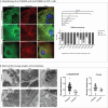
Fig.4:. TUBA4A variants disrupt microtubule morphology in cellular models and patient muscles.
A) Molecular physiology of cytoskeleton in COS1 cells overexpressed with TUBA4A-wildtype (wt) and TUBA4A variants. Morphology ranged from normal, mild to abnormal cytoskeleton. As compared to TUBA4A-wt, previously reported disease-causing variants Arg105Cys (ALS) and Arg320Cys (ALS) showed abnormal cytoskeleton structure. Additionally, the myopathic variants identified in our study, Thr179Ile, Leu227Phe, Ser241Phe and Gly354Asp showed abnormal cytoskeletal patterning. Conversely, Glu254Ala, Glu254Gly, Glu254Lys and Gln254Gln showed normal to mildly abnormal cytoskeletal patterning. B) Electron microscopy analysis of microtubules in control muscles and patient muscles showing statistically significant increase in microtubule diameter in patient muscles as compared to controls.

References

    1. Bahi-Buisson N, Maillard C. Tubulinopathies Overview. In: Adam MP, Feldman J, Mirzaa GM, Pagon RA, Wallace SE, Amemiya A, eds. GeneReviews((R)). University of Washington, Seattle
    2. Copyright © 1993–2024, University of Washington, Seattle. GeneReviews is a registered trademark of the University of Washington, Seattle. All rights reserved.; 1993.

    1. Desai A, Mitchison TJ. Microtubule polymerization dynamics. Annu Rev Cell Dev Biol. 1997;13:83–117. doi: 10.1146/annurev.cellbio.13.1.83 - DOI - PubMed
    1. Mitchison T, Kirschner M. Dynamic instability of microtubule growth. Nature. Nov 15-21 1984;312(5991):237–42. doi: 10.1038/312237a0 - DOI - PubMed
    1. Guzik BW, Goldstein LS. Microtubule-dependent transport in neurons: steps towards an understanding of regulation, function and dysfunction. Curr Opin Cell Biol. Aug 2004;16(4):443–50. doi: 10.1016/j.ceb.2004.06.002 - DOI - PubMed
    1. Romaniello R, Arrigoni F, Fry AE, et al. Tubulin genes and malformations of cortical development. Eur J Med Genet. Dec 2018;61(12):744–754. doi: 10.1016/j.ejmg.2018.07.012 - DOI - PubMed

Publication types

LinkOut - more resources