. 2025 Jul 1:16:1598902.
doi: 10.3389/fimmu.2025.1598902. eCollection 2025.

STING regulates NETs formation by activating GSDMD in influenza viral pneumonia

Affiliations

STING regulates NETs formation by activating GSDMD in influenza viral pneumonia

Rongrong Huang et al. Front Immunol. .

Abstract

Background: Viral pneumonia is the most common and lethal pandemic disease, but there are no broad-spectrum antiviral drugs with high genetic barriers to resistance. To elucidate the mechanisms of viral pneumonia progression and potential targets for its treatment.

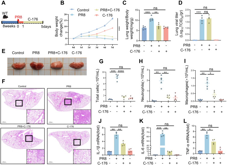
Methods: Viral pneumonia models were induced by the PR8 virus strain in wild-type (WT) and STING knockout (STING-KO) mice. Series of molecular biology techniques were used to evaluate the severity of pneumonia and cytokine levels.

Results: In this study, STING (stimulator of interferon genes) was activated in the lungs of virus-infected mice, leading to cytokine production and amplification of the immune response, thereby causing rapid deterioration of symptoms. Furthermore, excessive activation of innate immune response via STING was prevented by a STING inhibitor (C-176), which significantly reduced viral lung inflammation. The formation of neutrophil extracellular traps (NETs) was similarly suppressed during viral pneumonia treatment with STING inhibitors (C-176), and NETs formation and STING expression were positively correlated, indicating that STING plays an important role in NETs formation. Symptoms of pneumonia in STING-KO mice infected with PR8 were significantly milder than those in WT mice, and NETs were less likely to form in the lung tissue of STING-KO mice. Additionally, transcriptomic analysis revealed that STING-mediated regulation of NETs may be associated with gasdermin D (GSDMD), and immunoprecipitation experiments revealed that STING, GSDMD, and NETs-related proteins interact with each other. Immunofluorescence assays revealed that in neutrophils from WT mice, STING and GSDMD were colocalized on the membrane after viral infection, whereas in neutrophils from STING-KO mice, GSDMD expression was decreased after exposure to the virus.

Conclusions: Our study demonstrated that targeted intervention with STING alleviated pneumonia by inhibiting inflammation and NETs formation. The study also revealed that blocking STING could inhibit the activation of GSDMD to inhibit NETs formation, slowing the progression of viral pneumonia.

Keywords: GSDMD; NETs; STING; anti-inflammatory; viral pneumonia.


Conflict of interest statement

The authors declare that the research was conducted in the absence of any commercial or financial relationships that could be construed as a potential conflict of interest.

Figures

Figure 1
Figure 1
PR8-induced viral pneumonia is effectively inhibited in STING knockout mice. (A) Strategies for typing the STING gene. (B) Representative images of the identification of the STING gene. (C) Graphical representation of experimental models showing the effects of PR8 on WT and STING-KO mice. (D) Body weight change (%) (n = 10). (E) The lung weight index was calculated as the ratio of mouse lung weight (mg) to mouse body weight (g) (n = 5). (F-H) The mRNA levels of IL-1β, IL-6, and TNF-α were measured by RT-PCR. (I) Images of H&E staining showing notable lung pathology in WT and STING KO mice post-PR8 inhalation were acquired (scale bar: 100 μm). (J) Viral titration in the bronchoalveolar lavage fluid was conducted by the TCID50 assay (n = 4). Data are presented as the mean ± SD, *p < 0.05, **p < 0.01, ***p < 0.001, ****p < 0.0001, and ns, no significant difference between the two groups.
Figure 2
Figure 2
Induction of viral pneumonia by PR8 infection. Comparison of the severity of viral pneumonia according to the duration of viral action in mice. (A) Representative images of pathological changes in lung tissue from C57BL/6J mice stained with hematoxylin and eosin (scale bar: 100 μm). (B) The lung indices in mice exposed to the PR8 virus were calculated by the following equation: lung index = mouse lung weight (mg)/mouse body weight (g) (n = 8). (C) Body weight change (%) (n = 8). (D) Viral titration of homogenized lung samples was conducted by the TCID50 assay. The samples were analyzed in triplicate, and each dot corresponds to the viral titers of an individual mouse lung (n = 3). (E-G) The expression of the inflammatory cytokines IL-1β, IL-6, and TNF-α in lung tissue homogenates was measured by RT–qPCR (n = 3). Data are presented as the mean ± SD, *p < 0.05, **p < 0.01, ***p < 0.001, and ****p < 0.0001 vs. 0day.
Figure 3
Figure 3
STING was identified as a potential therapeutic target for viral pneumonia in histological analysis. (A) Volcano plot showing the expression profiles of the top ten upregulated and downregulated DEGs in the lung tissues of mice on the fifth day of exposure to PR8. The upregulated genes were identified using false discovery rate (FDR) correction with log2FC>1 as the criterion is highlighted in red. (B) Bubble plot illustrating the results of Reactome enrichment analysis of the significant DEGs in the lungs of mice on the fifth day of exposure to PR8, with log2 fold change >2 and adjusted p<0.01 as the criteria. (C) Venn diagram displaying the numbers and proportions of overlapping type I IFN, neutrophil degranulation, and immune system-related genes among the DEGs related to viral pneumonia. (D) Heatmap showing the results of correlation analysis of key genes identified in the Venn diagram between the Control group and model group. DEGs, differentially expressed genes. (E-G) RT–qPCR analysis of the mRNA expression of IFN-β, CXCL10, and ISG15 in mouse lung tissues (n = 3). Data are presented as the mean ± SD, *p < 0.05, **p < 0.01, ***p < 0.001, and ****p < 0.0001 vs. 0day.
Figure 4
Figure 4
The pharmacological blockade of STING has a protective effect against PR8-induced viral pneumonia. (A) The experimental protocol involved the induction of viral pneumonia in vivo using the PR8 strain (15 mice per group). (B) Body weight change (%) (n = 15). (C) The lung weight index was calculated as the ratio of lung weight (mg) to body weight (g). (D) Viral titration in the bronchoalveolar lavage fluid was conducted by the TCID50 assay (n = 4). (E) Representative gross images of lung tissue from each group were examined. (F) H&E-stained lung sections from the Control, PR8, PR8+C-176, and C-176 groups were analyzed (scale bar: 200 μm). The Control and C-176 groups exhibited a normal lung structure, characterized by an intact ciliated airway epithelium and alveoli. In contrast, influenza infection led to severe bronchiolitis, inflammation, and alveolar damage, which were alleviated by treatment with C-176. (G-I) The numbers of total cells, neutrophils, and macrophages in the bronchoalveolar lavage fluid were determined (n = 5). (J-L) The mRNA levels of IL-1β, IL-6, and TNF-α in the lung tissues of mice in the four groups were measured using RT-PCR (n = 4). Data are presented as the mean ± SD, *p < 0.05, **p < 0.01, ***p < 0.001, ****p < 0.0001, and ns, no significant difference between the two groups.
Figure 5
Figure 5
The production of NETs in PR8-induced viral pneumonia is affected by STING inhibition. (A) Lung tissue sections from mice in the four groups (Control, PR8, PR8+C-176, and C-176) were examined by immunofluorescence staining, as shown in the images (scale bar: 50 μm). MPO is labeled in green, and Cit-H3 is labeled in yellow. The concentrations of MPO-DNA complexes (expressed in pg/mL) in the BALF (B) and serum (C) of mice in all the experimental groups were quantified utilizing ELISA (n = 5). (D) Western blot analysis was conducted to evaluate the protein expression of MPO and Cit-H3 relative to that of GAPDH, which was used as a loading control (n = 3). (E) Band densities were quantified for graphical representation using ImageJ software and normalized to those of GAPDH. Data are presented as the mean ± SD, *p < 0.05, **p < 0.01, ***p < 0.001, ****p < 0.0001, and ns, no significant difference between the two groups. (F) Neutrophils were infected with 1ul of the PR8 virus strain (40 LD50/mL) and treated with C-176 (200 nM) in vitro. Subsequent visualization was performed using laser confocal microscopy, revealing the presence of MPO (green) and Cit-H3 (red) in the acquired images (scale bar: 10 µm).
Figure 6
Figure 6
STING regulates the generation of NETs in viral pneumonia. (A) PR8-induced formation of NETs in the bronchi of WT and STING-KO mice was identified through immunofluorescence analysis (scale bar: 50 μm). MPO is labeled in green, and Cit-H3 is labeled in yellow. (B) MPO and Cit-H3 protein expression levels were measured via Western blot analysis, utilizing Tubulin as an internal control (n = 3). (C, D) Quantification of B-figure was performed using ImageJ software with a sample size of three. (E) The concentration of MPO-DNA complexes in BALF, measured in picograms per milliliter (pg/mL), was assessed in each group of mice (n = 5) using an enzyme-linked immunosorbent assay (ELISA). Data are presented as the mean ± SD, *p < 0.05, **p < 0.01, ***p < 0.001, ****p < 0.0001, and ns, no significant difference between the two groups. (F) Following infection with PR8, neutrophils isolated from both WT and STING-KO mice were examined for the formation of NETs through immunofluorescence staining (scale bar: 10 μm). MPO is labeled in green, and Cit-H3 is labeled in red.
Figure 7
Figure 7
The regulation of NETs formation by STING is related to GSDMD in viral pneumonia. (A) Lung tissues from mice in the four groups (WT Control, WT+PR8, STING-KO Control, and STING-KO+PR8) were subjected to transcriptomic analysis. As determined by GSEA of the dataset, the vertical axis values on the “enrichment scores line graph” for the three groups were all above 0, indicating upregulation of the gene set (pathway) in the left group. (B) The heatmap shows the genes that directly corresponded to the gene set in panel (A). (C). A pairwise interaction analysis was conducted between NETs-related targets in the KEGG database and the genes identified by GSEA of the animal transcriptome. (D-F) In the in vivo experiments, the protein levels of GSDMD, PAD4, and PAD2 were measured via immunoblotting (n = 3). (G) An immunofluorescence assay was used to visualize the protein expression of STING and GSDMD on neutrophils in the lung tissue from the WT mice in the Control and PR8 groups (scale bar: 10 μm). MPO is labeled in purple, STING is labeled in green, and GSDMD is labeled in yellow.
Figure 8
Figure 8
The activation of GSDMD by STING promotes the upregulation of NETs formation in viral pneumonia. (A) The expression of STING and GSDMD proteins in neutrophils in the lung tissues of WT and STING-KO mice infected with PR8 on day 5 was visualized using an immunofluorescence assay. The images captured focused on the neutrophil structure along with the released net, indicated by a white arrow (scale bar: 10 μm). MPO is labeled in purple, STING is labeled in green, and GSDMD is labeled in yellow. (B) Lung tissue lysates were collected from mice in the Control and PR8 groups and subjected to immunoprecipitation with an anti-STING antibody, an anti-GSDMD antibody, and normal IgG followed by immunoblotting with the indicated antibodies. (C, D) Images of immunofluorescence staining for STING and GSDMD in the treated neutrophils in each experimental group were acquired using laser confocal microscopy at a magnification of 400× (scale bar: 10 μm). GSDMD is labeled in green, while STING is labeled in red.
Figure 9
Figure 9
Mechanisms by which STING regulates NETs formation via the activation of GSDMD in viral pneumonia. The STING signaling pathway and its downstream signaling molecules in lung epithelial cells are activated by the virus to produce type I interferon, which leads to the production of pro-inflammatory and chemokine factors and then recruit neutrophils. GSDMD activity is regulated by the activation of STING in neutrophils, leading to the formation of NETs and thereby exacerbating viral pneumonia.

Similar articles

References

    1. Rello J, Sabater-Riera J. Challenges in respiratory syncytial virus in adults with severe community-acquired pneumonia. Chest. (2022) 161:1434–5. doi: 10.1016/j.chest.2022.01.050 - DOI - PubMed
    1. Chousterman BG, Swirski FK, Weber GF. Cytokine storm and sepsis disease pathogenesis. Semin Immunopathol. (2017) 39:517–28. doi: 10.1007/s00281-017-0639-8 - DOI - PubMed
    1. Della Corte CM, Barra G, Ciaramella V, Di Liello R, Vicidomini G, Zappavigna S, et al. Antitumor activity of dual blockade of PD-L1 and MEK in NSCLC patients derived three-dimensional spheroid cultures. J Exp Clin Cancer Res CR. (2019) 38:253. doi: 10.1186/s13046-019-1257-1 - DOI - PMC - PubMed
    1. Teachey DT, Lacey SF, Shaw PA, Melenhorst JJ, Maude SL, Frey N, et al. Identification of predictive biomarkers for cytokine release syndrome after chimeric antigen receptor T-cell therapy for acute lymphoblastic leukemia. Cancer Discov. (2016) 6:664–79. doi: 10.1158/2159-8290.CD-16-0040 - DOI - PMC - PubMed
    1. Arabi YM, Deeb AM, Al-Hameed F, Mandourah Y, Almekhlafi GA, Sindi AA, et al. Macrolides in critically ill patients with Middle East Respiratory Syndrome. Int J Infect Dis IJID Off Publ Int Soc Infect Dis. (2019) 81:184–90. doi: 10.1016/j.ijid.2019.01.041 - DOI - PMC - PubMed

MeSH terms

LinkOut - more resources