. 2025 Jul 1:16:1513009.
doi: 10.3389/fimmu.2025.1513009. eCollection 2025.

CXCR5 engineered human and murine Tregs for targeted suppression in secondary and tertiary lymphoid organs

Affiliations

CXCR5 engineered human and murine Tregs for targeted suppression in secondary and tertiary lymphoid organs

Matteo Doglio et al. Front Immunol. .

Abstract

Introduction: Secondary and tertiary lymphoid structures are a critical target of suppression in many autoimmune disorders, protein replacement therapies, and in transplantation. Although antigen-specific regulatory T cells (Tregs), such as chimeric antigen receptor (CAR) Tregs, generally persist longer and localize to target tissues more effectively than polyclonal Tregs in animal models, their numbers still progressively decline over time. A potential approach to maximize Treg activity in vivo is the expression of chemokine receptors such as CXCR5, which would enable localization of a greater number of engineered cells at sites of antigen presentation. Indeed, CXCR5 expression on follicular T helper cells and follicular Tregs enables migration toward lymph nodes, B cell zones, and tertiary lymphoid structures that appear in chronically inflamed non-lymphoid tissues.

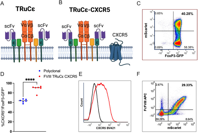
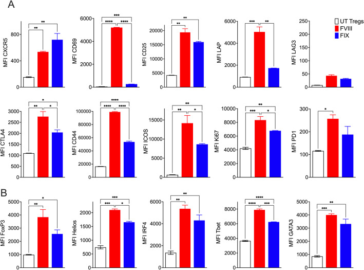
Methods: In this study, we generated human and murine CXCR5 co-expressing engineered receptor Tregs and tested them in preclinical mouse models of allo-immunity and hemophilia A, respectively. Additionally, we engineered a murine CXCR5 co-expressing clotting factor VIII (FVIII) specific T cell receptor fusion construct epsilon (FVIII TRuCe CXCR5) Treg to suppress anti-drug antibody development in a model of FVIII protein replacement therapy for hemophilia A.

Results: In vitro, anti-HLA-A2 CXCR5+ CAR-Tregs showed enhanced migratory and antigen-specific suppressive capacities compared to untransduced Tregs. When injected into an NSG mouse model of HLA-A2+ pancreatic islet transplantation, anti-HLA-A2 CXCR5+ CAR-Tregs maintained a good safety profile allowing for long-term graft survival in contrast to anti-HLA-A2 CXCR5+ conventional CAR-T (Tconv) cells that eliminated the graft. Similarly, FVIII TRuCe CXCR5 Treg demonstrated increased in vivo persistence and suppressive capacity in a murine model of hemophilia A.

Discussion: Collectively, our findings indicate that CXCR5 co-expression is safe and enhances in vivo localization and persistence in target tissues. This strategy can potentially promote targeted tolerance without the risk of off-target effects in multiple disease models.

Keywords: C-X-C chemokine receptor type 5 (CXCR5); TCR fusion construct epsilon (TRuCε); chimeric antigen receptor (CAR); hemophilia; pancreatic islet transplantation; regulatory T cells (Tregs); type 1 diabetes (T1D).


Conflict of interest statement

The authors declare that the research was conducted in the absence of any commercial or financial relationships that could be construed as a potential conflict of interest. The author(s) declared that they were an editorial board member of Frontiers, at the time of submission. This had no impact on the peer review process and the final decision.

Figures

Figure 1
Figure 1
HLA-A2-CAR CXCR5+ Tregs in vitro generation and validation. (A) Schematic representation of the bidirectional lentiviral vector (LV). Anti-HLA-A2 Chimeric Antigen Receptor (CAR) in sense under the control of a human phosphoglycerate kinase (hPGK) promoter, composed by single chain fragment variant (scFv), a truncated form of the neuron growth factor receptor (NGFR) as a spacer, the transmembrane/intracellular human CD28 domain fused to the intracellular portion of the human CD3ζ chain. In anti-sense, the human C-X-C motif chemokine receptor 5 (CXCR5) gene under the control of a minimal Cytomegalovirus (mCMV) promoter. Other essential components necessary for functionality of the LV are indicated. LTR long terminal repeat, SD splice donor, SA splice acceptor, GA gag-pol element, RRE REV responsive element, cPPT central polypurine tract, pA polyadenylation signal, CTE constitutive transport element, WPRE woodchuck hepatitis virus post-transcriptional regulatory element. (B) Frequency of transduced cells, evaluated as percentage of CAR+ and hCXCR5+ Tregs by flow cytometry at Day +14. Cell transduction was assessed as percentage of NGFR+ cells. (C) Percentage of CD4+CD25+CD127-FoxP3+ cells in UT and CAR-Tregs at Day +14, assessed by flow cytometry. (D) In vitro polyclonal suppressive capacities and (E) antigen-specific suppressive capacities of UT or anti-HLA-A2 CXCR5+ CAR-Tregs. Results are expressed as Suppression Index, calculated as [1-(PBMCs’ proliferation with Tregs)/(PBMCs’ proliferation alone)] * 100. (F) In vitro B cell maturation assay in the presence of UT or anti-HLA-A2 CXCR5+ CAR-Tregs with or without T follicular helper cells (Tfh), expressed as the number of IgG+ B cells. (G) Migration of UT and anti-HLA-A2 CXCR5+ CAR-Tregs in response to varying concentrations of human CXCL13. Results are expressed as the number of migrating Tregs. For all the experiments reported in panel (B-G), results are expressed as mean±SD (N = 3). 1 or 2-tailed Mann-Whitney test for (B, F, G) Two-way ANOVA with Tukey’s correction for (E) *p-value <0.05, **p-value < 0.01, ***p-value <0.001.
Figure 2
Figure 2
Anti-HLA-A2 CXCR5+ CAR-Tregs do not kill transplanted islets. (A) Experimental design. Five hundred HLA-A2+ pancreatic islets were isolated from HLA-A2 transgenic NSG mice and transplanted under the kidney capsule in HLA-A2- NSG mice treated with streptozotocin to induce diabetes. Once glycemic control was achieved, mice were injected with 2x10⁶ of anti-HLA-A2 CXCR5+ CAR-Tconv or CAR-Tregs or UT Tregs. Glycemia was monitored in the blood and when reached values >250 mg/dl, the graft was considered rejected. Mice treated with Tregs underwent nephrectomy 30 days after the cell injection. (B) Blood glucose monitoring in transplanted mice assessed with a glucometer. TX = graft. (C) Weight monitoring in transplanted mice expressed in grams. (D) Percentage of circulating human CD45+ cells assessed by flow cytometry at different time points. (E) Frequency of human CD45+ cells in the spleen (SPL) and the graft (TX) at euthanasia, assessed by flow cytometry. Frequency of CAR+ (F) and CXCR5+ (G) cells in the spleen (SPL) and the graft (TX) at euthanasia, assessed by flow cytometry. For this experiment we employed a total of 5 animals in each group in two independent experiments, indicated by the square and the round symbols, respectively.
Figure 3
Figure 3
FVIII TRuCε CXCR5 Treg ex vivo generation and validation. (A) Surface organization of FVIII TRuCε. The FVIII specific scFv is fused to CD3ε by a linker. The synthetic construct can integrate into the endogenous Treg CD3-TCR complex. (B) FVIII TRuCε CXCR5 co-expresses murine CXCR5 on the cell surface. (C) Representative density plot of FoxP3-GFP+ Tregs transduced with either (A) or (B) as indicated by mScarlet reporter protein expression. (D) Percentage of CXCR5+ expression in polyclonal and FVIII TRuCε CXCR5 transduced FoxP3-GFP+ Tregs. (E) Histogram overlay plots indicating overexpression of CXCR5 (red histogram) in FVIII TRuCε CXCR5 transduced Tregs as compared to polyclonal Tregs (black histogram). (F) Representative density plot indicating binding of FcFVIII by mScarlet+ FVIII TRuCε CXCR5 transduced Tregs as detected by APC conjugated α-IgG Fc secondary antibody. Data represents mean±SEM, ****p<0.0001 for (D) using unpaired t test.
Figure 4
Figure 4
FVIII TRuCε CXCR5 Tregs respond to FVIII stimulation in vitro. (A) In vitro upregulation of activation markers CXCR5, CD69, CD25, LAP, LAG3, CTLA4, CD44, ICOS, Ki67 and PD1 in recombinant FVIII or FIX stimulated engineered FVIII TRuCε CXCR5 Tregs at 48hrs. (B) Upregulation of transcription factors FoxP3, Helios, IRF4, TBet and GATA3 in recombinant FVIII or FIX stimulated engineered FVIII TRuCε CXCR5 Tregs at 48hrs. Changes in MFI are quantified. Data represents mean±SEM, p < 0.05, ∗∗p < 0.01, ***p<0.001, ****p<0.0001 using 1-way ANOVA with Sidak’s multiple comparisons analysis.
Figure 5
Figure 5
FVIII TRuCε CXCR5 Tregs display improved in vitro and in vivo persistence. (A) In vitro migration of FVIII CAR, FVIII CAR CXCR5, and FVIII TRuCε transduced Tregs through a transwell in response to either serum free media, CXCL12 or CXCL13 gradients. Number of migrated mScarlet+ cells at the bottom of the transwell are quantified by flow cytometry following 6hrs of incubation. (B) Kinetics of in vivo migration of adoptively transferred FVIII TRuCε or FVIII TRuCε CXCR5 Tconv cells to the spleen on days 1, 2, 4, and 7 following adoptive transfer. Mice received i.v. injections of recombinant FVIII on days 0, 3, and 6. Frequencies of mScarlet+ cells per total CD4+ T cells are quantified by flow cytometry. (C) Number of mScarlet+ FVIII TRuCε or FVIII TRuCε CXCR5 Tregs per 107 CD4+ T cells are quantified from spleens and (D) inguinal lymph nodes (ILN) on day 7 post adoptive transfer. Data represents mean±SEM, ****p<0.0001, ∗p < 0.05, ∗∗p < 0.01 using 2-way ANOVA with Tukey’s multiple comparisons analysis for (A), 2-way ANOVA with Sidak’s multiple comparisons analysis for (B), unpaired t test for (C, D).
Figure 6
Figure 6
In vivo suppression of ADAs to FVIII. (A) Schematic representing timeline for assessing in vivo prevention of ADA formation engineered Tregs. 0.5 × 10⁶ FVIII TRuCε or FVIII TRuCε CXCR5 Treg were adoptively transferred into BALB/c F8e16−/− (HA) recipient mice (n = 4−10/group). Mice received 8 weekly i.v. injections of 1IU recombinant FVIII, and plasma samples were analyzed after the 4th and 8th injection for (B) functional inhibitors by Bethesda assay, (C) α-FVIII IgG1 ELISA, and (D) α-FVIII IgG2a ELISA. FVIII Control mice received only BDD-FVIII injections without Treg transfer. Data represents mean±SEM. ∗p < 0.05 using 2-way ANOVA with Tukey’s multiple comparisons analysis for (A), 2-way ANOVA with Dunnett’s multiple comparisons analysis for (B, C).

Similar articles

References

    1. Ferreira LMR, Muller YD, Bluestone JA, Tang Q. Next-generation regulatory T cell therapy. Nat Rev Drug Discovery. (2019) 18:749–69. doi: 10.1038/s41573-019-0041-4 - DOI - PMC - PubMed
    1. Rana J, Biswas M. Regulatory T cell therapy: Current and future design perspectives. Cell Immunol. (2020) 356:104193. doi: 10.1016/j.cellimm.2020.104193 - DOI - PubMed
    1. Mancuso G, Bechi Genzano C, Fierabracci A, Fousteri G. Type 1 diabetes and inborn errors of immunity: Complete strangers or 2 sides of the same coin? J Allergy Clin Immunol. (2023) 151:1429–47. doi: 10.1016/j.jaci.2023.03.026 - DOI - PubMed
    1. Herold KC, Delong T, Perdigoto AL, Biru N, Brusko TM, Walker LSK. The immunology of type 1 diabetes. Nat Rev Immunol. (2024) 24:435–51. doi: 10.1038/s41577-023-00985-4 - DOI - PMC - PubMed
    1. Sherman A, Biswas M, Herzog RW. Innovative approaches for immune tolerance to factor VIII in the treatment of hemophilia A. Front Immunol. (2017) 8:1604. doi: 10.3389/fimmu.2017.01604 - DOI - PMC - PubMed

MeSH terms

LinkOut - more resources