

Lessons From Recurrent Dropped Head Syndrome After Inadequate Short Fixation: A Case Series
- PMID: 40666593
- PMCID: PMC12261008
- DOI: 10.7759/cureus.86041
Lessons From Recurrent Dropped Head Syndrome After Inadequate Short Fixation: A Case Series
Abstract
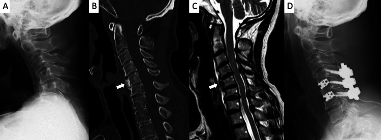
Dropped head syndrome (DHS) is a condition in which the head falls forward due to dysfunction or atrophy of the cervical extensor muscles. It is more commonly observed in elderly women and significantly affects horizontal gaze and activities of daily living (ADL). For cases in which conservative treatment is ineffective, surgical corrective fixation is considered; however, indications and standardized procedures have not yet been fully established. We retrospectively analyzed five cases of DHS treated surgically at our institution and examined the efficacy of corrective fixation and potential treatment strategies. Five patients (mean age: 82.6 years; all female) who underwent surgery for DHS between 2018 and 2024 were included. Three patients initially underwent short-segment fixation or laminoplasty, but DHS recurred. Eventually, all cases required long-segment fixation extending from C2 or the occiput to Th1/Th2. Radiological evaluations included measurements of the C2-C7 Cobb angles, sagittal vertical axis (SVA), and T1 slope before and after surgery. All patients exhibited cervical kyphosis and sagittal imbalance. Postoperatively, cervical lordosis was restored, and improvements were noted in SVA and T1 slope. In four cases, patients were able to maintain horizontal gaze for over 30 minutes, and improvements in ADL living were observed. One patient died from aspiration pneumonia, although horizontal gaze was maintained postoperatively. Long-segment corrective fixation within an appropriate range is considered a safe and effective treatment option.
Keywords: activities of daily living; cervical kyphosis; cervical sagittal alignment; corrective spinal fusion; dropped head syndrome; isolated neck extensor myopathy; long-segment fixation; sagittal vertical axis.
Copyright © 2025, Honda et al.
Conflict of interest statement
Human subjects: Informed consent for treatment and open access publication was obtained or waived by all participants in this study. Conflicts of interest: In compliance with the ICMJE uniform disclosure form, all authors declare the following: Payment/services info: All authors have declared that no financial support was received from any organization for the submitted work. Financial relationships: All authors have declared that they have no financial relationships at present or within the previous three years with any organizations that might have an interest in the submitted work. Other relationships: All authors have declared that there are no other relationships or activities that could appear to have influenced the submitted work.
Figures


Similar articles
-
Systemic pharmacological treatments for chronic plaque psoriasis: a network meta-analysis.Cochrane Database Syst Rev. 2021 Apr 19;4(4):CD011535. doi: 10.1002/14651858.CD011535.pub4. Cochrane Database Syst Rev. 2021. Update in: Cochrane Database Syst Rev. 2022 May 23;5:CD011535. doi: 10.1002/14651858.CD011535.pub5. PMID: 33871055 Free PMC article. Updated.
-
Systemic pharmacological treatments for chronic plaque psoriasis: a network meta-analysis.Cochrane Database Syst Rev. 2017 Dec 22;12(12):CD011535. doi: 10.1002/14651858.CD011535.pub2. Cochrane Database Syst Rev. 2017. Update in: Cochrane Database Syst Rev. 2020 Jan 9;1:CD011535. doi: 10.1002/14651858.CD011535.pub3. PMID: 29271481 Free PMC article. Updated.
-
Comparison of Two Modern Survival Prediction Tools, SORG-MLA and METSSS, in Patients With Symptomatic Long-bone Metastases Who Underwent Local Treatment With Surgery Followed by Radiotherapy and With Radiotherapy Alone.Clin Orthop Relat Res. 2024 Dec 1;482(12):2193-2208. doi: 10.1097/CORR.0000000000003185. Epub 2024 Jul 23. Clin Orthop Relat Res. 2024. PMID: 39051924
-
Morphological, functional and neurological outcomes of craniectomy versus cranial vault remodeling for isolated nonsyndromic synostosis of the sagittal suture: a systematic review.JBI Database System Rev Implement Rep. 2015 Sep;13(9):309-68. doi: 10.11124/jbisrir-2015-2470. JBI Database System Rev Implement Rep. 2015. PMID: 26470674
-
Tri-cortical pedicle screw fixation in the most cranial instrumented segment to prevent proximal junctional kyphosis.Spine J. 2025 Aug;25(8):1698-1707. doi: 10.1016/j.spinee.2025.02.002. Epub 2025 Feb 22. Spine J. 2025. PMID: 39993503
References
-
- The dropped head syndrome. Suarez GA, Kelly JJ Jr. Neurology. 1992;42:1625–1627. - PubMed
-
- Isolated neck extensor myopathy: a common cause of dropped head syndrome. Katz JS, Wolfe GI, Burns DK, Bryan WW, Fleckenstein JL, Barohn RJ. Neurology. 1996;46:917–921. - PubMed
-
- Dropped head syndrome: etiology and management. Sharan AD, Kaye D, Charles Malveaux WM, Riew KD. J Am Acad Orthop Surg. 2012;20:766–774. - PubMed
-
- Spinal sagittal alignment in patients with dropped head syndrome. Murata K, Kenji E, Suzuki H, et al. Spine (Phila Pa 1976) 2018;43:0–73. - PubMed
Publication types
LinkOut - more resources
Full Text Sources
Miscellaneous