[Preprint]. 2025 Jun 18:2025.06.18.660419.
doi: 10.1101/2025.06.18.660419.

Evidence that Entomophthora muscae controls the timing of host death via its own circadian clock

Affiliations

Evidence that Entomophthora muscae controls the timing of host death via its own circadian clock

Leslie Torres Ulloa et al. bioRxiv. .

Abstract

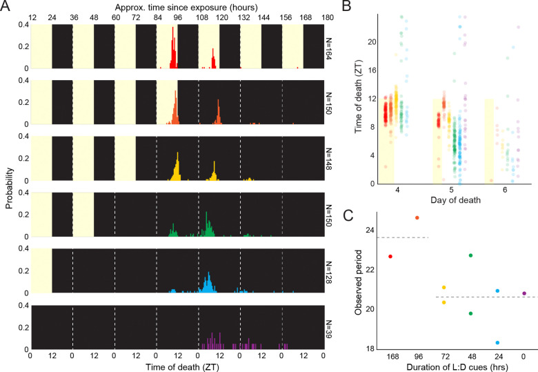
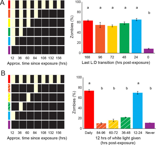
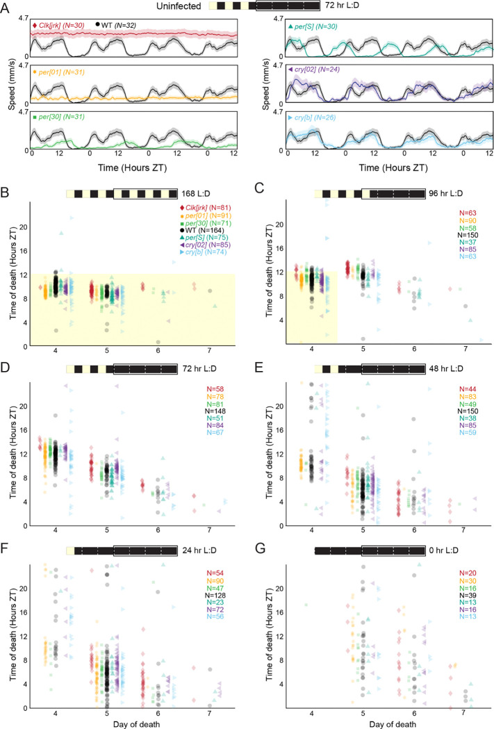
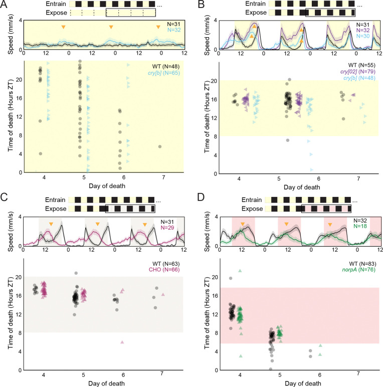
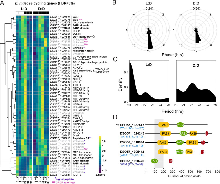
Timing in the natural world is a matter of life or death, consequently, nearly all life on Earth has evolved internal circadian clocks. Many behavior-manipulating parasites exhibit striking daily timing, but whether this is clock-driven has remained unclear. Here, we leveraged the laboratory-tractable zombie fruit fly model, Drosophila melanogaster infected by the behavior manipulating fungus Entomophthora muscae, to tackle this long standing mystery. Using an automated behavioral paradigm, we found that the timing of death of wild-type flies continues to occur with ~21 hour periodicity in the absence of environmental cues. Surprisingly, we also discovered that light is required within the first 24 hours of exposure for E. muscae to infect and kill flies. Experiments with circadian and photoreception mutants revealed that death is independent of host genotype, suggesting the fungus-not the host-drives this rhythm. Transcriptomic analysis of in vitro grown fungus revealed that E. muscae maintains rhythmic gene expression independent of the fly host that peaks at sunset and has a free-running period of ~22 hours. Among cycling genes, we identified a transcript encoding a protein with high homology to white collar-1, the blue light sensor and core component of the molecular oscillator in the model ascomycete fungus Neurospora crassa. Altogether, our findings suggest that E. muscae has an endogenous circadian clock that it uses to control the timing of host death. This study provides evidence that a fungal clock can influence fly outcomes, pointing to a new mechanism by which parasites temporally coordinate host manipulation.

Keywords: Drosophila melanogaster; Entomophthora muscae; behavior manipulation; circadian clock.


Figures

Figure 1.
Figure 1.. E. muscae-infected WT flies die in a gated fashion in the absence of proximal light cues.
A) Time of last movement distributions for WT flies exposed to E. muscae and housed until varying durations of L:D cues before release into free-running conditions (D:D). Yellow background indicates presence of white light cues during experiment; black indicates darkness. All flies were reared on the same 12:12 L:D cycle (ZT0 = 12:00 AM EST). B) Data from A presented as a scatter plot. C) Observed death period lengths (in hours) based on all observations in A. For every day in which at least 5 flies were observed to die, a mean time of death was calculated for that day; the difference between mean times of death across adjacent days is plotted. Dashed line at left reflects mean observed period for experiments where L:D cues were provided for experiments in which flies had at least one death day with L:D cues (i.e., 168 and 96 hours after E. muscae exposure; 23.7 hours); at right, experiments with D:D cues on all death days (i.e., 72, 48, 24, and 0 hours after E. muscae exposure), dashed line is mean for these experiments (20.6 hours). Two-tailed t-test shows significant difference between L:D and D:D periods (p = 0.0251).
Figure 2.
Figure 2.. E. muscae infection is promoted by light cues in the first 24 hours after exposure.
A) Right: Percentage of zombies (flies that died and then sporulated) within seven days of E. muscae exposure under indicated light conditions at left. B) Right: percentage of zombies subjected to lighting cues diagrammed at left. White hash marks reflect conditions diagrammed at left. Letters above bars indicate distinct significance groups (p < 0.05) as determined by pairwise comparisons (two-tailed t-test) for each pair of conditions.
Figure 3.
Figure 3.. E. muscae-infected mutant flies die with similar circadian timing under free-running conditions, regardless of host circadian phenotype.
A) Free-running activity patterns of uninfected flies of each of our six tested genotypes under D:D conditions. WT = CantonS, per[30] = per.T610A.S613A; all other genotypes are named exactly according to their alleles. Flies were raised on a 12:12 white light L:D cycle (ZT0 = 12:00 AM EST) and maintained under this cycle for 72 hours before releasing into D:D and tracking locomotion. B) Time of death for individual flies of six circadian mutants (colors) and WT (black) when kept under L:D conditions throughout infection and tracking. Each point represents the death of a single fly. C-G) As in B, but flies were released into free running D:D conditions after C) 96, D) 72, E) 48, F) 24, or G) 0 hours after exposure to E. muscae. WT data are the same as those presented in Figure 1. For all panels, the schematic at top shows lighting conditions since exposure (time 0, at left), with black box indicating when fly behavior was tracked and corresponding to the plot’s x-axis.
Figure 4.
Figure 4.. E. muscae-infected flies die with similar timing under all tested lighting conditions, regardless of host genetic background.
For all panels, the schematic at top summarizes entrainment and experimental conditions. The black outline indicates the period of time during which flies were tracked (corresponding to the middle and bottom panels below). Middle plot shows free-running activity patterns of uninfected flies during the ~96 hour window outlined in black. Bottom plot shows time of death for E. muscae-exposed flies under these experimental lighting conditions. All flies were entrained on the same 12:12 white light L:D cycle (ZT0 = 12:00 AM EST) prior to experiment. Conditions and genotypes are as follows: A) WT (CantonS) and cry[b] flies were exposed to E. muscae and housed with constant light (L:L); B) WT, cry[b] and cry[02] flies were exposed to E. muscae under 12:12 L:D cues then phase advanced by eight hours on the fourth day after exposure; C) WT and CHO flies were exposed to E. muscae under 12:12 L:D cues then phase advanced with dim white light by eight hours on the fourth day after exposure; D) WT and norpA flies were exposed to E. muscae under 12:12 L:D cues then phase advanced with dim red light by six hours on the fourth day after exposure. Each dot reflects the time of death for an individual fly. Yellow shading indicates presence of bright (>1000 lux) white light; tan, dim (40–50 lux) white light; red, dim (<10 lux) red light. Activity data are the mean (solid lines) +/− SEM (shaded regions). Golden arrowheads highlight deviations in activity of uninfected mutant flies relative to uninfected WT flies.
Figure 5.
Figure 5.. In vitro E. muscae shows rhythmic gene expression under both L:D and D:D conditions.
A) Gene expression heatmap of Z-scored rhythmic transcripts from in vitro-grown E. muscae that cycle under both L:D and D:D conditions (MetaCycle, false discovery rate (FDR) = 5%, min/max period = 20/28 hr). Genes are clustered according to expression patterns. Scale bar for Z-scores given at bottom right. Star = putative wc-1 homolog; purple asterisks = gene has predicted signal peptide, pink squiggle = gene product has predicted G-protein coupled receptor seven transmembrane (GPCR 7TM) topology. B) Phase and C) period distribution of cycling genes shown in A. D) Predicted E. muscae proteins with high homology to Neurospora crassa White Collar 1 (WC-1) and White Collar 2 (WC-2), as determined by BLASTp (query coverage 79%-87%). Multiple isoforms were identified for each protein; domain architecture of the longest isoform is shown. Percent positive matches to N. crassa homologs given in parenthesis, along with BLASTp E-values. PFAM domain architecture is shown to the right. Star denotes putative wc-1 homolog observed to cycle under both L:D and D:D conditions at FDR 5 (same as panel A).

Similar articles

References

    1. Alejevski F, Saint-Charles A, Michard-Vanhée C, Martin B, Galant S, Vasiliauskas D, Rouyer F. 2019. The HisCl1 histamine receptor acts in photoreceptors to synchronize Drosophila behavioral rhythms with light-dark cycles. Nat Commun 10:252. doi: 10.1038/s41467-018-08116-7 - DOI - PMC - PubMed
    1. Allada R, White NE, So WV, Hall JC, Rosbash M. 1998. A mutant Drosophila homolog of mammalian Clock disrupts circadian rhythms and transcription of period and timeless. Cell 93:791–804. - PubMed
    1. Aschoff J. 1979. Circadian rhythms: influences of internal and external factors on the period measured in constant conditions. Z Tierpsychol 49:225–249. - PubMed
    1. Baker CL, Loros JJ, Dunlap JC. 2012. The circadian clock of Neurospora crassa. FEMS Microbiol Rev 36:95–110. doi: 10.1111/j.1574-6976.2011.00288.x - DOI - PMC - PubMed
    1. Ballario P, Vittorioso P, Magrelli A, Talora C, Cabibbo A, Macino G. 1996. White collar-1, a central regulator of blue light responses in Neurospora, is a zinc finger protein. EMBO J 15:1650–1657. - PMC - PubMed

Publication types

LinkOut - more resources