[Preprint]. 2025 Jun 23:2025.06.23.661109.
doi: 10.1101/2025.06.23.661109.

Heterosynaptic interactions between dorsal and ventral hippocampus in individual medium spiny neurons of the nucleus accumbens ventromedial shell

Affiliations

Heterosynaptic interactions between dorsal and ventral hippocampus in individual medium spiny neurons of the nucleus accumbens ventromedial shell

Ashley E Copenhaver et al. bioRxiv. .

Abstract

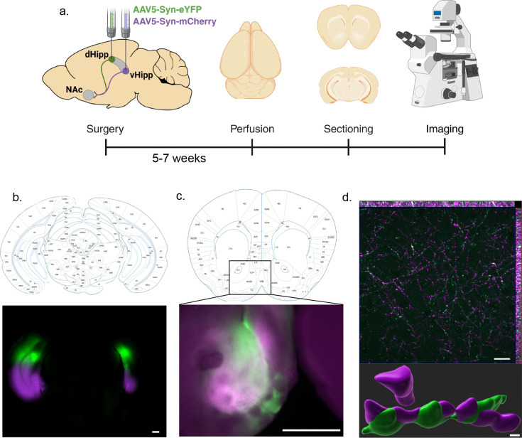
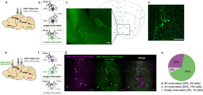
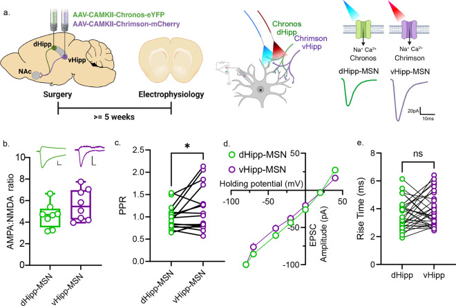
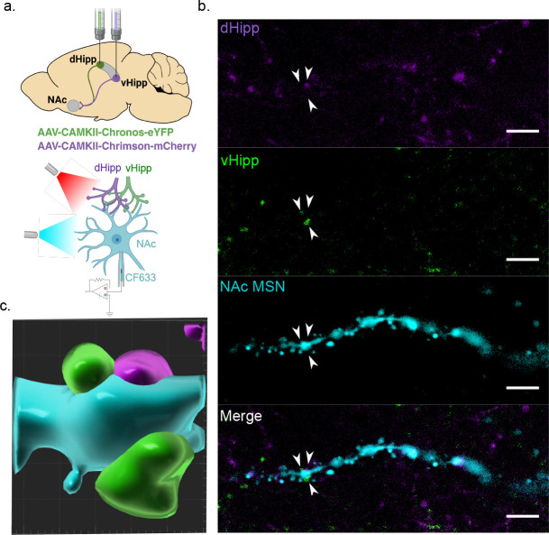
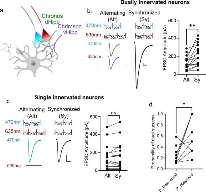
Establishing learned associations between rewarding stimuli and the context under which those rewards are encountered is critical for survival. Hippocampal input to the nucleus accumbens (NAc) is a key connection involved in integrating environmental information and reward processing to facilitate goal-directed behaviors. This connection consists of two independent pathways originating from the dorsal (dHipp) or ventral (vHipp) hippocampus, which have previously been considered functionally and anatomically distinct. Here, we show overlap in dHipp and vHipp terminal fields in the NAc, which led us to reconsider this view and raise new questions regarding the potential interactions between dHipp and vHipp pathways in the NAc. Using optogenetics, electrophysiology, and transsynaptic labeling in adult male and female mice, we investigated anatomical and functional convergence of dHipp and vHipp in the NAc. We identified a subpopulation of dually innervated cells in the NAc medial shell where dHipp and vHipp inputs are located near one another along dendritic branches. We independently manipulated dHipp and vHipp inputs via two-color optogenetic manipulation during whole-cell electrophysiology recordings to confirm functional dual innervation of individual neurons and revealed heterosynaptic interactions between the two pathways. Altogether, these results demonstrate that dHipp and vHipp dually innervate a subset of neurons in the NAc, suggesting integration of these inputs at the level of individual neurons. Exploring the physiological and behavioral implications of this convergence will offer new insights into how individual neurons incorporate information from distinct inputs and how this integration may shape learning.


Conflict of interest statement

Conflict of interest statement: The authors declare no competing financial interests.

Figures

Figure 1.
Figure 1.. Overlapping dHipp and vHipp axon terminal fields in the NAc.
a) Viral injection strategy and experimental timeline. b) Injection sites in the hippocampus. Scale bar=500μm. Atlas image from (Paxinos & Franklin, 2019) c) Overlap of dHipp and vHipp axon terminal fields in the NAc. Scale bar=500μm. Atlas image from (Paxinos & Franklin, 2019) d) Close-up image of overlapping dHipp and vHipp axon terminal fields within the NAc ventromedial shell (top). Scale bar=20μm. With 3-dimensional reconstruction of overlapping axon terminals (bottom). Scale bar= 0.5μm
Figure 2.
Figure 2.. dHipp+/vHipp+-innervated neurons in the NAc ventromedial shell.
a) Viral targeting strategy to selectively label dually innervated neurons b) Cartoon representation of singly vs dually innervated neurons. c) Image of NAc shell depicting GCaMP-expressing cells in the NAc ventromedial shell. Scale bar=100μm. Atlas image from (Paxinos & Franklin, 2019) d) Close-up image of GCaMP-expressing cells in the NAc ventromedial shell, where neuron morphology is consistent with medium spiny neurons. Scale bar=100μm. e) Viral targeting strategy and experimental timeline to label cells innervated by dHipp and vHipp. f) Cartoon representation of singly vs dually innervated neurons. g) Image of cells innervated by dHipp (green) and vHipp (magenta). Dually innervated neurons express both reporters and appear white. Arrowheads point out examples of dually innervated neurons. Scale bar = 100μm h) Quantification of cells innervated single and dually innervated neurons in the NAc medial shell across 4 slices.
Figure 3.
Figure 3.. Individual NAc neurons are functionally innervated by dHipp and vHipp.
a) Viral targeting and optogenetic approach. Electrophysiological recording strategy and representative traces from a dually innervated NAc MSN. b) Similar AMPA:NMDA receptor ratios between dHipp- and vHipp-MSN synapses (n=8 cells/4 mice, p=0.1953, Wilcoxon test). Scale bars on representative traces = 40pA/10ms c) Rectification curve reveals linear relationship, indicating a lack of calcium-permeable AMPA receptors (n=8 cells/4 mice). d) PPRs recorded from dHipp-MSN and vHipp-MSN synapses on dually innervated neurons (n=16 cells/10 mice, *p=0.0394, Paired t-test). e) Rise time was calculated for dHipp- and vHipp-evoked EPSCs, showcasing similar rise times (n=31 cells/12 mice, p=0.2877, Wilcoxon test).
Figure 4.
Figure 4.. Spatial proximity of dHipp and vHipp puncta along distal dendrites of MSN.
a) Cartoon depicting optogenetic, electrophysiology, and cell-filling strategy. b) Combined and separated images depicting an MSN filled with CF633 confirmed to have functional dHipp and vHipp synapses, dHipp puncta (green), vHipp puncta (red). c) Three-dimensional reconstruction of an MSN dendritic segment (cyan) with overlapping/merged dHipp (red) and vHipp (green) afferents.
Figure 5.
Figure 5.. Heterosynaptic potentiation of dHipp-MSN and vHipp-MSN synapses.
a) Viral expression, optogenetic, and electrophysiology strategy. b) Recording from dually innervated MSNs, sequential and synchronous stimulation assay reveals non-linear summation of responses (n=16 cells/10 mice, **p=0.0027, Wilcoxon test). c) Sequential and synchronous stimulation assay performed in singly innervated MSNs reveals similar responses to a single wavelength of light or both wavelengths of light (n=16 cells/10 mice, p=0.4037, Wilcoxon test). d) Theoretical probability of dual success, Pexp, compared with observed dual response success, Pobs, during sync period (n=9 cells/5 mice, *p=0.0273, Wilcoxon test).

Similar articles

References

    1. Amaral D. G., & Witter M. P. (1989). The three-dimensional organization of the hippocampal formation: A review of anatomical data. Neuroscience, 31(3), 571–591. 10.1016/0306-4522(89)90424-7 - DOI - PubMed
    1. Bailey C. H., Giustetto M., Huang Y. Y., Hawkins R. D., & Kandel E. R. (2000). Is heterosynaptic modulation essential for stabilizing Hebbian plasticity and memory? Nature Reviews. Neuroscience, 1(1), 11–20. 10.1038/35036191 - DOI - PubMed
    1. Bannerman D. M., Deacon R. M. J., Offen S., Friswell J., Grubb M., & Rawlins J. N. P. (2002). Double dissociation of function within the hippocampus: Spatial memory and hyponeophagia. Behavioral Neuroscience, 116(5), 884–901. 10.1037/0735-7044.116.5.884 - DOI - PubMed
    1. Biane J. S., Ladow M. A., Stefanini F., Boddu S. P., Fan A., Hassan S., Dundar N., Apodaca-Montano D. L., Zhou L. Z., Fayner V., Woods N. I., & Kheirbek M. A. (2023). Neural dynamics underlying associative learning in the dorsal and ventral hippocampus. Nature Neuroscience, 26(5), 798–809. 10.1038/s41593-023-01296-6 - DOI - PMC - PubMed
    1. Boxer E. E., Kim J., Dunn B., & Aoto J. (2023). Ventral Subiculum Inputs to Nucleus Accumbens Medial Shell Preferentially Innervate D2R Medium Spiny Neurons and Contain Calcium Permeable AMPARs. The Journal of Neuroscience, 43(7), 1166–1177. 10.1523/JNEUROSCI.1907-22.2022 - DOI - PMC - PubMed

Publication types

LinkOut - more resources