[Preprint]. 2025 Jun 17:2025.06.12.659373.
doi: 10.1101/2025.06.12.659373.

Nucleotide-dependent conformational changes direct peptide export by the transporter associated with antigen processing

Affiliations

Nucleotide-dependent conformational changes direct peptide export by the transporter associated with antigen processing

James Lee et al. bioRxiv. .

Abstract

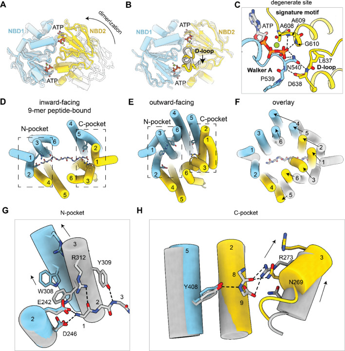
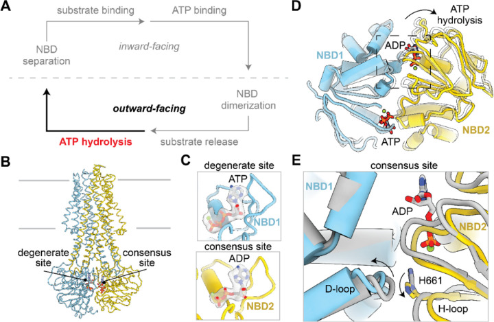
The transporter associated with antigen processing (TAP) is essential for adaptive immunity, delivering peptide antigens from the cytoplasm into the endoplasmic reticulum (ER) for loading onto MHC-I molecules. Previous studies have revealed the mechanism by which TAP selectively binds peptides while allowing for sequence diversity, but how the bound peptides are transported and released into the ER is not yet fully understood. Here, we report cryo-electron microscopy structures of human TAP in multiple functional states along the transport cycle. In the inward-facing conformation, ATP binding strengthens intradomain assembly. The transition to the outward-facing conformation is highly temperature-dependent and leads to a complete reconfiguration of the peptide-binding site, facilitating peptide release. ATP hydrolysis opens the consensus site, and the subsequent separation of the NBDs resets the transport cycle. These findings establish a comprehensive structural framework for understanding the mechanisms of peptide transport, vanadate trapping, and trans-inhibition.

Keywords: ABC transporter; MHC-I; adaptive immunity; antigen presentation; nucleotide binding domain; transporter associated with antigen processing.


Conflict of interest statement

Declaration of interests The authors declare no competing interests.

Figures

Figure 1:
Figure 1:. ATP binding stabilizes TAP nucleotide binding domain 1 (NBD1). See also Figure S1.
(A) Schematic of the TAP transport cycle. ATP binding stabilizes the inward-facing state. (B) The coupling helices (CHs) connect the soluble nucleotide binding domains (NBDs) to the transmembrane domains (TMDs). TAP is represented as white ribbon. The TAP CHs are represented as colored tubes. The boundaries of the membrane are shown as silver lines. (C) Cryo-EM density of TAP bound to a 9-mer peptide has a flexible NBD1 (EMD-41029). Density corresponding to the TAP TMDs, NBD1, and NBD2 are colored as silver, sky blue, and gold, respectively and contoured to 0.125 standard deviations (SDs). (D) Cryo-EM density of TAP bound to a 9-mer peptide and ATP has a rigid NBD1. Density is contoured to 0.8 SDs. (E) TAP1 CH1 and TAP2 CH3 interact with NBD1 near the ATP binding site at the interface between TMD and NBD1. TAP is shown as cartoon tubes and ATP is shown as sticks. (F-G) Zoomed-in view of TMD/NBD1 interface as boxed in (E) in the absence (PDB: 8T4F) (F) or presence (G) of ATP. The dotted line represents the unresolved CH1. (H) ATP binding is associated with a rotation of NBD1 that brings the two NBDs to face each other. Superposition of TAP in the absence (white) and presence (colored) of ATP. The arrow indicates the movement of NBD1 upon ATP binding.
Figure 2:
Figure 2:. NBD dimerization captures TAP in the outward-facing state. See also Figures S2–S4.
(A) NBD dimerization stabilizes the outward-facing state. (B) Molecular model of TAP (EQ) in the NBD-dimerized outward-facing ATP-bound state. TAP1 and TAP2 are shown as ribbon and colored in sky blue and gold, respectively. The boundaries of the membrane are shown as silver lines. (C-D) TAP2 TM3 is flexible and adopts two conformations in the outward-facing state: an outward-facing open (C) and kinked (D) state. The molecular model of TAP1 and TM3 are represented as cartoons with the side chains represented as sticks. Cryo-EM density corresponding to TAP1 and TAP2 TM3 are colored in transparent sky blue and gold, respectively and contoured to 0.12 and 0.196 SDs, respectively. The N- and C-pockets of the peptide binding sites are labeled. (E) The translocation pathway is open to the membrane (top) and endoplasmic reticulum (ER) lumen (bottom). TAP is represented as molecular surface.
Figure 3:
Figure 3:. Conformational changes upon NBD dimerization enable substrate release. See also Figures S2–S4.
(A) Global conformational changes upon NBD dimerization as viewed from the cytoplasm. Superposition of NBD1 in the NBD-separated (white) and NBD-dimerized (colored) conformations. The arrow indicates the movement of NBD2. (B) Local conformational changes in the NBDs upon NBD dimerization. Superposition of NBD1 and NBD2 individually in the NBD-separated (white) and NBD-dimerized (colored) conformations. The D-loop is highlighted and the arrow indicates movement of the D-loop to interact with ATP in the degenerate site. (C) Zoom-in view of the degenerate nucleotide-binding site in NBD1. Hydrogen bonds are represented as dashed lines. (D-E) The TAP translocation pathway in the 9-mer peptide-bound inward-facing (D) or outward-facing (E) conformation as viewed from the ER lumen. The helices of TAP are numbered and shown as colored cartoon tubes. The main chain backbone of the peptide substrate is shown as silver sticks. The N-pocket and C-pocket are boxed as indicated and residues that comprise each pocket are shown as sticks. (F) Superposition of the TAP translocation pathway in the inward-facing peptide-bound (silver) and outward-facing (colored) conformations. Arrows indicate movement of the TAP helices. (G-H) Superposition of the N- (G) and C- (H) pocket of the TAP substrate-binding site as shown in (D). The main chain of the peptide substrate is represented as silver sticks and numbered by alpha-carbon. TAP residues important for interacting with substrate are shown as sticks and hydrogen bonds are shown as dashed lines. Arrows indicate movement of TM3 upon NBD dimerization.
Figure 4:
Figure 4:. Asymmetric NBD separation after ATP hydrolysis and phosphate release. See also Figure S5.
(A) The post-hydrolytic state can adopt an outward-facing state. (B) Molecular model of wild-type TAP in the NBD-dimerized outward-facing post-hydrolytic state. The degenerate and consensus ATP binding sites are labeled. (C) ATP is bound in the NBD1 degenerate site and ADP is bound in the NBD2 consensus site. ATP is shown as sticks and the magnesium atom is shown as spheres. Density corresponding to bound nucleotide is shown as gray surface and contoured to 0.22 SDs. (D) Superposition of NBD1 before (white) and after (colored) after ATP hydrolysis as viewed from the cytoplasm. Arrows indicate conformational changes in NBD2 after ATP hydrolysis. (E) Zoom-in view of NBD2 consensus site as boxed in (D). Arrows indicate local conformational changes in the D-loop of NBD1 and the H-loop of NBD2 after ATP hydrolysis
Figure 5:
Figure 5:. Structure of TAP in the NBD-separated post-hydrolytic conformation. See also Figure S5.
(A) NBD separation resets the transport cycle. (B) Molecular model of wild-type TAP in the NBD-separated outward-facing post-hydrolytic state. Bound nucleotide is shown as sticks and the magnesium atom is shown as spheres. The degenerate and consensus ATP binding sites are labeled. (C) ATP is bound in the NBD1 degenerate site (top) while ADP is bound in the NBD2 consensus site (bottom). Density corresponding to bound nucleotide is shown as gray surface and contoured to 0.33 SDs.

Similar articles

References

    1. Blum J. S., Wearsch P. A. & Cresswell P. Pathways of Antigen Processing. Annu Rev Immunol 31, 443–473 (2013). - PMC - PubMed
    1. Pishesha N., Harmand T. J. & Ploegh H. L. A guide to antigen processing and presentation. Nat Rev Immunol 22, 751–764 (2022). - PubMed
    1. Raghavan M. Immunodeficiency due to defective antigen processing: the molecular basis for type 1 bare lymphocyte syndrome. The Journal of clinical investigation 103, 595–6 (1999). - PMC - PubMed
    1. Spies T. et al. Presentation of viral antigen by MHC class I molecules is dependent on a putative peptide transporter heterodimer. Nature 355, 644–646 (1992). - PubMed
    1. Kelly A. et al. Assembly and function of the two ABC transporter proteins encoded in the human major histocompatibility complex. Nature 355, 641–644 (1992). - PubMed

Publication types