[Preprint]. 2025 Jun 26:2025.06.23.661150.
doi: 10.1101/2025.06.23.661150.

Human Protein Synthesis Requires aminoacyl-tRNA Pivoting During Proofreading

Affiliations

Human Protein Synthesis Requires aminoacyl-tRNA Pivoting During Proofreading

Divya Sapkota et al. bioRxiv. .

Update in

Abstract

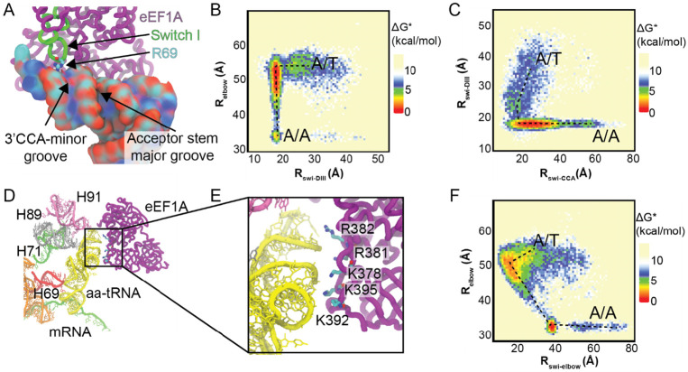
Rigorous studies have characterized the aa-tRNA selection mechanism in bacteria, which is essential for maintaining translational fidelity. Recent investigations have identified critical distinctions in humans, such as the requirement of subunit rolling and a tenfold slower proofreading step. Although these studies captured key intermediates involved in tRNA selection, they did not elucidate the transitions of aa-tRNA between intermediates. Here, we simulated 1,856 aa-tRNA accommodation events into the human ribosomal A site, revealing the requirement of a distinct ~30° pivoting of aa-tRNA about the anticodon stem within the accommodation corridor. This pivoting is crucial for navigating the crowded accommodation corridor, which becomes more constrained due to subunit rolling. Subunit rolling-dependent crowding increases the steric contributions of the accommodation corridor during aa-tRNA accommodation, consistent with the 10-fold reduction in the rate of proofreading. The pivoting of the aa-tRNA enables precise alignment within the accommodation corridor, allowing it to traverse the narrower passage. Furthermore, we found that domain III of eEF1A interacts with the accommodating aa-tRNA through conserved basic residues, providing a steric block to prevent dissociation from the A site. Together, these findings provide a structural framework for understanding the distinctions between bacterial and human aa-tRNA selection and demonstrate that the alignment of the aa-tRNA relative to the ribosomal catalytic sites is a critical determinant of translational fidelity.


Figures

Figure 1.
Figure 1.. Conformations of ribosomes during eukaryotic aa-tRNA accommodation.
(A) All-atom structural representation of the pre-accommodated GTPase activated state with aa-tRNA in the A/T position. (B) Simplified model of the GTPase activated state with aa-tRNA in the A/T position. (C) All-atom structural representation of the accommodated state with aa-tRNA in the A/A position. (D) Simplified model of the accommodated state with aa-tRNA in the A/A position. In these models aa-tRNA (yellow), mRNA (green), peptidyl-tRNA (orange), 28S, 5S, 5.8S rRNA (white), 18S (grey), ribosomal proteins (blue) and eEF1A (purple) are represented.
Figure 2.
Figure 2.. Dynamics of human aa-tRNA accommodation.
(A) Distance measured between O3’ atoms of U60 and U8 of the aa-tRNA and peptidyl-tRNA, respectively, to characterize the elbow accommodation dynamics of the aa-tRNA (Relbow). (B) Distance measurement between the O3’ atoms of A76 of the aa-tRNA and peptidyl tRNA during accommodation into the A site of the ribosome (RCCA). (C) Representative distance of Relbow during simulation using potential 2. (D) Representative distance of RCCA during a simulation using potential 2. (E) Representative distance of Relbow during a simulation using potential 1, showing reversible fluctuations. (F) Representative distance of RCCA during a simulation using potential 1, showing reversible fluctuations.
Figure 3.
Figure 3.. Human aa-tRNA accommodation occurs through a 3’-CCA end dependent pathway.
(A) Approximate free energy (ΔG) landscape of tRNA accommodation as measured by comparison of the Relbow and RCCA distance measurements from simulations using potential 1. (B) Representative structure of EA-1. Complete 80S structure of EA-1 (i). Representation of the accommodation corridor of the ribosome with aa-tRNA engaging H89 in EA-1 (ii). Zoom in of accommodation corridor with aa-tRNA engaging H89 in EA-1 (iii). Representation of the accommodation corridor in EA-1 from the LSU perspective (iv). Zoom in of the 3’ CCA end of the aa-tRNA interacting with H71 in EA-1 (v). (C) Representative structure of the accommodation corridor during transition path from EA-1 to EA-2 (Path). Complete 80S structure of Path (i). Representation of the accommodation corridor of the ribosome with aa-tRNA engaging H71 in Path (ii). Zoom in of the Path position with the aa-tRNA interacting with H89 (iii). Representation of the accommodation corridor in Path from the LSU perspective (iv). Zoom in of the 3’ CCA end of the aa-tRNA interacting with H71 in Path (v). (D) Representative structure of EA-2. Complete 80S structure of EA-2 (1). Representation of the accommodation corridor of the ribosome wiith aa-tRNA engaging H71 after passing H89 (ii). Zoom in of the EA-2 position with the aa-tRNA interacting with H89 (iii).Representation of the accommodation corridor in EA-2 from the LSU perspective (iv). Zoom in of the 3’ CCA end of the aa-tRNA interacting with H71 in EA-2(v). In these models Relbow highlighted in blue and RCCA highlighted in red, while the rRNA (white and grey), ribosomal proteins (blue), peptidyl-tRNA (orange), aa-tRNA (yellow), H89 (grey), H90 (mauve), H71 (green), h18 (purple), H44 (red), and mRNA (green) are represented.
Figure 4.
Figure 4.. Human aa-tRNA pivoting during accommodation into the ribosomal A site.
(A) Structural representation of aa-tRNA pivoting (θtRNA) during accommodation into the A site. Perspective from the central protuberance (left), LSU (middle), and P site (right) are represented to highlight the pivoting of the aa-tRNA. Peptidyl-tRNA (orange), mRNA (green), pre-pivot aa-tRNA (yellow), and post-pivot aa-tRNA (blue) are represented and the axis of the pivoting along the anticoding stem to the elbow domain is highlighted in red. θtRNA is the angle of the tRNA as it moves into the ribosomal A site as measured by changes in the vector perpendicular to the plane of the tRNA (Methods). (B) Cartoon representations of the aa-tRNA pivoting into the ribosomal A site. aa-tRNA begins accommodation into the ribosome in the bent non-pivoted position (left), the tRNA then needs to pivot and accommodate through a 3’CCA-end first mechanism (middle), then the tRNA returns to a non-pivoted position once the elbow of the tRNA enters the A site. (C) Approximate free energy landscape of human tRNA accommodation into the ribosomal A site generated by comparing the Relbow distance and θtRNA angle, generated from potential 1 simulations. (D) Approximate free energy landscape of human tRNA accommodation into the ribosomal A site generated by comparing the RCCA distance and θtRNA angle, generated from potential 1 simulations. (E) Representative structure of the open A site of the ribosome in the GA complex, prior to accommodation (F) Representative structure of the closed A site of the ribosome in the AC complex, after accommodation. (G) Approximate free energy landscape of human ribosome rolling during tRNA accommodation generated by comparing Relbow and Rrolling, generated from potential 2 simulations. (H) Approximate free energy landscape of human ribosome rolling during tRNA accommodation generated by comparing RCCA and Rrolling, generated from potential 2 simulations. (I) Histogram of distances between atoms A35 and C61 of the accommodating H. sapiens and E. coli aa-tRNA, representing the compression of tRNA during accommodation (Rcompression), generated from potential 2 simulations. (J) Solvent accessible surface area (SASA) of aa-tRNA during H. sapiens and E. coli during accommodation, generated from potential 2 simulations.
Figure 5
Figure 5. aa-tRNA interactions with the accommodation corridor.
(A) Regions of the 28S rRNA that are within 4 Å of the aa-tRNA during accommodation simulations of the tRNA into the ribosomal A site. (B) Regions of the 18S rRNA that are within 4 Å of the aa-tRNA during accommodation simulations of the tRNA into the ribosomal A site. (C) Structural representation of rRNA and ribosomal protein uL14 that are in proximity to aa-tRNA during accommodation. (D) Histogram of θtRNA during accommodation of tRNA, captured from N ≥ 5 potential 2 simulations composed of full ribosome or with no H69/71 or no H89.
Figure 6
Figure 6. eEF1A interacts with accommodating aa-tRNA through R69 and conserved basic amino acids in Domain III.
(A) R69 of switch I of eEF1A interacts with the minor groove of the aa-tRNA adjacent to the 3’CCA end. R69 is highlighted interacting with the phosphodiester backbone of the tRNA. (B) Approximate free energy landscape of Relbow in comparison to the distance between eEF1A R69 and A425 of domain III (Rswi-DIII), generated from potential 2 simulations. This landscape indicates that for aa-tRNA to accommodate, R69 of switch I needs to pass through the 3’CCA-minor groove of the aa-tRNA and dock on domain III. (C) Approximate free energy landscape of Rswi-DIII with respect to the distance of R69 and A76 of aa-tRNA, generated from potential 2 simulations. This landscape highlights that only the path where R69 passes by the 3’CCA-minor groove is available. (D) Interactions of eEF1A domain III with in proximity to the accommodating aa-tRNA. (E) Zoomed in image of domain III of eEF1A interacting with the accommodating aa-tRNA. Basic amino acids are highlighted that could interact with the aa-tRNA. (F) Approximate free energy landscape of Relbow distance with respect to K378 to U55 O3’ distance, generated from potential 2 simulations. This demonstrates that domain III approaches the accommodating aa-tRNA in all simulations at an Relbow of ~52 Å.
Figure 7.
Figure 7.. Model of aa-tRNA pivoting into the ribosomal A-site.
(A) Crowded environment of the human accommodation corridor enables aa-tRNA accommodation to be 10-fold slower. Additional eEF1A stays in complex with the ribosome long enough form transient interactions between domain III of eEF1A and the elbow domain of the accommodating aa-tRNA (B) aa-tRNA in human approaches the accommodation corridor until it reaches the H89 steric barrier. It then requires pivoting of the aa-tRNA to properly align with all of the elements of the accommodation corridor to enter the A site.

Similar articles

References

    1. Bouadloun F., Donner D. & Kurland C. G. Codon-specific missense errors in vivo. The EMBO Journal 2, 1351–1356 (1983). 10.1002/j.1460-2075.1983.tb01591.x - DOI - PMC - PubMed
    1. Edelmann P. & Gallant J. Mistranslation in E. coli. Cell 10, 131–137 (1977). 10.1016/0092-8674(77)90147-7 - DOI - PubMed
    1. Kramer E. B. & Farabaugh P. J. The frequency of translational misreading errors in E. coli is largely determined by tRNA competition. RNA 13, 87–96 (2007). 10.1261/rna.294907 - DOI - PMC - PubMed
    1. Laughrea M., Latulippe J., Filion A. M. & Boulet L. Mistranslation in twelve Escherichia coli ribosomal proteins. Cysteine misincorporation at neutral amino acid residues other than tryptophan. Eur J Biochem 169, 59–64 (1987). 10.1111/j.1432-1033.1987.tb13580.x - DOI - PubMed
    1. Ogle J. M. & Ramakrishnan V. Structural insights into translational fidelity. Annu Rev Biochem 74, 129–177 (2005). 10.1146/annurev.biochem.74.061903.155440 - DOI - PubMed

Publication types

LinkOut - more resources