[Preprint]. 2025 Jun 17:2025.06.11.659108.
doi: 10.1101/2025.06.11.659108.

Modeling the role of urokinase plasminogen activator, uPA, and circulating Cancer-Associated Fibroblasts (cCAFS) in breast cancer cell extravasation

Affiliations

Modeling the role of urokinase plasminogen activator, uPA, and circulating Cancer-Associated Fibroblasts (cCAFS) in breast cancer cell extravasation

Angela Spartz et al. bioRxiv. .

Abstract

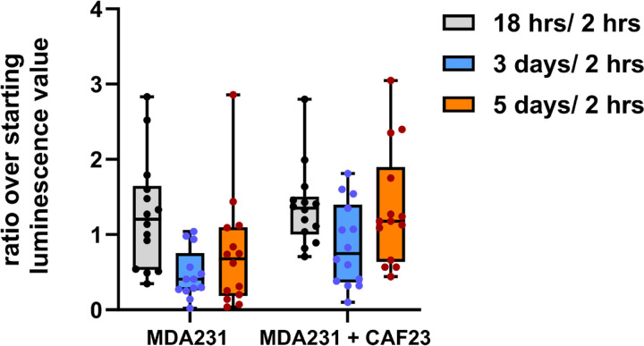
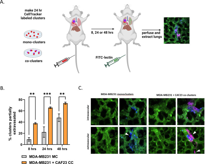
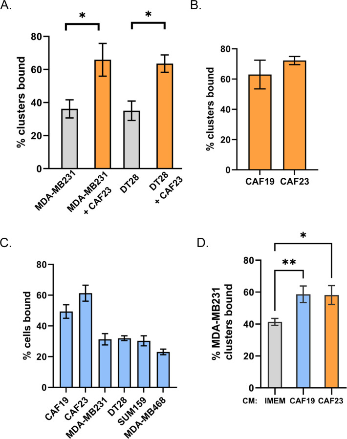
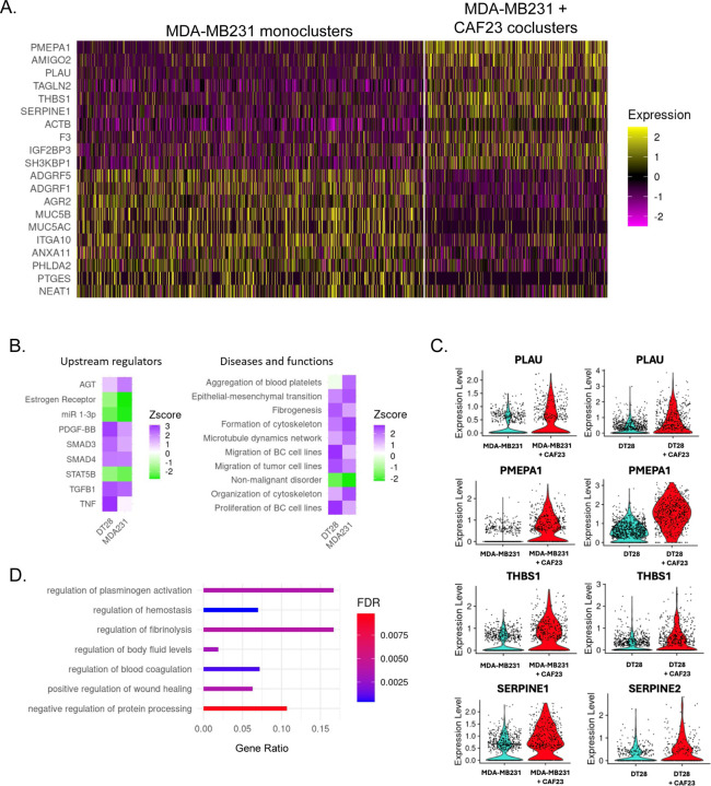
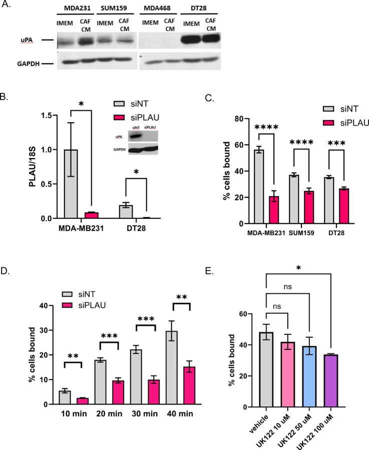
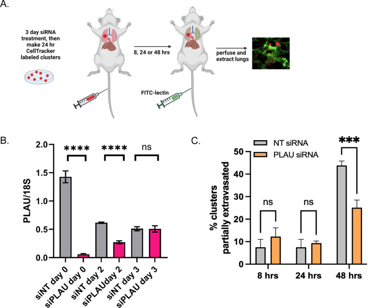
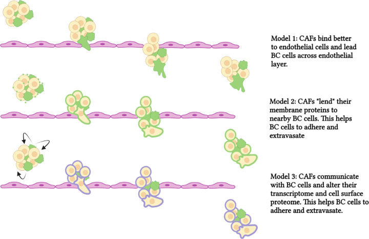
Circulating Cancer-Associated Fibroblasts (cCAFs) have been discovered in circulating tumor cell clusters from all stages of disease progression in breast cancer patients. We have shown that CAFs promote lung metastases in the mouse tail vein model when they are clustered with triple negative breast cancer (TNBC) MDA-MB231 cells. Following on this observation, we saw that MDA-MB231-luciferase labeled cells persist at higher levels when present in CAF23/MDA-MB231 co-clusters compared to MDA-MB231 mono-clusters within the first 3 days after tail vein injection. This prompted us to investigate whether CAFs aid cancer cell extravasation from capillary venules into the lung parenchyma, which would impart better survival and faster seeding of metastases. Ex vivo lung extravasation assays showed that within the first 8-24 hrs after tail vein injection, more cells from CAF23/MDA-MB231 co-clusters extravasated than cells from MDA-MB231 mono-clusters. Using in vitro endothelial binding assays, we determined that CAF/TNBC co-clusters bind to HUVEC endothelial cells better than TNBC mono-clusters. Single Cell RNA-seq identified several genes in the fibrinolysis pathway whose expression increases in TNBC cells when they are clustered with CAFs. One of these genes is PLAU, which encodes the urokinase-type plasminogen activator, uPA. siRNA knockdown of PLAU decreased in vitro TNBC-endothelial cell interactions and ex vivo extravasation of MDA-MB231 mono-clusters, revealing a role for uPA/PLAU in breast cancer cell extravasation. Our data helps to define the role of CAFs in breast cancer extravasation and highlights the importance of our previous work showing that CAFs promote tumor cell dissemination and metastasis.

Keywords: breast cancer; cancer-associated fibroblasts; circulating tumor cell clusters; extravasation; metastasis; urokinase-type plasminogen activator.


Conflict of interest statement

Competing interests: The authors have no competing interests to declare that are relevant to the content of this article.

Figures

Figure 1.
Figure 1.. CAF23/MDA-MB231 co-clusters survive and grow faster than MDA-MB231 mono-clusters in the tail vein assay.
MDA-MB231-luciferase mono-clusters and CAF23/ MDA-MB231-luciferase co-clusters were made for 24 hrs in Mammocult media (75,000 cells of each cell type per mouse). Clusters were collected into PBS, injected into the tail vein of NSG mice, and luminescence was followed over the first 5 days using live animal imaging. The luminescence observed at each time point (18 hrs, 3 days, and 5 days) was normalized to the amount that was present 2 hrs after injection. Each dot represents a single animal. N = 14 mice per group.
Figure 2.
Figure 2.. CAF23/MDA-MB231 co-clusters extravasate better than MDA-MB231 mono-clusters in the ex vivo lung extravasation assay.
A. Schematic of experimental design. CAF23 cells were labeled with Cell Tracker Deep Red and MDA-MB231 cells were labeled with CellTracker Red. MDA-MB231 mono-clusters and CAF23/MDA-MB231 co-clusters were made for 24 hrs. Clusters were collected and injected into the tail vein of FVB mice. 30 minutes prior to perfusion, FITC-lectin was injected into the tail vein to visualize the vasculature. Lung perfusion and fixation was performed at 8 hrs, 24 hrs, and 48 hrs. Pieces of lung were cut and imaged with confocal microscopy to visualize and quantify cell clusters inside or outside of the vasculature. B. Quantitation of ex vivo assay using 3 mice per time point per treatment. At least 20 clusters per animal were counted and scored as being intravascular or partially extravascular. C. Example images of mono-clusters or co-clusters that are intravascular or partially extravascular. Vasculature is green. MDA-MB231 cells are pseudo-colored blue. CAFs are pink. Arrowheads indicate cells that have extravasated. ** p ≥ 0.005, *** p ≥ 0.0005
Figure 3.
Figure 3.. CAFs and CAF/BC co-clusters bind to HUVEC endothelial cells better than BC cells and BC mono-clusters.
Assays were performed with CellTracker Deep Red labeled CAFs and CellTracker Red labeled breast cancer cells. Single cell suspensions or 24-hr clusters were made and pipetted over a lawn of HUVEC cells for 30 minutes before being washed. Wells were scanned on a TiE fluorescent microscope and bound cells or clusters were counted. A. Comparison of co-clusters and mono-clusters. B. Comparison of CAF19 and CAF23 mono-clusters. C. CAF cells compared with 4 different TNBC cells. D. Comparison of MDA-MB231 mono-clusters made in CAF conditioned media vs IMEM media. The endothelial binding assay was performed in IMEM. * p ≥ 0.05, ** p ≥ 0.005
Figure 4:
Figure 4:. Single Cell RNA seq analysis of MDA-MB231 cells from MDA-MB231 mono-clusters vs CAF23/MDA-MB231 co-clusters.
A. Heatmap showing the top 10 up and down DEG in breast cancer cells from MDA-MB231 mono-clusters vs breast cancer cells from CAF23/MDA-MB231 co-clusters. Plot shows only cells that were in the G1 phase of the cell cycle; each line represents an individual cell. Number of MDA-MB231 cells analyzed: 902 cells from MDA-MB231 mono-clusters and 474 cells from CAF23/MDA-MB231 co-clusters. B. Heatmap showing top upstream regulators and top diseases/functions identified by Ingenuity Pathway Analysis (IPA) of DEG lists of breast cancer cells from mono-clusters vs co-clusters for MDA-MB231 and DT28 cell lines. The Z score indicates the likely activation (positive) or inhibition (negative) of a regulator or function; a score of 2 is considered significant. C. Violin plots from the top 4 genes upregulated in both CAF23/MDA-MB231 co-clusters and CAF23/DT28 co-clusters. Each dot represents a cell. D. Gene Ontology analysis of a ranked list of DEG from MDA-MB231 mono-clusters vs CAF23/MDA-MB231 co-clusters. Gene ratio indicates the fraction of DEG genes over the number of genes in each gene set. FDR is the adjusted p value.
Figure 5.
Figure 5.. Reduction of uPA/PLAU inhibits BC-endothelial cell binding.
A. Western blot showing that CAF23 conditioned media increases uPA protein in MDA-MB231 cells. B. qPCR of PLAU in MDA231 or DT28 cells treated for 3 days with non-targeting siRNAs (siNT) or PLAU siRNAs (siPLAU). PLAU was normalized to the 18S ribosomal RNA. Inset: Western blot of siNT or siPLAU MDA-MB231 cells. C. Endothelial binding assay of MDA-MB231, DT28 and SUM159 cells treated for 3 days with siNT or siPLAU. D. Time course of endothelial binding assay using MDA-MB231 cells treated with siNT or siPLAU showing % of cells bound for the indicated periods. E. Endothelial binding assay using MDA-MB231 cells pre-treated with vehicle or uPA inhibitor, UK122, for 24 hrs at increasing concentrations. * p ≥ 0.05, ** p ≥ 0.005, *** p ≥ 0.0005
Figure 6.
Figure 6.. Reduction of uPA/PLAU in MDA-MB231 cells inhibits extravasation in the ex-vivo lung assay.
A. Schematic of ex vivo assay. MDA-MB231 cells were treated with siNT or siPLAU for 2 days, then cells were stained with CellTracker Red and made into 24 hr mono-clusters in the presence of siNT or siPLAU (3 days total siRNA exposure). Clusters were collected and injected by tail vein into FVB mice. 30 minutes prior to lung perfusion, mice were injected by tail vein with FITC-lectin to stain the vasculature. Lung perfusion and fixation was performed at 8 hrs, 24 hrs, and 48 hrs. Pieces of lung were cut and imaged with confocal microscopy to visualize and quantify BC clusters inside or outside of the vasculature. B. qPCR experiment showing persistence of siRNA effect. MDA-MB231 cells were treated with siRNAs for 3 days as before. Cells were then washed of siRNA, cultured in regular media, and qPCR was performed at the given times after removal of siRNA. 18S was used as a control for the qPCR. C. Results from ex vivo assay described in A using 2 mice per time point, per treatment. 20–40 clusters were counted for each animal and scored as being completely intravascular or partially extravascular.
Figure 7.
Figure 7.. Schematic of models showing CAF-aided BC extravasation.
In the top panel, CAFs bind to vascular endothelium better than breast cancer cells. CAFs then lead BC cells across the endothelium. In the middle panel, CAFs lend ECM proteins to BC cells (by secreted extracellular vesicles) and this increases endothelial binding and promotes extravasation. In the bottom panel, CAFs communicate with BC cells and alter the BC transcriptome. This increases proteins, such as uPA, that promote extravasation. CAFs are depicted in green, breast cancer cells are depicted in beige/yellow. ECM proteins on CAFs are in green. Proteins induced by CAFs and expressed in breast cancer cells in purple. *** p ≥ 0.0005. **** p ≥ 0.00005

Similar articles

References

    1. Giaquinto AN, et al. (2024) Breast cancer statistics 2024. CA Cancer J Clin. 10.3322/caac.21863 - DOI
    1. Santi A, Kugeratski FG, Zanivan S (2018) Cancer Associated Fibroblasts: The Architects of Stroma Remodeling. Proteomics 18: e1700167. 10.1002/pmic.201700167 - DOI - PMC - PubMed
    1. D’Arcangelo E, et al. (2020) The life cycle of cancer-associated fibroblasts within the tumour stroma and its importance in disease outcome. Br J Cancer 122: 931–42. 10.1038/s41416-019-0705-1 - DOI - PMC - PubMed
    1. Joshi RS, et al. (2021) The Role of Cancer-Associated Fibroblasts in Tumor Progression. Cancers (Basel) 13 10.3390/cancers13061399 - DOI - PMC - PubMed
    1. Sahai E, et al. (2020) A framework for advancing our understanding of cancer-associated fibroblasts. Nat Rev Cancer 20: 174–86. 10.1038/s41568-019-0238-1 - DOI - PMC - PubMed

Publication types

LinkOut - more resources