[Preprint]. 2025 Jun 29:2025.06.28.661837.
doi: 10.1101/2025.06.28.661837.

Inhibition of nonsense-mediated decay in TDP-43 deficient neurons reveals novel cryptic exons

Affiliations

Inhibition of nonsense-mediated decay in TDP-43 deficient neurons reveals novel cryptic exons

Irika R Sinha et al. bioRxiv. .

Abstract

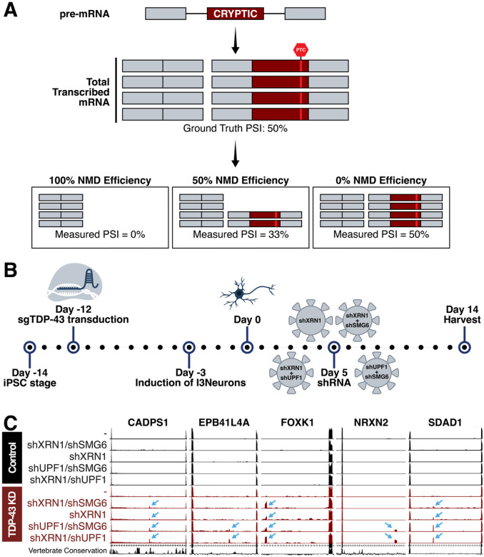
TAR DNA-binding protein 43 kDa (TDP-43) is an essential splicing repressor whose loss of function underlies the pathophysiology of amyotrophic lateral sclerosis and frontotemporal dementia (ALS-FTD). Nuclear clearance of TDP-43 disrupts its function and leads to the inclusion of aberrant cryptic exons. These cryptic exons frequently introduce premature termination codons resulting in the degradation of affected transcripts through nonsense-mediated mRNA decay (NMD). Conventional RNA sequencing approaches thus may fail to detect cryptic exons that are efficiently degraded by NMD, precluding identification of potential therapeutic targets. We generated a comprehensive set of neuronal targets of TDP-43 in human iPSC-derived i3Neurons (i3N) by combining TDP-43 knockdown with inhibition of multiple factors essential for NMD, revealing novel cryptic targets. We then restored expression of selected NMD targets in TDP-43 deficient i3Ns and determined which genes improved neuronal viability. Our findings highlight the role of NMD in masking cryptic splicing events and identify novel potential therapeutic targets for TDP-43-related neurodegenerative disorders.


Figures

Figure 1.
Figure 1.. Nonsense-mediated decay (NMD) was inhibited to identify TDP-43 gene targets which undergo efficient degradation.
(A) Pre-mRNA transcribed in a cell includes cryptic exons when TDP-43 is dysfunctional. This pre-mRNA is processed into mRNA and the cryptic exons often encode PTCs that are targeted by NMD. Efficiency of NMD impacts measured cryptic exon PSI after RNA sequencing or RT-PCR. (B) Schematic of experimental timeline. i3Ns with a CRISPRi module were transduced with sgTDP-43 12 days prior to differentiation to create a stable TDP-43 knockdown cell line. Five days post-differentiation, we conducted shRNA-mediated knockdown of NMD factors XRN1, UPF1, and SMG6. i3Ns were harvested 14 days after differentiation and RNA extracted for RNA sequencing. (C) Cryptic exons in CADPS1, EPB41L4A, FOXK1, NRXN2, and SDAD1 are measurable by RNA sequencing after NMDi. These cryptic exons are not identifiable in cells with only TDP-43 knockdown or NMDi and are specific to TDP-43 knockdown conditions.
Figure 2.
Figure 2.
(A) 421 total CEs were identified in i3Ns and categorized as cassette exons, exon extensions, early polyadenylation events, or alternative start sites. 26 CEs were “leaky” in the control conditions. (B) Comparison of CEs measurable with and without NMDi based on given cut-offs. Each cut-off defines the minimum avgPSI needed for a CE to be considered measurable without NMDi. (C) Comparison of median cryptic exon avgPSIs between conditions. (D-F) Frequency of [UG]n dinucleotide repeats near CE splice sites. TDP-43 CEs often have [UG]n after the 3’ end of the CE. (D) Full compendium of 421 CEs. (E) Split by type of CE (cassette, exon extension, polyA, alternative splice site). Exon extension CEs are enriched for [UG]n repeats after the 3’ end of the cryptic exon compared to the overall average and other types. PolyA cryptic exons have a slight enrichment for the [UG]n before the 5’ end of the cryptic exon. Due to lack of 3’ and 5’ exon-exon junctions for polyA and alternative start site CEs, respectively, they only have the repeat mapped near one splice site. (F) Split by measurability without NMDi. Cut-offs are the same as in Figure 2C. CEs with PSI less than 1% in TDP-43 knockdown conditions have less pronounced [UG]n repeats at the 3’ end of the CE. (G) UCSC RepeatMasker annotations present at least twice between 600 bp external each splice site and 50 bp internal of the sites. Alu family repeats are most common near cryptic exons. (H) UCSC RepeatMasker annotations present at least twice between 600 bp external each splice site and 50 bp internal of the sites of high expressor CEs. Alu family repeats are most still common.
Figure 3.
Figure 3.. PSI and Gene Expression Levels of Cryptic Exons
(A) avgPSI in different conditions of top 30 cryptic exon gene targets, as ranked by maximum avgPSI in any TDP-43 knockdown condition. Many targets are transcribed at a higher frequency in at least one NMDi condition than expected from non-NMDi TDP-43 knockdown condition. (B-C) Gene expression levels as measured by normalized area under the curve (NAUC) extracted from ASCOT for high expressor CEs. Different tissues are represented by rows and different CEs by the columns. (B) (Left) All high expressor CEs (Right) Top 30 CE gene targets as per 3A. (C) All CEs with less than 1% PSI after only TDP-43 knockdown. (D) CEs with less than 1% PSI after only TDP-43 knockdown and ubiquitous expression across different tissue types.
Figure 4.
Figure 4.. Connections to ALS-FTD and differential gene expression.
(A) ATXN1 and CHMP2B are ALS-associated risk genes that contain cryptic exons (B) Biological pathways including multiple ALS-associated risk genes and cryptic exon-containing genes include autophagy (C-E) Differential gene expression after TDP-43 knockdown of cryptic exon-containing genes (C) Schematic describing transcript count correction for functional transcripts. I3N were treated and harvested. Prepared RNA library was sequenced and STAR and Salmon were used to align and quantify transcripts. Transcript counts for CE-including transcripts were corrected for functional gene expression by multiplying total count by the percentage of transcripts without CE. (D) Normal method without any adjustment (E) Transcript counts adjusted for average PSI of cryptic exon which would decrease the number of transcripts encoding functional protein
Figure 5.
Figure 5.. shRNA-mediated knockdown of cryptic exon-containing genes.
(A) Sample images of shRNA-treated i3Ns co-stained for DAPI and propidium iodide (PI), which stains dying cells; shCtrl is a non-targeting control. Scale bar = 200μm. (B) Ranked quantification of shRNA toxicity normalized to shCtrl and shTDP-43. Min-max normalization was used with maximum toxicity set to shTDP-43 and minimum toxicity to shCtrl. Most shRNA treatment leads to intermediate toxicity and does not recapitulate TDP-43 toxicity. (C) Comparison of cryptic exon gene adjusted downregulation in i3Ns after TDP-43 knockdown with toxicity of shRNA-mediated knockdown. Genes with at least a two-fold downregulation and toxicity at least 50% of shTDP-43 are highlighted and labeled.
Figure 6.
Figure 6.. Rescue experiments in i3Ns with TDP-43 knockdown.
(A) Schematic of rescue experiments in i3N where cells are transduced with shTDP-43 or shCtrl (non-targeting control) on day 4 post-differentiation and then target genes are overexpressed on day 7 post-differentiation. (B) Quantification and ranking of normalized cell toxicity after over-expression of genes after shTDP-43 treatment. Min-max normalization was used with minimum survival set to shTDP-43 and maximum to shCtrl. Values less than 0 correspond with decreased survival compared to shTDP-43 with GFP overexpression and greater than zero correspond with increased survival. Conditions with survival significantly different from shTDP-43+GFP treatment are marked. (C) Sample images from rescue experiments of negative control (shCtrl with GFP overexpression) and three conditions of shTDP-43-treated i3N. ZDHHC11 overexpression led to the highest survival in comparison to shTDP-43 with GFP overexpression while RAP1GAP overexpression led to the lowest. Scale bar = 100μm. (D) ZDHHC11 cryptic exon is leaky in the control but increased significantly after TDP-43 knockdown.

Similar articles

References

    1. Boer E. M. J. de et al. TDP-43 proteinopathies: a new wave of neurodegenerative diseases. J. Neurol., Neurosurg. Psychiatry 92, 86–95 (2021). - PMC - PubMed
    1. Ke H., Liu K., Jiao B. & Zhao L. Implications of TDP-43 in non-neuronal systems. Cell Commun. Signal. 21, 338 (2023). - PMC - PubMed
    1. Zeng J. et al. Decoding TDP-43: the molecular chameleon of neurodegenerative diseases. Acta Neuropathol. Commun. 12, 205 (2024). - PMC - PubMed
    1. Neumann M. et al. Ubiquitinated TDP-43 in Frontotemporal Lobar Degeneration and Amyotrophic Lateral Sclerosis. Science 314, 130–133 (2006). - PubMed
    1. Arai T. et al. TDP-43 is a component of ubiquitin-positive tau-negative inclusions in frontotemporal lobar degeneration and amyotrophic lateral sclerosis. Biochem. Biophys. Res. Commun. 351, 602–611 (2006). - PubMed

Publication types

LinkOut - more resources