[Preprint]. 2025 Jun 20:2025.06.16.660009.
doi: 10.1101/2025.06.16.660009.

A Human Tumor-Immune Organoid Model of Glioblastoma

Affiliations

A Human Tumor-Immune Organoid Model of Glioblastoma

Shivani Baisiwala et al. bioRxiv. .

Abstract

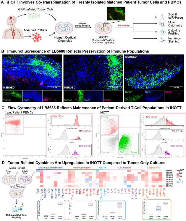
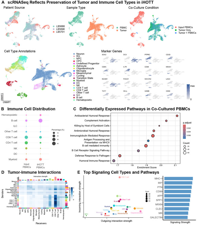
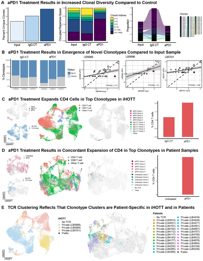
A major obstacle to identifying effective therapies for the aggressive brain tumor glioblastoma is the lack of human-specific, immunocompetent models that reflect the human tumor microenvironment. To address this, we developed the immune-Human Organoid Tumor Transplantation (iHOTT) model. This is an autologous co-culture platform that integrates patient-derived tumor cells and matched peripheral blood mononuclear cells (PBMCs) within human cortical organoids, enabling the study of the patient-specific immune response to the tumor and tumor-immune interactions. This platform preserves tumor and immune populations, immune signaling, and cell-cell interactions observed in patient tumors. Treatment of iHOTT with pembrolizumab, a checkpoint inhibitor, mirrored cell type shifts and cell interactions observed in patients. TCR sequencing further revealed pembrolizumab-driven expansion of stem-like CD4-T-cell clonotypes exhibiting patient-specific repertoires. These findings establish iHOTT as a physiologically relevant platform for exploring autologous tumor-immune interactions and underscore the critical need for antigen-targeted strategies to enhance immunotherapy in glioblastoma.


Conflict of interest statement

DECLARATION OF INTERESTS We (SB, EF, AB) are in the process of filing a provisional patent for the iHOTT system described in this manuscript.

Figures

Figure 1:
Figure 1:. iHOTT Enables Tumor and Immune Co-Culture and Reflects Patient Tumor Cytokine Profiles
A) Human cortical organoids derived from embryonic stem cells were cultured for 8–12 weeks before co-transplantation. Patient-derived glioblastoma cells were dissociated, transduced with GFP-expressing lentivirus, and co-transplanted with matched freshly isolated CellTrace-labeled patient PBMCs onto the organoid surface. Cultures were maintained for 7 days before processing. The inset shows representative fluorescence imaging of GFP+ tumor cells and CellTrace+ PBMCs within the organoid. After 7 days, organoids were collected for downstream flow cytometry, immunostaining, cytokine profiling, and single-cell RNA sequencing of tumor and immune compartments. B) iHOTT organoids were fixed, embedded in OCT, sectioned, and stained for GFP, CD3, CD14, and CD19. Immunofluorescence revealed robust infiltration of CD3+ T cells with minor populations of CD14+ myeloid and CD19+ B cells. GFP+ tumor cells were observed invading into the organoid. C) Flow cytometry of input PBMCs prior to culture revealed a predominantly CD45+ population composed primarily of CD3+ T cells. Analysis of dissociated iHOTT samples after 7 days showed maintenance of tumor (GFP+), organoid (GFP and CD45 negative), and immune (CD45+) compartments, with the immune population again composed primarily of CD3+ T cells, mirroring the original patient PBMC composition. D) Cytokine profiling of conditioned media from iHOTT, tumor-only, and PBMC-only cultures was performed at day 7. The heatmap shows log2 fold change in iHOTT compared to the tumor alone. iHOTT samples showed elevated secretion of tumor-associated cytokines, including G-CSF, IL-6, and IL-10, relative to both tumor-only and PBMC-only controls. Notably, PBMC-only cultures failed to produce these cytokines at comparable levels, indicating a tumor-immune interaction–dependent response within the iHOTT system.
Figure 2:
Figure 2:. scRNA-seq Confirms Preservation of Tumor and Immune Compartments and Recapitulates Cell-Cell Interactions
A) UMAPs from scRNA-seq of three patient samples comparing PBMC input, tumor-only cultures, and tumor + PBMC co-cultures. Top Left: cells annotated by patient source. Top Middle: cells annotated by tumor vs. PBMC origin. Top Right: cells annotated by co-culture condition. Bottom Left: Cells were annotated by reference projection onto the GBM meta-atlas and a PBMC reference object. Bottom Right: Feature plots show expression of representative marker genes validating tumor and immune cell type identities, activation, and exhaustion across conditions. B) Comparison of immune cell distributions between input PBMCs and PBMCs recovered from iHOTT after 7 days. All major immune populations, including CD4+ and CD8+ T cells, B cells, NK cells, and myeloid subsets, were preserved in co-culture. C) Gene Set Enrichment Analysis (GSEA) comparing input PBMCs and iHOTT PBMCs showed enrichment of immune activation pathways in the co-culture condition, suggesting engagement of functional immune programs. D) CellChat was used to analyze tumor and immune cell-cell interactions. We noted interactions between all cell types, with CD8 T cells as a notable receiver. E) CellChat analysis of ligand-receptor interactions between tumor and immune cells. Left: Heatmap of sender-receiver interactions showing CD8+ T cells as major communicators. Oligodendrocytes and astrocytes displayed strong outgoing signals. Right: Co-culture specifically induced upregulation of MHC-I and MIF signaling pathways, indicating active tumor-immune cross-talk within the iHOTT system.
Figure 3:
Figure 3:. Pembrolizumab Treatment in iHOTT Recapitulates Cell Type and Interaction Changes Observed in Treated GBM Patients
A) UMAPs from scRNA-seq of iHOTT samples annotated by patient source (left), treatment condition (middle), and cell type (right). All major tumor and immune cell populations were preserved across IgG control and pembrolizumab-treated samples. B) Cytokine profiling of iHOTT culture supernatants from three tumors treated with pembrolizumab or IgG control. Heatmap depicts relative cytokine expression in pembrolizumab-treated samples, with notable increases in IL-15, IL-25, and IL-17 following treatment. C) UMAPs from scRNA-seq of freshly isolated recurrent GBM patient tumor samples treated with either placebo or pembrolizumab. Left: annotated by patient source. Middle: annotated by treatment condition. Right: annotated by immune cell type, showing consistent detection of all major immune subsets observed in iHOTT, including CD4+ and CD8+ T cells, NK cells, B cells, and myeloid cells. D) Comparative analysis of cell type abundance post-treatment revealed parallel trends in iHOTT and patient samples, including increases in CD4+ T cells, B cells, innate lymphoid cells (ILCs), and non-canonical “Other T” populations in response to pembrolizumab. E) CellChat analysis of ligand-receptor communication networks post-treatment reflects that in iHOTT, pembrolizumab induced increased signaling activity involving ILCs and Other T cells (left). This was consistent with patterns observed in pembrolizumab-treated patient tumors (right). Strong interactions with CD8+ T cells and NK cells were maintained across all treatment conditions.
Figure 4:
Figure 4:. TCR Sequencing Reveals Pembrolizumab-Induced Clonal Remodeling of T Cells in iHOTT and Patient Samples
A) TCR sequencing of iHOTT samples across three patients. Left: Quantification of unique TCR clonotypes revealed a stepwise increase in clonal diversity from input PBMCs to IgG control, and a further increase in pembrolizumab-treated samples. Middle: Repertoire occupancy analysis showed that pembrolizumab treatment resulted in greater representation of rare clonotypes, shifting the overall clonal architecture. Right: Comparison of the top 20 clones in each condition demonstrated the emergence of new clonotypes in pembrolizumab and control samples, alongside persistence of some input-derived dominant clones. B) Left: Assessment of novel clonotype composition showed a significant increase in the proportion of the repertoire composed of new clonotypes in pembrolizumab-treated samples compared to both input and control conditions. Right: Scatter plot comparing clone frequencies between pembrolizumab-treated and input samples showed emergence of new clones and partial overlap with the input repertoire. C) Left: UMAPs of reclustered T cells from iHOTT showing treatment condition (input, IgG control, pembrolizumab) and TCR detection. All samples retained robust TCR coverage. Middle: Cells labeled by treatment and by the top 5 expanded clonotypes within control and pembrolizumab conditions. Right: Top clonotypes in the pembrolizumab condition were enriched for CD4+ T cells, suggesting clonal expansion and reinvigoration mediated by CD4+ populations following PD-1 blockade. D) Analogous TCR analysis in recurrent GBM patient samples treated with pembrolizumab or placebo. While TCR coverage was approximately 30%, consistent trends were observed. Pembrolizumab-treated tumors demonstrated the emergence of novel clonotypes, with top clones reflecting an increase in CD4+ T cells. E) GLIPH2 analysis of TCR sequences was used to group clonotypes based on predicted shared antigen specificity. In both iHOTT (left) and patient samples (right), clonotype clusters were predominantly patient-specific, with limited sharing across individuals. This suggests antigenic heterogeneity in GBM.

Similar articles

References

    1. Stupp R, Mason WP, van den Bent MJ, Weller M, Fisher B, Taphoorn MJB, et al. Radiotherapy plus concomitant and adjuvant temozolomide for glioblastoma. N Engl J Med. 2005. Mar 10;352(10):987–96. - PubMed
    1. Stupp R, Taillibert S, Kanner A, Read W, Steinberg DM, Lhermitte B, et al. Effect of Tumor-Treating Fields Plus Maintenance Temozolomide vs Maintenance Temozolomide Alone on Survival in Patients With Glioblastoma: A Randomized Clinical Trial. JAMA. 2017. Dec 19;318(23):2306–16. - PMC - PubMed
    1. Klein E, Hau AC, Oudin A, Golebiewska A, Niclou SP. Glioblastoma Organoids: Pre-Clinical Applications and Challenges in the Context of Immunotherapy. Front Oncol. 2020. Dec 8;10:604121. - PMC - PubMed
    1. Yabo YA, Moreno-Sanchez PM, Pires-Afonso Y, Kaoma T, Nosirov B, Scafidi A, et al. Glioblastoma-instructed microglia transition to heterogeneous phenotypic states with phagocytic and dendritic cell-like features in patient tumors and patient-derived orthotopic xenografts. Genome Medicine. 2024. Apr 2;16(1):51. - PMC - PubMed
    1. Liu Y, Zhou F, Ali H, Lathia JD, Chen P. Immunotherapy for glioblastoma: current state, challenges, and future perspectives. Cell Mol Immunol. 2024. Dec;21(12):1354–75. - PMC - PubMed

Publication types