

This is a preprint.
Specific GPCRs Elicit Unique Extracellular Vesicle MiRNA Array Signatures: An Exploratory Study
- PMID: 40667070
- PMCID: PMC12262688
- DOI: 10.1101/2025.06.16.659918
Specific GPCRs Elicit Unique Extracellular Vesicle MiRNA Array Signatures: An Exploratory Study
Abstract
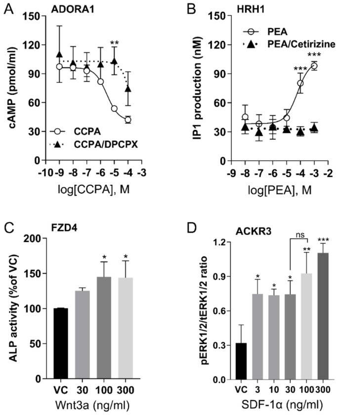
All cells secrete extracellular vesicles (EVs) containing nucleic acid cargo, including microRNAs (miRNAs), that regulate the function of receiving cells. G protein-coupled receptors (GPCRs) affect intracellular function via multiple signaling cascades. However, the mechanisms of GPCR intercellular signaling through EV miRNA activity are unknown. Human U2 osteosarcoma cells expressing native GPCRs were used to selectively stimulate distinct G protein signaling cascades (Gαi, Gαq, Gα12/13, and β-arrestin) by members of specific receptor subclasses including the adenosine receptor A1 (ADORA1), the histamine receptor H1 (HRH1), the frizzled class receptor 4 (FZD4), and the atypical chemokine receptor 3 (ACKR3), respectively. We hypothesized that stimulation of specific classes of GPCRs would cause the release of EVs containing miRNAs with receptor-specific up- or down-regulated expression, affecting unique pathological downstream signaling cascades. Receptor-specific agonists dose-dependently increased respective signaling cascade intermediates. We found no change in the quantity of EVs (~200nm diameter), but there were distinct EV miRNA signatures following stimulation of GPCRs. Network analyses of differentially expressed miRNA and their predicted targets validated the linkage between specific receptors and cell function and pathological states. The data can be used to reverse engineer mechanisms involving EV miRNAs for various physiological and pathological processes. GPCRs are major pharmacological targets, so understanding the mechanisms that stimulate or inhibit GPCR-mediated changes in extracellular miRNA signatures could improve long- and short-term therapeutic and unwanted drug effects.
Keywords: G protein-coupled receptor; U2OS; extracellular vesicle; miRNA; receptor signaling.
Conflict of interest statement
Competing Interests: The authors declare that they have no competing interests.
Figures


Similar articles
-
Sertindole for schizophrenia.Cochrane Database Syst Rev. 2005 Jul 20;2005(3):CD001715. doi: 10.1002/14651858.CD001715.pub2. Cochrane Database Syst Rev. 2005. PMID: 16034864 Free PMC article.
-
Diverse Populations of Extracellular Vesicles with Opposite Functions during Herpes Simplex Virus 1 Infection.J Virol. 2021 Feb 24;95(6):e02357-20. doi: 10.1128/JVI.02357-20. Print 2021 Feb 24. J Virol. 2021. PMID: 33361424 Free PMC article.
-
Drugs for preventing postoperative nausea and vomiting in adults after general anaesthesia: a network meta-analysis.Cochrane Database Syst Rev. 2020 Oct 19;10(10):CD012859. doi: 10.1002/14651858.CD012859.pub2. Cochrane Database Syst Rev. 2020. PMID: 33075160 Free PMC article.
-
Pharmacological intervention for irritability, aggression, and self-injury in autism spectrum disorder (ASD).Cochrane Database Syst Rev. 2023 Oct 9;10(10):CD011769. doi: 10.1002/14651858.CD011769.pub2. Cochrane Database Syst Rev. 2023. PMID: 37811711 Free PMC article.
-
Clinical symptoms, signs and tests for identification of impending and current water-loss dehydration in older people.Cochrane Database Syst Rev. 2015 Apr 30;2015(4):CD009647. doi: 10.1002/14651858.CD009647.pub2. Cochrane Database Syst Rev. 2015. PMID: 25924806 Free PMC article.
References
-
- Allison P. (2009). Fixed Effects Regression Models 10.4135/9781412993869 - DOI
-
- Alonso R., Mazzeo C., Rodriguez M. C., Marsh M., Fraile-Ramos A., Calvo V., Avila-Flores A., Merida I., & Izquierdo M. (2011). Diacylglycerol kinase α regulates the formation and polarisation of mature multivesicular bodies involved in the secretion of Fas ligand-containing exosomes in T lymphocytes. Cell Death & Differentiation, 18(7), 1161–1173. 10.1038/cdd.2010.184 - DOI - PMC - PubMed
Publication types
Grants and funding
LinkOut - more resources
Full Text Sources