[Preprint]. 2025 Jun 27:2025.06.24.661412.
doi: 10.1101/2025.06.24.661412.

Phosphoglycerate Kinase Can Adopt a Topologically Misfolded Form that is More Stable than its Native State

Affiliations

Phosphoglycerate Kinase Can Adopt a Topologically Misfolded Form that is More Stable than its Native State

Yingzi Xia et al. bioRxiv. .

Abstract

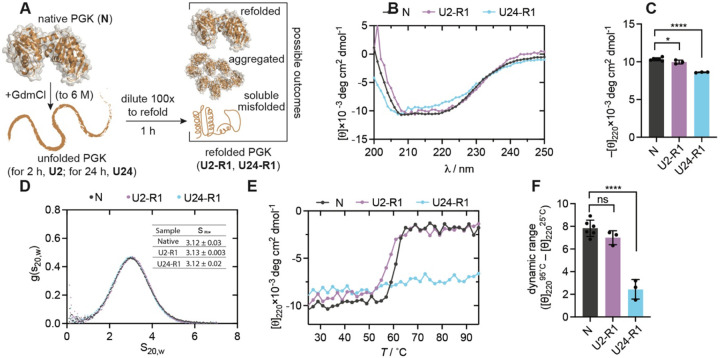
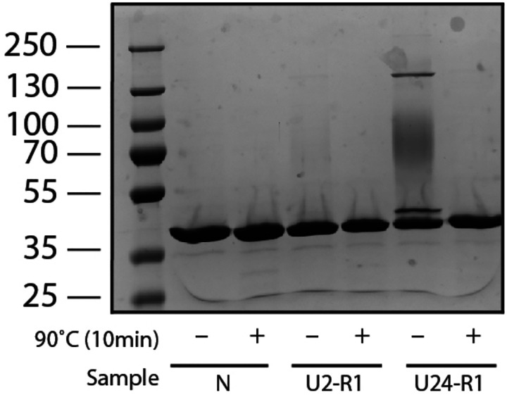
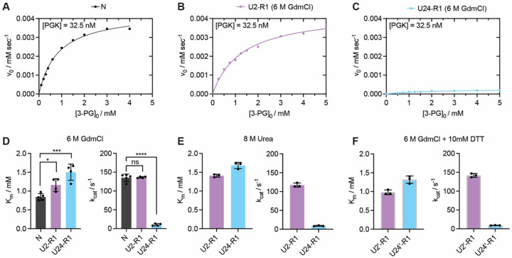
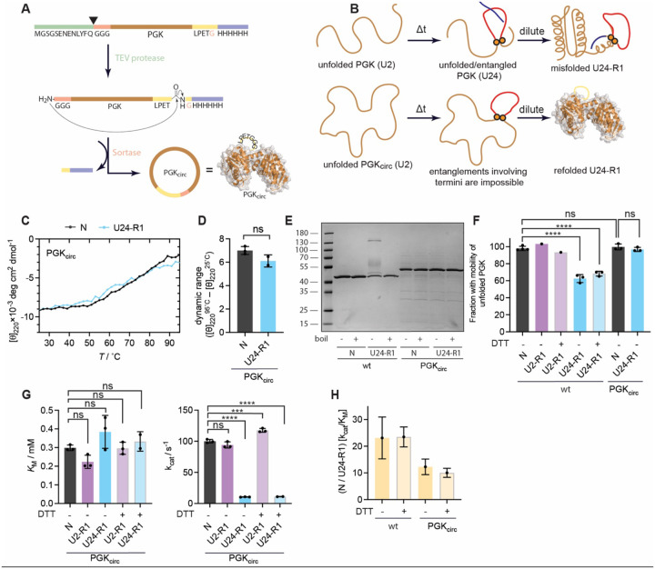
The native states of globular proteins are typically viewed as being the most stable conformations on their respective proteins' soluble free energy landscapes. This view, known as the Thermodynamic Hypothesis, explains why many proteins can reversibly refold after being denatured. Here we report an intriguing counterexample to this paradigm. When E. coli phosphoglycerate kinase (PGK) is stimulated to refold upon dilution from denaturant, instead of returning to its native state, it populates an unusual misfolded form that is monomeric and native-like, but which is even more kinetically stable than its native form, as based on its resistance to thermal and detergent-induced denaturation. Moreover, this misfolded form cannot self-correct, even for days. We show that the key structural feature of this misfolded form of PGK is topological in nature by demonstrating that kinetically stable misfolded forms do not form any longer if PGK is circularized, which prevents its termini from threading through other portions of the protein. Our findings demonstrate that a misfolded protein need not aggregate or form an amyloid to become stabilized with respect to the native state, and call attention to topologically-misfolded proteins as a potential Achilles heel to the cellular proteostasis network.


Conflict of interest statement

Competing interests: The authors declare no competing interests.

Figures

Figure 1.
Figure 1.. PGK Misfolding Produces a Soluble Kinetically Stable State.
(A) Scheme illustrating how PGK is chemically unfolded and refolded and nomenclature for unfolded states based on unfolding time (U2, U24) and their corresponding refolded states (U2-R1, U24-R1). (B) Representative far-UV circular dichroism (CD) spectra of native (N) and chemically unfolded-refolded forms of PGK. (C) Technical replicate (n ≥ 3) values for mean residual ellipticity (MRE) of forms of PGK at 220 nm. P-values calculated by one-way ANOVA with Bonferroni’s multiple comparison test. *, P< 0.05; ****, P < 0.0001. Error bars are std. devs. (D) Representative sedimentation velocity distributions for native and chemically unfolded-refolded forms of PGK. Inset table shows mean ± std. dev. for sedimentation velocity normalized to 20°C in pure water (S20,w) from technical replicates (n ≥ 2). (E) Representative thermal melts of PGK as monitored by CD signal at 220 nm either on native enzyme or on chemically unfolded-refolded forms, ramping at 1 °C/min upto 95°C. (F) Difference in MRE at 95°C endpoint and 25°C starting point for technical replicates (n ≥ 3) of the thermal melt. P-values calculated by one-way ANOVA with Bonferroni’s multiple comparison test. ns, not significant; ****, P < 0.0001. Error bars are std. devs.
Figure 2.
Figure 2.. Detergent Resistance Assay of Misfolded PGK.
Native (N) and chemically unfolded-refolded forms of PGK (U2-R1, U24-R1) were incubated in 0.6% SDS, boiled (or not), resolved by Tris-Glycine SDS-PAGE, and visualized with Coomassie stain. U24-R1 adopts conformations resistant to SDS denaturation.
Figure 3.
Figure 3.. Enzymatic Characterization of Native and Chemically Unfolded-Refolded PGK.
(A, B, C) Representative Michaelis-Menten curves showing initial rates as a function of initial substate concentration (3-phosphoglycerate) for (A) native PGK; (B) PGK unfolded for 2 h in 6 M GdmCl, diluted 100-fold and incubated for 1 h; (C) PGK unfolded for 24 h in 6 M GdmCl, diluted 100-fold and incubated for 1 h. (D) Michaelis constant (KM) and unimolecular rate constant (kcat) derived from fitting replicates (n ≥ 4) of datasets such as the one shown in panels A-C. Each dot represents the optimal value for KM and kcat based on a non-linear fit of the initial rates at 8–10 substrate concentrations. P-values calculated by one-way ANOVA with Bonferroni’s multiple comparison test. ns, not significant; *, P< 0.05; ***, P<0.001; ****, P < 0.0001. Error bars are std. devs. (E) KM and kcat derived from fitting replicates (n = 3) of Michaelis-Menten kinetics on chemically unfolded-refolded forms of PGK, except where PGK was unfolded in 8 M urea instead. Error bars are std. devs. (F) KM and kcat derived from fitting replicates (n = 3) of Michaelis-Menten kinetics on chemically unfolded-refolded forms of PGK, except where PGK was unfolded with 10 mM dithiothreitol (DTT) in addition to 6 M GdmCl. Error bars are std. devs.
Figure 4.
Figure 4.. Topological Restriction of PGK Alters its Refolding Outcome.
(A) Scheme illustrating the procedure to create a circularized form of PGK (PGKcirc). (B) Scheme illustrating how circularization could impact refolding by preventing particular conformations from populating in the unfolded ensemble. (C) Thermal melts of PGKcirc as monitored by CD signal at 220 nm either on native enzyme or on chemically unfolded-refolded form. (D) Difference in MRE at 95°C endpoint and 25°C starting point for technical replicates (n ≥ 2) of the thermal melt. Error bars are std. devs. (E) Native (N) and chemically unfolded-refolded forms of PGK and PGKcirc were incubated in 0.6 % SDS, boiled (or not), resolved by Tris-Glycine SDS-PAGE, and visualized with Coomassie stain. (F) Gel image quantification of the fastest-migrating unfolded PGK band in the non-boiled samples relative to the same band in boiled controls for the various forms of PGK and PGKcirc with the indicated number of replicates (n = 1 to n = 3). P-values calculated by one-way ANOVA with Bonferroni’s multiple comparison test. ns, not significant; ****, P < 0.0001. Error bars are std. devs. (G) KM and kcat derived from fitting replicates (n = 3) of Michaelis-Menten kinetics on natively expressed/circularized and chemically unfolded-refolded forms of PGKcirc. PGKcirc was denatured in 6 M GdmCl, with or without 10 mM DTT, for the time indicated. Error bars are std. devs. (H) Comparison of activity loss upon refolding for WT PGK and PGKcirc.
Figure 5.
Figure 5.. Biophysical and Structural Characterization of PGK’s Unfolded Ensemble.
(A) Far-UV circular dichroism (CD) spectra of unfolded PGK, incubated in 6 M GdmCl for the indicated periods of time. Native PGK (black trace) of same material shown as a reference. (B) Representative sedimentation velocity distributions for unfolded PGK, incubated in 8 M urea and 1 mM DTT for the indicated period of time prior to initiating sedimentation. Inset table shows extracted sedimentation velocity and estimated error from fit. (C) Scheme illustrating the crosslinking mass spectrometry (XL-MS) with isotopic labels to assess time-dependent structural evolution of the unfolded ensemble. (D) Connectograms illustrating crosslinks unique to the PGK samples that were unfolded for the indicated periods of time.
Figure 6.
Figure 6.. Topologically Misfolded PGK is Persistent.
(A) Michaelis-Menten curves showing initial rates as a function of initial substate concentration (3-phosphoglycerate) for (left) native PGK without peptidyl prolyl isomerase (PPIase) and (right) with 1 μM PPIase supplied. (B) Model for the energy landscape of E. coli PGK. (C) Far-UV CD spectra of native PGK and chemically unfolded-refolded forms of PGK that were first denatured in 6 M GdmCl, diluted 100-fold, and then incubated for the indicated period of time. (D) Initial enzymatic rates (with 5 mM 3-phosphoglycerate) of chemically unfolded-refolded forms of PGK that were first denatured in 6 M GdmCl, diluted 100-fold, and then incubated for the indicated period of time.

Similar articles

References

    1. Anfinsen C. B., Principles that Govern the Folding of Protein Chains. Sci. New Ser. 181, 223–230 (1973). - PubMed
    1. Dill K. A., Chan H. S., From Levinthal to pathways to funnels. Nat. Struct. Mol. Biol. 4, 10–19 (1997). - PubMed
    1. Karplus M., Behind the folding funnel diagram. Nat. Chem. Biol. 7, 401–404 (2011). - PubMed
    1. Hartl F. U., Hayer-Hartl M., Converging concepts of protein folding in vitro and in vivo. Nat. Struct. Mol. Biol. 16, 574–581 (2009). - PubMed
    1. Anfinsen C. B., Haber E., Sela M., White F. H., THE KINETICS OF FORMATION OF NATIVE RIBONUCLEASE DURING OXIDATION OF THE REDUCED POLYPEPTIDE CHAIN. Proc. Natl. Acad. Sci. 47, 1309–1314 (1961). - PMC - PubMed

Publication types

LinkOut - more resources