[Preprint]. 2025 Jun 28:2025.06.26.661770.
doi: 10.1101/2025.06.26.661770.

Four neurons pattern brain-wide developmental activity through neuropeptide signaling

Affiliations

Four neurons pattern brain-wide developmental activity through neuropeptide signaling

Jun Reichl et al. bioRxiv. .

Abstract

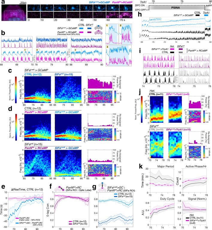
In both vertebrates and invertebrates, the developing brain becomes electrically active before it is ready to process sensory input1-4. During neural circuit maturation, developmental activity is thought to refine synaptic connections by driving neuronal co-activation in rhythmic patterns5. Here we describe cellular interactions that shape brainwide developmental activity and their molecular basis. In Drosophila, patterned stimulus independent neural activity (PSINA) engages the entire brain in highly stereotyped, globally coordinated cycles of activity6. A molecularly-defined population of ~2,000 neurons (Transient Receptor Potential Gamma, Trpγ+ neurons) act as an activity template for PSINA. We show that this activity template is patterned by four neurons expressing the neuropeptide SIFamide (SIFa)7. Signaling through the SIFa Receptor8, SIFa modulates the activity of both SIFa and Trpγ+ neurons to establish the brainwide activity cycles of PSINA. In turn, Trpγ+ neurons sustain SIFa neuron activity through a recurrent interaction. Neuropeptides modulate neuronal activity through synapse-free, or wireless, signaling9; a fitting mode of communication for a process tasked with refining on-going synapse formation. By placing neuropeptide signaling at the core of developmental activity, this work highlights the rich neurophysiological potential of the chemical connectome in shaping the developing brain.


Figures

Figure 1.
Figure 1.. SIFa and SIFaR are necessary for PSINA cycles
a. 65 hAPF D. melanogaster pupa. Cuticle around the head (boxed) has been removed for widefield imaging. Brain is outlined: CB, central brain; OL, optic lobe. Scale bar, 500μm. b. Montage of pupal heads from one widefield imaging session. Signal is fluorescence from pan-neuronally expressed GCaMP6s. Yellow rectangle (top-right) is typical region of interest (ROI) used to extract PSINA traces. All time axes in this and following figures are in units of hAPF. Scale bar, 500μm. c. Top: Wildtype PSINA recorded from enumerated pupae in (b); sampling rate is 0.5Hz. Bottom: Expanded view of PSINA from the blue highlighted 1h span above depicting the active (ON) and silent (OFF) phases of activity cycles, sweeps, and principal metrics used in analyses. PSD, power spectral density: PSINA traces were filtered to remove sweeps (coral overlaid profile), and PSD was estimated using the Discrete Fourier Transform (‘F’). d. Maximum intensity projection (MIP) of 72hAPF brain with SIFaRG4 driving mCherry.NLS (native fluorescence, cyan-hot look-up table (LUT)); brain also stained against SIFa (orange-hot LUT) and N-Cadherin (magenta). Green rectangle boxes SIFa soma. CB, central brain; OL, optic lobe; PI, Pars Intercerebralis. Scale bar, 100μm. e-g. SIFa mutant analysis e. Representative PSINA traces, color-matched to genotypes shown; numbers are sample sized used in (f) and (g). Df, deficiency; Dp, genomic duplication. f. Duty cycle and average signal, normalized to control genotype (black). Inset in duty cycle plots true values of metric. Shaded areas, standard deviation. Closed circles mark where experimental distribution differs from control (p<0.05) by two-sample KS test. g. Average PSDs from control (first column) and experimental (second column) animals. PSD for each PSINA trace was calculated at every hour within a 2h sliding window and normalized to remove contribution from changes in signal amplitude. Displayed PSDs are averages of replicates for each condition. Labels color-matched to (e,f); sample sizes indicated. PSD comparison of SIFa[1]/SIFa[2] to +/SIFa[1] omitted for brevity. In third column, bottom panel of each set maps p-values from KS tests of control and experimental PSD distributions at each frequency-time coordinate; p<0.05 displayed in magenta. Top panel is fraction of control signal power re-assigned to different (p<0.05) frequencies in the experimental data. h-j. SIFaR mutant analysis with SIFaRLoF (i.e., SIFaRGFSTF); data presented as in (e-g).
Figure 2.
Figure 2.. SIFa and SIFaR are necessary during PSINA
a. Temperature schedule used to temporally target RNAi knockdown and KiR2.1 expression to pupal development. b,c. Temporally-targeted knockdown of SIFa. b. PSINA traces, color-matched to genotypes shown; numbers are sample sized used in (c). c. Average PSDs from control and experimental animals; presented as in Fig.1g. d,e. Temporally-targeted KiR2.1 expression in SIFa neurons; presented as in (b,c). f,g. Temporally-targeted knockdown of SIFaR; presented as in (b,c). h. Temporally-targeted SIFaRG4-driven KiR2.1 expression; one control and three experimental traces shown.
Figure 3.
Figure 3.. SIFaR in the central brain is necessary for wildtype PSINA cycles
a. Spatial fractionation of GAL4 expression. Both schemes carry two variants of tubP-GAL80: GAL80ts and one of two FLP-responsive conditional alleles. In the optic lobes, OL-FLP (29C07-FLP) either turns on GAL80 expression by removing the interruption cassette (‘-STOP-’, top) or turns it off by locally excising the FRT-flanked ORF (bottom). Animals are reared at 18°C and shifted 29°C at in the early pupa to unmask these differential GAL80 expression domains ahead of PSINA onset; GAL4-driven effector expression is disinhibited in the complementary domains (blue). CB, central brain. OL, optic lobe. b. Anterior-posterior (A/P) and dorso-ventral (D/V) MIPs of 60hAPF brains with PanN-, Trpγ-, or SIFaR-GAL4 driving UAS-KiR2.1-T2A-tdTOM expression principally in the CB (top row) or the OLs (bottom row). PanN labeling reveals some posterior OL neurons in the CB-designated fraction and Kenyon cells in the OL-designated fraction. KiR2.1 expression domain detected through tdTOM native fluorescence (cyan-hot LUT); reference stain is N-Cadherin (magenta). Scale bar, 100μm. c,f. Representative PSINA traces from animals in which KiR2.1 (c) or SIFaR-RNAi (f) expression was targeted to the CB (top set) or OL (bottom set) with the indicated GAL4s. CB (left columns) or OL (right columns) recordings are from the same animal in each condition. d,g. Left: Normalized average signal from animals in which KiR2.1 (d) or SIFaR-RNAi (g) expression was spatially targeted to the CB and recorded from the CB (top) or OL (bottom). Genotypes color-matched to (c) and (f); sample sizes indicated in Right. Closed circles mark where experimental distributions differ from control (p<0.05) by two-sample KS test. Right: Average PSDs from control and experimental conditions described for Left; presented as in Fig.1g. e,h. Data from animals in which KiR2.1 (e) or SIFaR-RNAi (h) expression was spatially targeted to the OLs; presentation as in (d,g).
Figure 4.
Figure 4.. SIFa neurons pattern brainwide PSINA
a-g. PSINA v. SIFa neuron activity in wildtype and SIFa mutant animals. a. 2-channel recording of SIFa neuron activity (cyan) and brainwide PSINA (magenta). Left: Pan-neuronal>RCaMP and SIFa neuron>GCaMP overlay of pupal head at 69.5 hAPF. Right: Progressive frames from boxed span no.1 in (b). Color-matched ROIs from left shown; intensities adjusted to display fluorescence changes. b. Left: Representative traces for the indicated conditions. PSINA, as reported by pan-neuronal RCaMP, was recorded from the SIFa neuron ROI. Right: Expanded views for numbered boxes in left; SIFa neuron active phases demarcated in light blue. c. PSD comparison of SIFa neuron activity in control and SIFa mutant animals; sample sizes as indicated. d. PSD comparison of PSINA, as reported by pan-neuronal RCaMP, and SIFa neuron activity in control (top) and SIFa mutant (bottom) animals; sample sizes as indicated. e. Active phase rise time differences between SIFa neurons and central brain (blue) or left optic lobe and central brain (magenta). ~30 data points per hourly distribution compiled from 12 traces; shaded areas, s.d. Closed circles mark where the blue distributions differ from magenta (p<0.05) by two-sample KS test. f. Zero-lag correlation of SIFa-ROI pan-neuronal activity to left optic lobe in control and SIFa mutant animals. g. Zero-lag correlation of SIFa neuron to SIFa-ROI pan-neuronal activity in control and SIFa mutant animals. h-k. TrpA1 activation of SIFa neurons h. Top: Temperature schedule used. Bottom: SIFa neuron activity response to temperature shift, with (blue) and without (black) TrpA1. Dashed lines mark GCaMP6s baseline at 22°C. Inset expanded view of gray highlighted span. i. Representative PSINA traces at 29°C for the indicated genotypes. j. PSD comparison of PSINA at 29°C in control (top) and SIFa mutant (bottom) animals with and without TrpA1 expression in SIFa neurons; sample sizes as indicated. k. PSINA metrics at 29°C for the indicated genotypes. Major period corresponds to the dominant frequency in the PSD. Shaded areas, s.d. Closed circles mark where experimental distributions (magenta) differ from control (p<0.05) by two-sample KS test.
Figure 5.
Figure 5.. SIFa neurons pattern PSINA by modulating Trpγ+ neurons
a-c. Double mutant analysis of SIFa and Trpγ. a. Top: Representative traces for the indicated genotypes shown below long-pass filtered (<5.6×10⁻⁴ Hz, >30 min) derivates (i.e., swells). Bottom: Expanded view of the gray highlighted span in top. b. Average signal and sweeps per hour, both normalized to control, for the genotypes and sample sizes in (a). Magenta traces are scaled products of the single mutant average values. Shaded areas, s.d. Closed circles mark where experimental distributions differ from control (p<0.05) by two-sample KS test. c. Average PSDs from control and experimental animals; presented as in Fig.1g. d-f. PSINA v. Trpγ+ neuron activity in wildtype and SIFa mutant animals. d. Representative traces from 2-channel recordings of Trpγ+ neuron activity and brainwide PSINA in control and SIFa mutant animals. e. Zero-lag correlation of Trpγ+ neuron activity to brainwide PSINA in control and SIFa mutant animals. f. PSD comparison of Trpγ+ neuron activity and PSINA in control (top) and SIFa mutant (bottom) animals; sample sizes as indicated. g. Model of the core PSINA engine. In the absence SIFa signaling (left, ‘No SIFa’), neither SIFa (blue) nor Trpγ± (magenta) neurons have stereotyped cycles; however, the activity profiles remain coupled due to input from Trpγ± neurons. SIFa-SIFaR signaling patterns both activity profiles into cycles (right, ‘With SIFa’), and brainwide PSINA follows the Trpγ± neuron activity template. SIFaR expression (light gray gradient ramp) and activity levels (dark gray triangles) control for how long Trpγ± neurons can sustain an active phase. See Discussion for more details.

References

    1. Blankenship A. G. & Feller M. B. Mechanisms underlying spontaneous patterned activity in developing neural circuits. Nature Reviews Neuroscience 11, 18–29 (2010). - PMC - PubMed
    1. Wu M. W., Kourdougli N. & Portera-Cailliau C. Network state transitions during cortical development. Nat. Rev. Neurosci. 25, 535–552 (2024). - PMC - PubMed
    1. Martini F. J., Guillamón-Vivancos T., Moreno-Juan V., Valdeolmillos M. & López-Bendito G. Spontaneous activity in developing thalamic and cortical sensory networks. Neuron 109, 2519–2534 (2021). - PMC - PubMed
    1. Akin O. & Zipursky S. L. Activity regulates brain development in the fly. Current Opinion in Genetics & Development 65, 8–13 (2020). - PubMed
    1. Willshaw D. J., Von Der Malsburg C. & Longuet-Higgins H. C. How patterned neural connections can be set up by self-organization. Proceedings of the Royal Society of London. Series B. Biological Sciences 194, 431–445 (1976). - PubMed

Publication types

LinkOut - more resources