[Preprint]. 2025 Jun 24:2025.06.18.660406.
doi: 10.1101/2025.06.18.660406.

Cetuximab increases LGR5 expression and augments LGR5-targeting antibody-drug conjugate efficacy in patient-derived colorectal cancer models

Affiliations

Cetuximab increases LGR5 expression and augments LGR5-targeting antibody-drug conjugate efficacy in patient-derived colorectal cancer models

Peyton C High et al. bioRxiv. .

Update in

Abstract

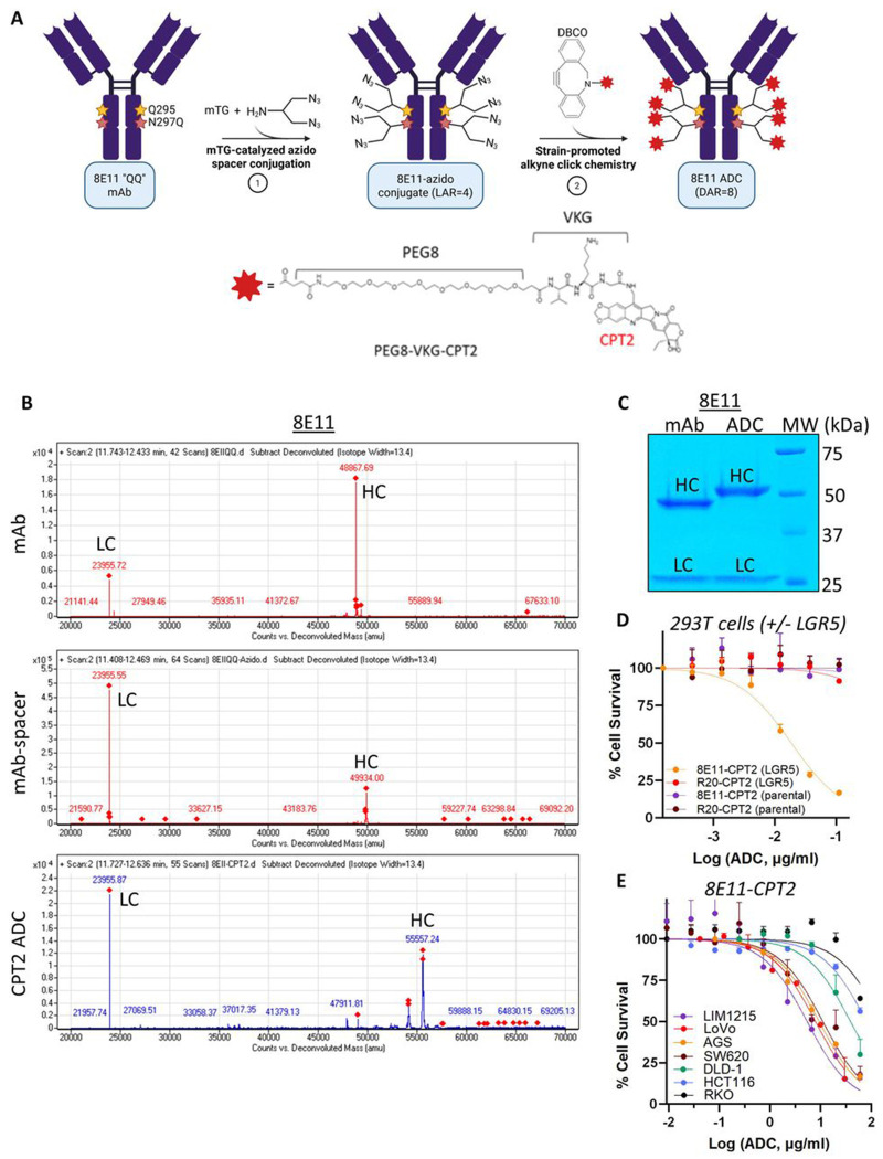
Colorectal cancer (CRC) remains the second-leading cause of cancer-associated deaths, indicating an urgent need for improved therapeutic options. We previously generated antibody-drug conjugates (ADCs) targeting the cancer stem-like cell marker leucine-rich repeat-containing G protein-coupled receptor 5 (LGR5). However, tumor relapse due to LGR5 downregulation and suboptimal payload selection warranted strategies to improve ADC efficacy. Here we report cetuximab, an EGFR-targeting monoclonal antibody indicated for RAS WJ metastatic CRC, augments LGR5 expression independent of RAS/PIK3CA mutation status and promotes EGFR-LGR5 interactions. Furthermore, we describe the development of LGR5 ADCs incorporating a camptothecin-derived payload that is well-tolerated and significantly inhibits tumor growth. Importantly, cetuximab in combination with LGR5 ADCs results in enhanced tumor inhibition or regression versus single-agent treatment and extends survival in RAS MUT patient-derived xenografts. These findings support growing evidence that ADC combination therapies may be more effective than monotherapies and suggests a broader clinical use for cetuximab in treating RAS MUT CRC.

Keywords: Antibody-drug conjugate; EGFR; LGR5; cetuximab; colorectal cancer; combination treatment.


Conflict of interest statement

DECLARATION OF INTERESTS K.S.C serves on an advisory board for Merus, NV.

Figures

Figure 1.
Figure 1.. EGFR inhibitors reversibly increase LGR5 expression in CRC cells independent of mutation status.
(A) Western blot of endogenous EGFR and LGR5 expression in a panel of CRC cell lines and patient-derived xenograft (PDX) models XST-GI-010 and CRC-001. (B) EGFR and LGR5 RNA-seq expression data from Cancer Cell Line Encyclopedia (CCLE). Values are read per kilobase of transcript per million (RPKM). Western blots and quantification of LGR5 protein fold-changes normalized to actin in (C) LIM1215, LoVo, and SW620 cells following 48 h treatment with a panel of EGFR- and HER2-targeting mAbs (3 μg/ml) and TKIs (1 μΜ), (D) 5 μg/ml CTX over indicated time-course, and (E) LIM1215, LoVo, and DLD-1 cells following 15 min CTX treatment and subsequent washout for indicated time-course. (F) Immunocytochemistry staining of total (permeabilized) and surface (non-permeabilized) LGR5 in LoVo cells treated for 15 min with vehicle or 5 μg/ml CTX. (G-H) Western blot and quantification of LGR5 protein fold-changes normalized to actin in (G) cycloheximide (CHX, 30 μg/ml) chase for 4 or 24 h performed in LoVo cells pre-treated for 15 min with vehicle or 5 μg/ml CTX (s, short exposure; I, long exposure) and (H) LoVo cells pre-treated for 15 min with 0.1 μM Bafilomycin A1 or 10 μM MG132 to inhibit lysosomal or proteasomal degradation, respectively, in the presence or absence of 5 μg/ml CTX.
Figure 2.
Figure 2.. LGR5 levels are reduced simultaneously with loss of EGFR expression in CRC cells.
Western blot and quantification following (A) 72 h treatment with 100 nM EGFR siRNA or non-targeting control siRNA in LIM1215, LoVo, and DLD-1 cells, (B) treatment with 30 ng/ml EGF for 8 or 24 h in LoVo cells, (C) 48 h treatment with 1 ng/ml EGF in LIM1215 and DLD-1 cells, and (D) 48 h treatment of LoVo, DLD-1, DLD-1 shLGR5, and SW620 cells with increasing doses of MEK1/2 inhibitor trametinib. EGFR and LGR5 protein fold-changes were normalized to actin and p-ERK/2 protein fold-change was normalized to total ERK1/2 for western blot quantification.
Figure 3.
Figure 3.. EGFR interaction with LGR5 is enhanced by CTX.
Co-immunoprecipitation (co-IP) experiments with EGFR antibody or IgG isotype control shows (A) recombinant LGR5 in 293T-LGR5 cells and (B) endogenous LGR5 in LoVo cells specifically interacts with endogenous EGFR. (C) Co-IP experiment with LGR5 mAb in LoVo cells shows EGFR pulls down with endogenous LGR5. (D) Co-IP shows enhanced LGR5 interaction with EGFR in LoVo cells after 15 min treatment with 5 μg/ml CTX. TL, total lysate. Proximity ligation assays and quantification performed in (E) LIM1215 and (F) LoVo cells treated with vehicle or 5 μg/ml CTX. Statistical analysis was performed using two-tailed unpaired t-test; *p<0.05; **p<0.01. Data presented as mean +/− SD.
Figure 4.
Figure 4.. Generation and characterization of LGR5-targeting ADC 8E11-CPT2
(A) Schematic of microbial transglutaminase (mTG)-mediated conjugation of a branched linker to Q295 and N297Q of the Fc region of 8E11 mAb followed by strain promoted alkyne-azide cycloaddition of camptothecin 2 (CPT2) payload attached to a cleavable tripeptide valine-lysine-glycine (VKG) linker to generate 8E11-CPT2 ADC (DAR = 8). (B) LC-MS/MS analysis of 8E11 mAb, 8E11-azido spacer conjugate, and 8E11-CPT2 ADC under reducing conditions. LC, light chain; HC, heavy chain. (C) Coomassie staining of 8E11 mAb and 8E11-CPT2 ADC under reducing conditions shows increase in HC molecular weight, indicating linker-payload conjugation. (D) 8E11-CPT2 and R20-CPT2 cytotoxicity in LGR5-negative 293T parental and LGR5-overexpressing 293T-LGR5 cells. (E) Comparison of 8E11-CPT2 cytotoxicity in a panel of gastrointestinal cancer cell lines with different levels of LGR5 expression. Data presented as mean +/− SD.
Figure 5.
Figure 5.. CTX enhances the efficacy of 8E11-CPT2 ADC in vitro.
Confocal images and quantification of rat 8F2 LGR5 mAb internalization and co-localization with lysosome marker LAMP1 after 45 min incubation at 37° C in (A) LIM1215 and (B) LoVo cells pre-treated with vehicle or 5 μg/ml CTX for 15min. Statistical analysis was performed using two-tailed unpaired t-test; *p<0.05; **p<0.01. Data presented as mean +/− SD. Loewe synergy score heat maps and associated cell survival plots for (C) LIM1215, (D) AGS, (E) LoVo, and (F) DLD-1 cells co-treated with increasing concentrations of CTX and 8E11-CPT2 for 5 days. Example experiments showing % cell survival values for CTX and 8E11-CPT2 combinations with marked additive effects for (G) LIM1215 and (H) AGS cells and synergistic effects for (I) LoVo and (J) DLD-1 cells. Statistical significance was performed using ANOVA. *p<0.05; **p<0.01; ***p<0.001; ****p<0.0001.
Figure 6.
Figure 6.. CTX treatment increases LGR5 expression in CRC models in vivo.
Western blot and quantification of LGR5 expression in (A) LoVo cell line-derived xenograft models treated with 2 or 20 mg/kg CTX or PBS vehicle (N=4/group) once every three days for a total of two doses (tumors were harvested on day 4), (B) DLD-1 cell line-derived xenograft models from a previously reported survival study treated weekly for 2 doses of 10 mg/kg CTX or PBS vehicle (N=5/group), and (C) KRASMUT XST-GI-010 PDOs treated for 72 h with vehicle or 5 μg/ml CTX. LGR5 protein fold-change was normalized to actin. Data presented as mean +/− SD. (D) LGR5 mRNA microarray expression data following acute (24 to 72 h) or chronic (6-week) CTX treatment (20 mg/kg twice per week) in RASWT mCRC PDX models (GSE108277; N=21). Statistical analyses performed using ANOVA. *p<0.05; **p<0.01.
Figure 7.
Figure 7.. CTX enhances 8E11-CPT2 ADC antitumor efficacy and extends survival in patient-derived CRC models.
Safety assessment shows (A) bodyweights, (B) ALT, (C) AST, (D) CRE, and (E) WBC plasma levels of C57BL/6J mice (N=5/group) following 2-week treatment with single-dose 8E11-CPT2 as indicated. (F) Anti-tumor efficacy of CTX or 8E11-CPT2 ADC compared to combination therapy in XST-GI-010 PDX models up to day 24 when first vehicle-treated animal reached maximal tumor burden. All treatments were performed at 5 mg/kg by intraperitoneal injection weekly for a total of 3 doses (N=7/group). Statistical significance performed using ANOVA. ***p<0.001; ****p<0.0001. Data presented as mean +/− SD. (G) Percent tumor growth inhibition (TGI) at day 24 in XST-GI-010 PDX models. Data presented as mean +/− SD. (H) Kaplan–Meier survival plot and log-rank test for the XST-GI-010 study. (I) Anti-tumor efficacy of CTX, 8E11-CPT2, and R20-CPT2 monotherapies and combination therapies in an NRASMUTCRC-001 PDX model up to day 24 when first vehicle-treated animal reached maximal tumor burden. All treatments were performed at 5 mg/kg by intraperitoneal injection weekly for a total of 3 doses (N=5 for all groups except 8E11-CPT2 and 8E11-CPT2+CTX, N=6). Statistical significance performed using ANOVA. *p<0.05; **p<0.01. Data presented as mean +/− SD. (J) TGI at day 24 in CRC-001 PDX models. Data presented as mean +/− SD. (K) Kaplan–Meier survival plot and log-rank test for the CRC-001 study.

References

    1. Gong X., Azhdarinia A., Ghosh S.C., Xiong W., An Z., Liu Q., and Carmon K.S. (2016). LGR5-Targeted Antibody-Drug Conjugate Eradicates Gastrointestinal Tumors and Prevents Recurrence. Mol. Cancer Ther. 15, 1580–1590. 10.1158/1535-7163.MCT-16-0114. - DOI - PubMed
    1. Jang B.G., Kim H.S., Chang W.Y., Bae J.M., Kim W.H., and Kang G.H. (2018). Expression Profile of LGR5 and Its Prognostic Significance in Colorectal Cancer Progression. Am. J. Pathol. 188, 2236–2250. 10.1016/j.ajpath.2018.06.012. - DOI - PubMed
    1. Liu W., Zhang J., Gan X., Shen F., Yang X., Du N., Xia D., Liu L., Qiao L., Pan J., et al. (2018). LGR5 promotes epithelial ovarian cancer proliferation, metastasis, and epithelial–mesenchymal transition through the Notch1 signaling pathway. Cancer Med. 7, 3132–3142. 10.1002/cam4.1485. - DOI - PMC - PubMed
    1. Cao W., Li M., Liu J., Zhang S., Noordam L., Verstegen M.M.A., Wang L., Ma B., Li S., Wang W., et al. (2020). LGR5 marks targetable tumor-initiating cells in mouse liver cancer. Nat. Commun. 11, 1961. 10.1038/s41467-020-15846-0. - DOI - PMC - PubMed
    1. Yamamoto Y, Sakamoto M., Fujii G., Tsuiji H., Kenetaka K., Asaka M., and Hirohashi S. (2003). Overexpression of orphan G-protein-coupled receptor, Gpr49, in human hepatocellular carcinomas with beta-catenin mutations. Hepatol. Baltim. Md 37, 528–533. 10.1053/jhep.2003.50029. - DOI - PubMed

Publication types