[Preprint]. 2025 Jun 17:2025.06.12.658902.
doi: 10.1101/2025.06.12.658902.

The soluble HIV-1 Vpu protein interacts with calmodulin in a Ca2+-dependent manner

Affiliations

The soluble HIV-1 Vpu protein interacts with calmodulin in a Ca2+-dependent manner

Olamide Ishola et al. bioRxiv. .

Abstract

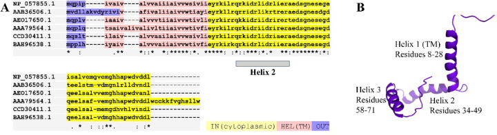
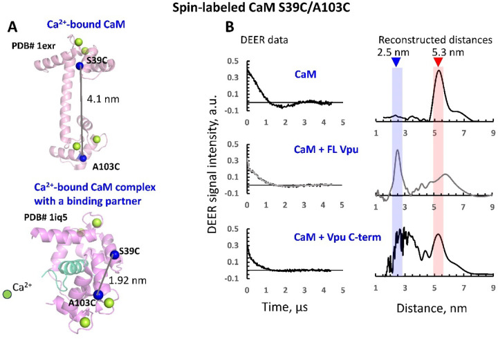
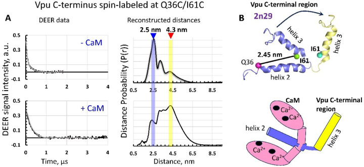
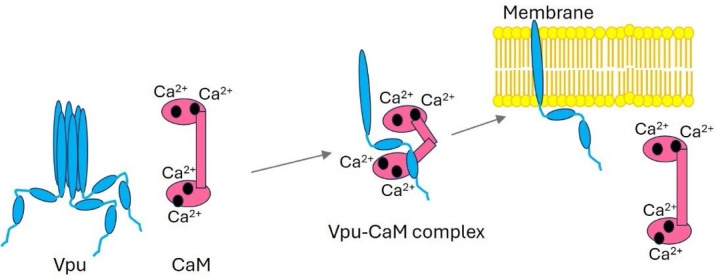
The HIV-1-encoded membrane protein Vpu plays key roles in virus lifecycle. Our lab recently revealed a soluble form of Vpu, and we strived to determine its possible physiological function. Here, we provide solid experimental proof that soluble Vpu interacts with Ca2+-bound calmodulin (Ca2+-CaM). A putative CaM-binding motif in Vpu was predicted, but there was no experimental evidence of the Vpu-CaM association. We applied double electron electron-resonance (DEER) and protein spin labeling to detect the soluble Vpu-CaM complex. We found that soluble full-length and truncated C-terminal region of Vpu directly interact with Ca2+-CaM. DEER results from the spin-labeled double cysteine mutant S39C/A103C of CaM showed that upon association with Vpu Ca2+-CaM adopts a more closed conformation compared to those in the absence of Vpu. This restructuring is in agreement with previously observed Ca2+-CaM association with cellular and other HIV-1 proteins. Our results indicate that soluble Vpu and CaM form an equimolar complex. DEER results from doubly spin-labeled at residues Q36C/I61C in Vpu C-terminal region suggest that Vpu's helices 2 and 3 move away from each other to facilitated CaM binding. These observations tell that under physiological conditions the soluble Vpu-CaM complex may provide Vpu with a trafficking pathway to membrane destination.

Keywords: DEER spectroscopy to study protein-protein complexes; protein conformational changes in protein-protein interaction; soluble HIV-1 Vpu protein; soluble Vpu-calmodulin interaction.


Figures

Figure 1.
Figure 1.. Amino acid (aa) sequence and membrane-bound structure of the HIV-1 Vpu protein:
(A) The alignment of aa sequences of Vpu protein of randomly selected diverse HIV-1 strains, as follow: NP_057855.1 (strain for reference annotation); AAB36506.1 (an HIV-1 isolate of genetic subtype C); AEO17650.1 (HIV-1 sero-prevalent individual, subtype B); AAA79564.1 (isolate “cntrl 1”, clone=“3”; CCD30411.1 (group M, subgroup G); and BAH96538.1 (multidrug-resistant HIV-1 subtype B). In addition to the high conservation of the TM helix 1 (highlighted in tan), the composition of the loop between helix 1 and helix 2, and the helix 2 also demonstrate high sequence identity. The helix 2 is emphasized with gray bar under the aa sequences. (B) The solid-state NMR structure of full-length Vpu in lipid (PDB # 2N28) is shown with the helices 1, 2 and 3 labeled—helix 1 traverses membrane bilayer (not shown), whereas helix 2 is nearly parallel to the membrane surface. The unstructured loops between helices 1 and 2, and helices 2 and 3 are shown for clarity. The T-COFFEE software was used to align the Vpus’ sequences, and PyMOL was used to visualize the ssNMR structure of Vpu.
Figure 2.
Figure 2.. Analysis of the CaM-binding sites in the Vpu aa sequence.
(A) The aligned Vpu aa sequences corresponding to the protein region containing the partial TM helix 1, helix 2 and the loops between helices 1/2 and 2/3 are shown. The aa sequence of the previously predicted CaM-binding motif and its positive charge (+6) are shown above the aligned aa sequences. (B) The aligned Vpu aa sequences corresponding to the protein region containing the helix 2 and the loops between helices 1/2 and 2/3 are shown. The positive charge of this aa region (+8 or +9, depending on the HIV-1 strain) is shown. For both (A) and (B) the Vpu aa are from the same HIV-1 staring shown in Figure 1. (C) The aa sequences of the common CaM-binding motifs, which have resemblance with the aa sequence of the putative Vpu’s CaM-binding region are shown. The “x” in these sequences is an any aa. Proteins with these motives bind CaM in Ca2+-dependent manner (except IQ-like motif, which could bind with and without Ca2+). In all aa sequences, the basic residues, which are like those found in the basic 1-5-10 motif of CaM-binding proteins, are highlighted in magenta. The hydrophobic residues, which are like those found in the 1-8-14 and/or 1–14 motifs in CaM-binding proteins are highlighted in yellow. The conserved Gln (Q) residue in Vpu’s aa sequences, which could be like the Gln residue found in the IQ-like motif of CaM-binding proteins is highlighted in cyan. This, it may well be that Vpu possesses a hybrid motif to associate with CaM via both electrostatic and hydrophobic interactions.
Figure 3.
Figure 3.. Ca2+-bound CaM interacts with the FL Vpu and truncated C-terminal of Vpu.
(A) The crystal structures of Ca2+-bound CaM (PDB # 1EXR) and Ca2+-bound CaM in the complex with a calmodulin dependent kinase fragment (cyan) (PDB # 1IQ5) illustrate extended and closed conformations, respectively. The Cβ-atoms of residues S39 and A103 are shown as blue spheres. In the structures shown, these residues are S38 and A102, since these CaM forms are shorter by 1 aa compared to the one, which we used. The estimated Cβ-Cβ distances are also shown. (B) Baseline-corrected DEER data (left) accompanied by the reconstructed inter-spin label distances (right) for the following cases: (B, top) CaM doubly spin-labeled at residues S39C/A103C alone; (B, mid) CaM in the presence of excess of SUMO-FL Vpu; and (B, bottom) CaM with SUMO-C terminal region of Vpu. In the absence of these Vpu variants, a distinct DEER distance of 5.3 nm was obtained between MTSL labels at positions 39 and 103 in Ca2+-bound CaM alone. However, with the addition of unlabeled Vpu variants shifts the average distance between residues 39/103 to a much shorter value of about 2.5 nm, thereby revealing a major restructuring of CaM. In the middle B-panel, reconstructed distance distribution is from the denoised DEER signal. This distance distribution is close to one obtained from original signal (Figure S6).
Figure 4.
Figure 4.. DEER on spin-labeled residues Q36C/I61C in the Vpu C-terminal indicate significant restructuring of this protein region upon binding to Ca2+-CaM:
(A) The baseline corrected DEER data (left) and reconstructed distances (right) for Vpu C-terminal spin-labeled at residues Q36C/I61C in the absence (upper panels) and presence (lower panels) of CaM. Significant shift in the main distances from about 2.5 nm to 3.5–5 nm was observed suggesting significant restructuring in the Vpu C-terminus upon binding to CaM. (B) The proposed conformation rearrangements taking place in Vpu C-terminus upon binding of CaM—helix 3 moves further apart from helix 2. The ribbon-represented structure in blue in the upper panel is that of Vpu C-terminus, which was determined by NMR (PDB # 2N29).
Figure 5.
Figure 5.. A hypothetical model of soluble Vpu-CaM physiological role:
Soluble Vpu binds to Ca2+-bound CaM in 1:1 stoichiometry to facilitate Vpu’s trafficking to the membrane. Once Vpu is inserted into the membrane to adopt its functional state, CaM is released. The Ca2+ ions bound to CaM are shown as black circles.

Similar articles

References

    1. Chin D.; Means A. R. Calmodulin: a prototypical calcium sensor. Trends Cell Biol 2000, 10 (8), 322–328. DOI: 10.1016/s0962-8924(00)01800-6 - DOI - PubMed
    1. Yang C. F.; Tsai W. C. Calmodulin: The switch button of calcium signaling. Tzu Chi Med J 2022, 34 (1), 15–22. DOI: 10.4103/tcmj.tcmj_285_20 - DOI - PMC - PubMed
    1. O’Neil K. T.; DeGrado W. F. How calmodulin binds its targets: sequence independent recognition of amphiphilic alpha-helices. Trends Biochem Sci 1990, 15 (2), 59–64. DOI: 10.1016/0968-0004(90)90177-d - DOI - PubMed
    1. Andrews C.; Xu Y.; Kirberger M.; Yang J. J. Structural Aspects and Prediction of Calmodulin-Binding Proteins. Int J Mol Sci 2020, 22 (1). DOI: 10.3390/ijms22010308 - DOI - PMC - PubMed
    1. Miller M. A.; Mietzner T. A.; Cloyd M. W.; Robey W. G.; Montelaro R. C. Identification of a calmodulin-binding and inhibitory peptide domain in the HIV-1 transmembrane glycoprotein. AIDS Res Hum Retroviruses 1993, 9 (11), 1057–1066. DOI: 10.1089/aid.1993.9.1057 - DOI - PubMed

Publication types

LinkOut - more resources