[Preprint]. 2025 Jun 17:2025.06.17.660080.
doi: 10.1101/2025.06.17.660080.

Extracellular Vesicle-Mediated Delivery of Genetic Material for Transformation and CRISPR/Cas9-based Gene Editing in Pneumocystis murina

Affiliations

Extracellular Vesicle-Mediated Delivery of Genetic Material for Transformation and CRISPR/Cas9-based Gene Editing in Pneumocystis murina

Steven G Sayson et al. bioRxiv. .

Abstract

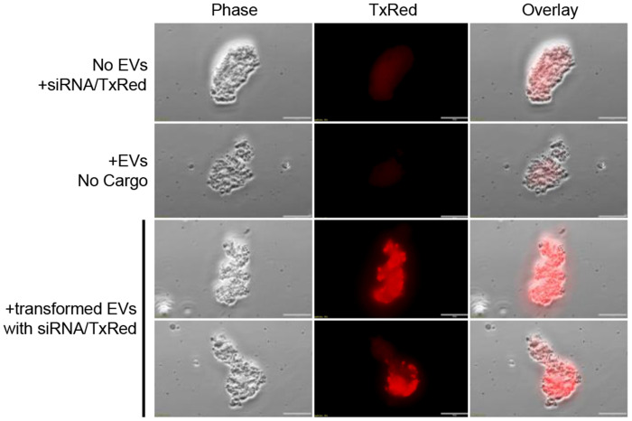
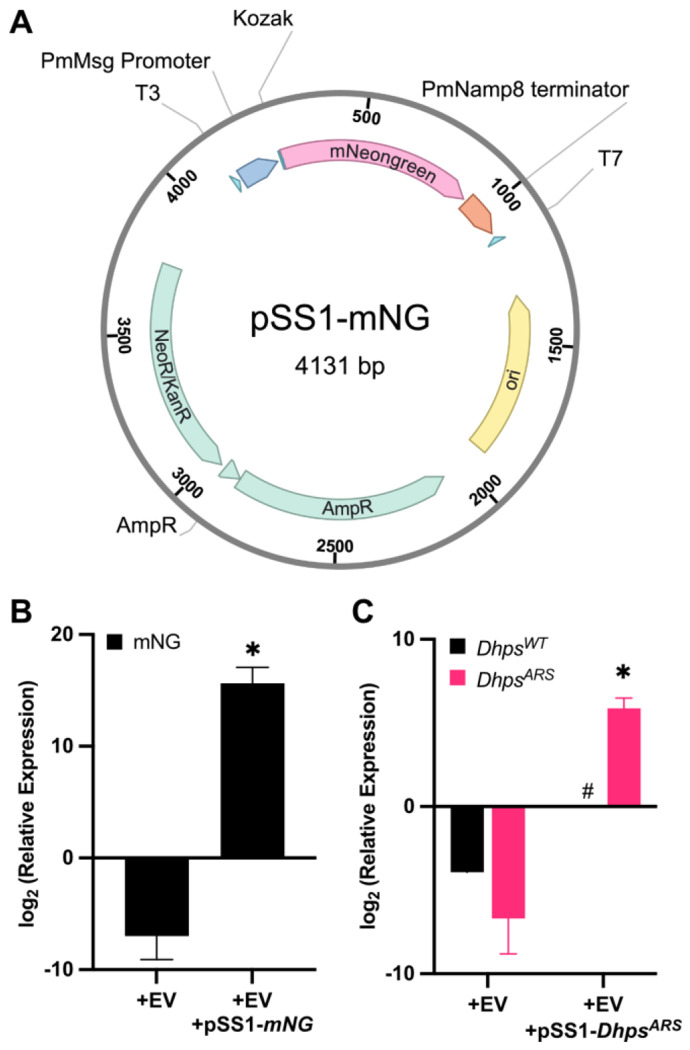
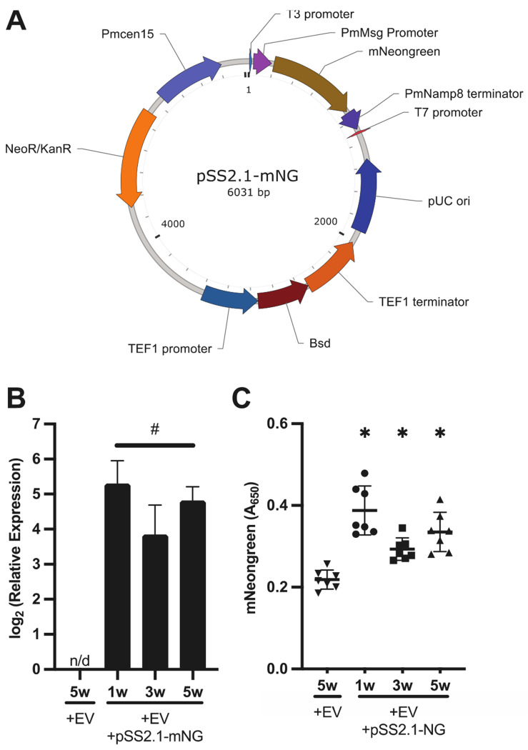
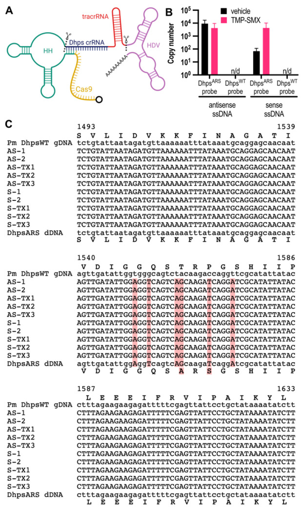
Pneumocystis species are obligate fungal pathogens that cause severe pneumonia, particularly in immunocompromised individuals. The absence of robust genetic manipulation tools has impeded our mechanistic understanding of Pneumocystis biology and the development of novel therapeutic strategies. Herein, we describe a novel method for the stable transformation and CRISPR/Cas9-mediated genetic editing of Pneumocystis murina utilizing extracellular vesicles (EVs) as a delivery vehicle. Building upon our prior investigations demonstrating EV-mediated delivery of exogenous material to Pneumocystis, we engineered mouse lung EVs to deliver plasmid DNA encoding reporter genes and CRISPR/Cas9 components. Our initial findings demonstrated successful in vitro transformation and subsequent expression of mNeonGreen and DhpsARS in P. murina organisms. Subsequently, we established stable in vivo expression of mNeonGreen in mice infected with transformed P. murina for a duration of up to 5 weeks. Furthermore, we designed and validated a CRISPR/Cas9 system targeting the P. murina Dhps gene, confirming its in vitro cleavage efficiency. Ultimately, we achieved successful in vivo CRISPR/Cas9-mediated homologous recombination, precisely introducing a DhpsARS mutation into the P. murina genome, which was confirmed by Sanger sequencing across all tested animals. Here, we establish a foundational methodology for genetic manipulation in Pneumocystis, thereby opening avenues for functional genomics, drug target validation, and the generation of genetically modified strains for advanced research and potential therapeutic applications.


Figures

Figure 1.
Figure 1.. Pneumocystis murina uptake BALF EVs containing exogenous nucleotides.
P. murina were treated with TxRed-conjugated siRNA (top row), EVs alone (middle row), or EVs-loaded with siRNA-TxRed (bottom row) for 16 hours. Scale, 20 μm.
Figure 2.
Figure 2.. Pneumocystis murina cultured in vitro transcribes plasmid-encoded genes following delivery by extracellular vesicles.
(A) Plasmid map of pSS1-mNG, which encodes mNeongreen driven by the Pm Msg promoter, and a Namp8 terminator. P. murina (1 × 10⁶ organisms) were treated with EVs (2-μg protein equivalent) loaded with either pSS1-mNeongreen (mNG) or pSS1-DhpsARS for 16 hours. Total RNA was extracted from the cells and synthesized to cDNA. RT-qPCR was performed and relative expression was calculated using the 2−ΔCt method, normalized to LSU. Plasmid-transformed (B) mNeongreen and (C) DhpsARS groups (n=9) showed significant transcriptional activity, confirming successful expression of the plasmid-delivered genes. Native DhpsWT was notably absent in plasmid transformed P. murina. T-test; * , p < 0.05 against control. #, unable to perform comparison due to the lack of detectable expression.
Figure 3.
Figure 3.. Pneumocystis murina expresses mNeonGreen in vivo following in vitro extracellular vesicle-mediated gene delivery.
(A) Plasmid map of pSS2.1-mNG, which includes a second expression cassette encoding blasticidin S deaminase (Bsd) and PmCen15, a truncated centromeric sequence. P. murina (1 × 10⁶ organisms) were incubated for 16 hours with extracellular vesicles (2μg protein equivalent) either lacking cargo or loaded with pSS2.1-mNeonGreen (mNG). Total RNA was extracted, synthesized to cDNA and RT-qPCR was performed. Relative expression was calculated using the 2−ΔCt method, normalized to LSU. (B) Pm displayed sustain expression of mNeongreen mRNA expression after 1-, 3-, and 5- weeks post-inoculation (n=3). (C) mNeongreen protein was detected in P. murina lysates by ELISA after 1-, 3-, and 5- weeks post-inoculation (n=3). ANOVA followed by Sidak’s multiple comparisons post hoc test; n/d, no detectable expression. #, unable to perform comparison due to the lack of detectable expression. *, p < 0.05 against control.
Figure 4.
Figure 4.. CRISPR RNA (crRNA) sequences mediate efficient cleavage of Dhps amplicons in vitro.
A full-length Dhps amplicon was generated from P. murina genomic DNA. Annealed crRNA and tracrRNA were complexed with S. pyogenes Cas9 nuclease to form ribonucleoprotein (RNP) complexes, which were then incubated with the Dhps amplicons for 1 hour. (A) Agarose gel electrophoresis showed cleavage of Dhps by both sense and antisense RNPs, yielding two fragments of ~1560 bp and ~660 bp, as predicted. Scramble (scr) RNPs did not induce cleavage. (B) Densitometric analysis confirmed significantly increased cleavage by sense and antisense RNPs compared to scr-RNPs (n=3). ANOVA followed by Sidak’s multiple comparisons post hoc test; †, p < 0.05 vs. uncleaved control; *, p < 0.05 vs. cleaved control.
Figure 5.
Figure 5.. pSS2.1 effectively delivers Cas9 and gRNA for genetic editing of the Dhps locus.
(A) Schematic of the Cas9-gRNA expression cassette, which encodes Cas9 and a guide RNA flanked by a hammerhead (HH) ribozyme and a hepatitis delta virus (HDV) ribozyme. Created in BioRender. Sayson, S. (2025) https://BioRender.com/dp0twrz. (B) After 5 weeks of infection and 2 weeks of treatment, quantitative PCR detects DhpsARS in DNA extracted from P. murina in both antisense- and sense- ssDNA-treated groups (n=5). In the same P. murina populations, DhpsWT was not detected. (C) Alignment of the Dhps [1493-1633] regions from untreated organisms (Pm DhpsWT gDNA) were compared to P. murina organisms treated with EVs containing pSS2.1-Cas9-HH-gRNADhps-HDV and antisense (AS) or sense (S) ssDNA. Donor DNA for DhpsARS is displayed in bottom row of alignment. Treated Pm organisms all display precise genetic editing to include the associated nucleotide changes for an amino acid change from TRP to ARS, as well as silent point mutations included on the donor dDNA. TX, TMX-SMX groups. Red, change in sequence from DHPSWT isolated from Pm.

Similar articles

References

    1. Kanj A., Samhouri B., Abdallah N., Chehab O. & Baqir M. Host Factors and Outcomes in Hospitalizations for Pneumocystis jirovecii Pneumonia in the United States. Mayo Clin Proc 96, 400–407 (2021). 10.1016/j.mayocp.2020.07.029 - DOI - PubMed
    1. Kolbrink B. et al. Evolving epidemiology of Pneumocystis pneumonia: Findings from a longitudinal population-based study and a retrospective multi-center study in Germany. Lancet Reg Health Eur 18, 100400 (2022). 10.1016/j.lanepe.2022.100400 - DOI - PMC - PubMed
    1. Brown G. D. et al. Hidden killers: human fungal infections. Sci Transl Med 4, 165rv113 (2012). 10.1126/scitranslmed.3004404 - DOI - PubMed
    1. Xue T., Kong X. & Ma L. Trends in the Epidemiology of Pneumocystis Pneumonia in Immunocompromised Patients without HIV Infection. J Fungi (Basel) 9, 812 (2023). 10.3390/jof9080812 - DOI - PMC - PubMed
    1. Cushion M. T., Tisdale-Macioce N., Sayson S. G. & Porollo A. The Persistent Challenge of Pneumocystis Growth Outside the Mammalian Lung: Past and Future Approaches. Front Microbiol 12, 681474 (2021). 10.3389/fmicb.2021.681474 - DOI - PMC - PubMed

Publication types

LinkOut - more resources