. 2025 Jul 1:16:1563040.
doi: 10.3389/fneur.2025.1563040. eCollection 2025.

A data-mining analysis of host solute carrier family proteins in SARS-CoV-2 infection with reference to brain endothelial cells and the blood-brain barrier in COVID-19

Affiliations

A data-mining analysis of host solute carrier family proteins in SARS-CoV-2 infection with reference to brain endothelial cells and the blood-brain barrier in COVID-19

Talia Fradkin et al. Front Neurol. .

Abstract

Background: The brain vasculature is a key player in neurological manifestations of COVID-19. Infection of brain endothelial cells with SARS-CoV-2 along with circulating cytokines may cause dysfunction of the blood-brain barrier (BBB). Solute carrier transporters (SLCs) in brain endothelial cells regulate substrate transport across the BBB. Here, it was hypothesized that transport functions of SLCs will be impaired by interactions with viral proteins, and subsequently, data-mining studies were performed.

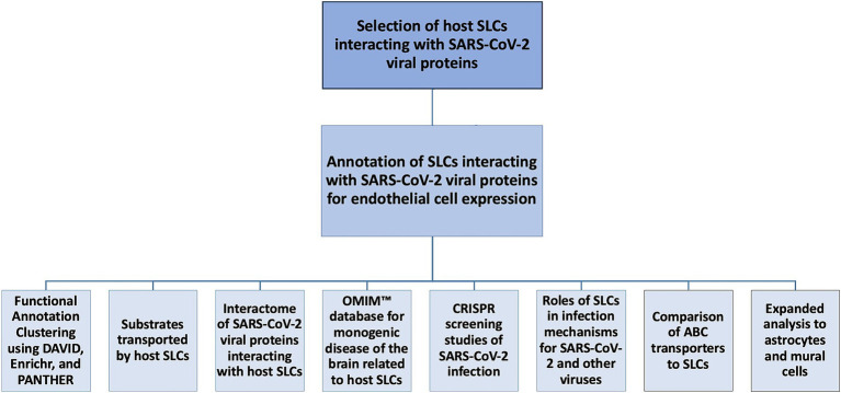
Methods: Virus-host protein-protein interaction data for SARS-CoV-2 infection were retrieved from the BioGRID database, filtered for SLCs, and then annotated for relevant expression in brain endothelial cells using a mouse brain transcriptomics database. Host SLCs expressed in brain endothelial cells were further explored using publicly available databases and information in the literature. Functional Annotation Clustering was performed using DAVID, and Enrichr served for pathway analysis. Substrates were retrieved from NCBI Gene. Links to monogenic disorders were retrieved from Online Mendelian Inheritance in Man™ and screened for disorders of the nervous system. Interactome data for viral proteins of SARS-CoV-2 were retrieved from BioGRID. Reports for host SLCs in viral receptor functions, viral entry mechanisms, and other major roles in the viral cycle were explored in databases (VThunter) and literature. ATP-binding cassette transporters (ABCs) were studied in parallel.

Results: N = 80 host SLCs showed relevant expression in brain endothelial cells whereby amino acid transporter stood out. N = 24/80 host SLCs were linked to monogenic disorders of the nervous system. N = 9/29 SARS-CoV-2 viral proteins had strong links to SLCs and key functions in viral infection (e.g., interferon response). SLCs serving as viral receptors and with closely associated functions were significantly enriched among all known listed viral receptors (chi-square test, p = 0.001). Literature searches for host SLCs revealed involvement of a subset of SLCs in infection mechanisms for SARS-CoV-2 and more broadly for other viruses. N = 17 host ABCs were found in brain endothelial cells where they may serve as efflux transporters.

Discussion: This hypothesis-generating work proposes a set of N = 80 host SLCs expressed in endothelial cells as contributors to BBB impairment after SARS-CoV-2 infection. Theoretically, persistent dysfunction of SLCs at the BBB, in particular insufficient transport of amino acids, could be one of many reasons for cognitive changes in long-COVID. Functions of SLCs in viral entry and associated roles deserve close attention.

Keywords: COVID-19; SARS-CoV-2; amino acid transport; blood-brain barrier; brain endothelial cells; protein-protein interactions; solute carrier proteins; virus-host interactions.


Conflict of interest statement

The authors declare that the research was conducted in the absence of any commercial or financial relationships that could be construed as a potential conflict of interest.

Figures

Figure 1
Figure 1
Flow chart of the data-mining process. Solute carrier proteins (SLCs) interacting with Severe acute respiratory syndrome coronavirus 2 (SARS-CoV-2) viral proteins were identified in the virus-host protein–protein interaction (VH-PPI) data. Several analyses were conducted, including a Functional Annotation Clustering analysis using the Database for Annotation, Visualization, and Integrated Discovery (DAVID); substrate classification using the National Center for Biotechnology Information (NCBI) Gene database; viral protein interactome identification using Biological General Repository for Interaction Datasets (BioGRID); diseases of the brain using Online Mendelian Inheritance in Man (OMIM™); Clustered Regularly Interspaced Short Palindromic Repeats (CRISPR) studies; roles of infection based on current literature; a comparison to ATP-binding cassette (ABC) proteins, and an expanded analysis to astrocytes and mural cells.
Figure 2
Figure 2
Counts of SLCs in virus-host protein–protein interaction data versus mRNA expression values. The chart displays the counts of each SLC-H-E in the virus-host protein–protein interaction data plotted against its brain endothelial expression value. The dotted line for linear fit indicates low correlation (r = 0.2186).
Figure 3
Figure 3
Relative distribution of substrate classes among selected SLCs. The pie chart demonstrates the relative contribution (in percentage) for classes of substrates with at least two members as identified by NCBI Gene for SLC-H-E. SLCs with unknown substrates were excluded. The key to the left shows the color coding for substrate classes. Distribution from highest to lowest were amino acids, zinc, sodium/potassium/chloride/hydrogen, ADP/ATP, bicarbonate, monocarboxylates, phosphate, choline, carnitine, sulfate, and nucleosides. For a complete list, see Table 3.
Figure 4
Figure 4
Analysis of individual viral proteins of SARS-CoV-2 for interactions with selected SLCs. (A) The plot displays the counts for host SLCs expressed in endothelial cells (SLC-H-E) after removing duplicates, as found in the interactome for each individual SARS-CoV-2 viral protein. (B) The plot displays the relative contribution of host SLCs expressed in endothelial cells (SLC-H-E) compared to the total count of host proteins in VH-PPI for each SARS-CoV-2 viral protein. Fisher’s exact tests were carried out for over- or underrepresentation of SLCs relative to the total count of host proteins interacting with each viral protein (* p < 0.002, corrected for multiple comparisons).
Figure 5
Figure 5
Venn diagram of SLCs in endothelial cells, VLMC, and astrocytes. The Venn diagram displays the number of SLCs belonging to each category, including endothelial cells (red), VLMC (blue), and astrocytes (yellow) as well as SLCs shared between multiple categories. Details are provided in Supplementary File 6.

Similar articles

  • The effect of sample site and collection procedure on identification of SARS-CoV-2 infection.
    Davenport C, Arevalo-Rodriguez I, Mateos-Haro M, Berhane S, Dinnes J, Spijker R, Buitrago-Garcia D, Ciapponi A, Takwoingi Y, Deeks JJ, Emperador D, Leeflang MMG, Van den Bruel A; Cochrane COVID-19 Diagnostic Test Accuracy Group. Davenport C, et al. Cochrane Database Syst Rev. 2024 Dec 16;12(12):CD014780. doi: 10.1002/14651858.CD014780. Cochrane Database Syst Rev. 2024. PMID: 39679851 Free PMC article.
  • Rapid, point-of-care antigen tests for diagnosis of SARS-CoV-2 infection.
    Dinnes J, Sharma P, Berhane S, van Wyk SS, Nyaaba N, Domen J, Taylor M, Cunningham J, Davenport C, Dittrich S, Emperador D, Hooft L, Leeflang MM, McInnes MD, Spijker R, Verbakel JY, Takwoingi Y, Taylor-Phillips S, Van den Bruel A, Deeks JJ; Cochrane COVID-19 Diagnostic Test Accuracy Group. Dinnes J, et al. Cochrane Database Syst Rev. 2022 Jul 22;7(7):CD013705. doi: 10.1002/14651858.CD013705.pub3. Cochrane Database Syst Rev. 2022. PMID: 35866452 Free PMC article.
  • Signs and symptoms to determine if a patient presenting in primary care or hospital outpatient settings has COVID-19.
    Struyf T, Deeks JJ, Dinnes J, Takwoingi Y, Davenport C, Leeflang MM, Spijker R, Hooft L, Emperador D, Domen J, Tans A, Janssens S, Wickramasinghe D, Lannoy V, Horn SRA, Van den Bruel A; Cochrane COVID-19 Diagnostic Test Accuracy Group. Struyf T, et al. Cochrane Database Syst Rev. 2022 May 20;5(5):CD013665. doi: 10.1002/14651858.CD013665.pub3. Cochrane Database Syst Rev. 2022. PMID: 35593186 Free PMC article.
  • Antibody tests for identification of current and past infection with SARS-CoV-2.
    Fox T, Geppert J, Dinnes J, Scandrett K, Bigio J, Sulis G, Hettiarachchi D, Mathangasinghe Y, Weeratunga P, Wickramasinghe D, Bergman H, Buckley BS, Probyn K, Sguassero Y, Davenport C, Cunningham J, Dittrich S, Emperador D, Hooft L, Leeflang MM, McInnes MD, Spijker R, Struyf T, Van den Bruel A, Verbakel JY, Takwoingi Y, Taylor-Phillips S, Deeks JJ; Cochrane COVID-19 Diagnostic Test Accuracy Group. Fox T, et al. Cochrane Database Syst Rev. 2022 Nov 17;11(11):CD013652. doi: 10.1002/14651858.CD013652.pub2. Cochrane Database Syst Rev. 2022. PMID: 36394900 Free PMC article.
  • Laboratory-based molecular test alternatives to RT-PCR for the diagnosis of SARS-CoV-2 infection.
    Arevalo-Rodriguez I, Mateos-Haro M, Dinnes J, Ciapponi A, Davenport C, Buitrago-Garcia D, Bennouna-Dalero T, Roqué-Figuls M, Van den Bruel A, von Eije KJ, Emperador D, Hooft L, Spijker R, Leeflang MM, Takwoingi Y, Deeks JJ. Arevalo-Rodriguez I, et al. Cochrane Database Syst Rev. 2024 Oct 14;10(10):CD015618. doi: 10.1002/14651858.CD015618. Cochrane Database Syst Rev. 2024. PMID: 39400904

References

    1. Hu B, Guo H, Zhou P, Shi ZL. Characteristics of SARS-CoV-2 and COVID-19. Nat Rev Microbiol. (2021) 19:141–54. doi: 10.1038/s41579-020-00459-7, PMID: - DOI - PMC - PubMed
    1. Carabelli AM, Peacock TP, Thorne LG, Harvey WT, Hughes J, Peacock SJ, et al. SARS-CoV-2 variant biology: immune escape, transmission and fitness. Nat Rev Microbiol. (2023) 21:162–77. doi: 10.1038/s41579-022-00841-7, PMID: - DOI - PMC - PubMed
    1. Burki T. WHO ends the COVID-19 public health emergency. Lancet Respir Med. (2023) 11:588. doi: 10.1016/S2213-2600(23)00217-5, PMID: - DOI - PMC - PubMed
    1. Huang C, Wang Y, Li X, Ren L, Zhao J, Hu Y, et al. Clinical features of patients infected with 2019 novel coronavirus in Wuhan, China. Lancet. (2020) 395:49. doi: 10.1016/S0140-6736(20)30183-5 - DOI - PMC - PubMed
    1. Zhu N, Zhang D, Wang W, Li X, Yang B, Song J, et al. A novel coronavirus from patients with pneumonia in China, 2019. N Engl J Med. (2020) 382:727–33. doi: 10.1056/NEJMoa2001017, PMID: - DOI - PMC - PubMed

LinkOut - more resources