. 2025 Jul 16;21(7):e1013318.
doi: 10.1371/journal.pcbi.1013318. eCollection 2025 Jul.

Cardiogenic and chronobiological mechanisms in seizure-induced sinus arrhythmias

Affiliations

Cardiogenic and chronobiological mechanisms in seizure-induced sinus arrhythmias

Pan Li et al. PLoS Comput Biol. .

Abstract

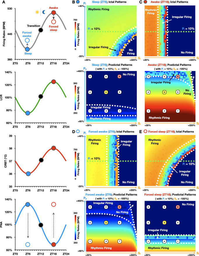
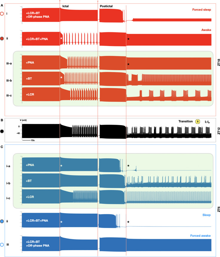
Seizure-induced cardiac arrhythmias, such as ictal (during seizure) or postictal (post-seizure) sinus arrhythmias, are potential triggers for sudden unexpected death in epilepsy. Traditionally, these arrhythmias have been attributed to changes in autonomic balance during ictal or postictal phases, as per the neurogenic mechanism. However, it remains unclear if these arrhythmias may involve intrinsic cardiogenic mechanisms. Furthermore, while circadian and sleep-wake patterns influence both neurogenic and cardiogenic mechanisms, a direct mechanistic link to seizure-induced arrhythmias remains to be established. In this study, we utilized a mathematical model of mouse sinoatrial nodal cell pacemaking and an autonomic clamping protocol, to dissect neurocardiogenic mechanisms in seizure-induced sinus arrhythmias and to test the hypothesis that circadian and sleep-wake rhythms directly modulate cellular susceptibility to these arrhythmias. Our simulations revealed that, in the context of altered autonomic levels associated with seizure progression, diverse seizure-induced sinoatrial nodal cell firing patterns during ictal or postictal phases can be triggered directly by intrinsic cardiac dynamics, without the need for dynamical changes in within-phase autonomic activities. This finding highlights the distinct roles of neurogenic and cardiogenic mechanisms in shaping sinoatrial nodal cell firing patterns, challenging the predominance of the neurogenic mechanism. This neurocardiogenic framework also successfully captures distinct circadian and vigilance state patterns of seizure-induced arrhythmias. Specifically, while daytime sleep predisposed sinoatrial nodal cells to postictal sinus arrhythmias, nighttime wakefulness promotes ictal sinus arrhythmias. However, these circadian patterns can be disrupted when sleep-wake cycles are decoupled from circadian rhythms, supporting the hypothesis that sleep-wake patterns can directly be a key determinant of seizure-induced sinus arrhythmias. Our findings may facilitate the development of novel therapeutic strategies for managing the risk of sudden unexpected death in epilepsy.


Conflict of interest statement

The authors have declared that no competing interests exist.

Figures

Fig 1
Fig 1. Clinical and experimental measurements of seizure-induced sinus arrhythmias and mortality.
(A-B) Representative clinical heart rate recordings from patients with epilepsy, capturing patterns before, during (pink blocks), and after seizures. (A) Ictal heart rate patterns in infants experiencing apneic seizures, showing mixed sinus tachycardia and bradycardia (left), or tachycardia alone (right) [16]. The red arrow marks the onset of progressive sinus bradycardia during the seizure. Data from Maruyama et al, Pediatric Neurology, 127: p52, 2022. (B) Postictal low-frequency heart rate oscillations were observed in adult patients with partial epilepsy [22]. Before the seizure, heart rate maintains a respiratory sinus rhythm at 0.3Hz. During the seizure, heart rate elevates, and it subsequently transitions into transient low-frequency heart rate oscillations at 0.13Hz (left) and 0.07Hz (right) (blue blocks). Data from Al-Aweel et al, Neurology, 53: p1591-1592, 1999. (C) Circadian and sleep-wake patterns of mortality rates in adult mice following maximal electroshock-induced seizures [28]. Fatalities are predominantly observed in seizures that occur during sleep, both under dark and light conditions. Data from Purnell et al, Journal of Neurophysiology, 118 [5]: p2594, 2017.
Fig 2
Fig 2. Schematic of a mouse SANC model incorporating circadian and vigilance state variations.
(A) Each heartbeat is initiated by an action potential (AP) generated in a SANC. The firing patterns of SANC APs are shaped over a 24-hour cycle by circadian variations in the ANS, body temperature (BT), and LCR (green dot). The ANS, regulated by the master circadian clock located in the suprachiasmatic nucleus, finely adjusts the balance between SNA (yellow dot) and PNA (blue dot). This autonomic balance modulates SANC firing rates (FR) via SNA (e.g., IHCN, L-type (ICaL) and T-type Ca currents (ICaT)) and PNA-dependent (e.g., muscarinic K current (IKACh)) regulatory targets, adapting to diverse physiological demands throughout the day. The circadian rhythm of body temperature (CRBT) further regulates this balance, impacting the kinetics and/or conductance of ion channels, exchangers, and pumps in a SANC. Moreover, vigilance state (sleep/wake) changes, e.g., high PNA during sleep and low PNA during wakefulness, also modulate the autonomic balance in the mouse SANC. (B) During an epileptic seizure event, the well-maintained autonomic balance can be disrupted, potentially leading to distinct ictal and postictal outcomes, influenced by both circadian and vigilance state variations. The CRBT icon is derived from https://openclipart.org/detail/231080/thermometer, while the circadian clock icon is modified based on https://openclipart.org/detail/198766/mono-tool-timer.
Fig 3
Fig 3. Transient autonomic changes are not required to trigger ictal and postictal sinus arrhythmias.
(A) Illustration of the autonomic clamping protocol used to induce epileptic seizures in the SANC model without within-phase autonomic changes. A seizure event with the duration of τ is simulated by a dominant level of ictal SNA (S×(1 + Si)) over ictal PNA (P×(1 + Pi)) (Si > Pi). After such an event, postictal levels of SNA and PNA are linearly ramped towards S×(1 + Sp) and P×(1 + Pp), respectively, with a ramping duration of τp. Green triangles indicate the locations where ictal and postictal SANC FR are sampled. (B-C) Resulting ictal (B) and ictal-postictal parameter space maps (C) at the sleep-wake transition phase (light off at zeitgeber time (ZT) 12 in a 12h:12h lighting regime [41]). (B) When Si and Pi are varied, the borders between rhythmic (Ri), irregular (Ii), and no-firing (Ni) regions of ictal SANC firing patterns are indicated by white dashed lines. (C) After setting Pi to 10% (B; green dashed line and dots) and Sp to -100%, Si and Pp are varied to obtain the postictal parameter space map. Green and white dashed lines delineate the borders between rhythmic (R), irregular (I), and no-firing (N) regions of ictal (i) and postictal (p) SANC firing patterns, respectively. This results in a total of nine distinct regions in the parameter space map (labeled 1 to 9). (D) Simulation traces of SANC membrane potentials (black traces) sampled from the nine parameter regions, demonstrating a wide range of seizure-induced SANC excitation patterns. These include ictal sinus rhythm [–3], mixed ictal tachycardia and bradycardia [–6], ictal asystole [–9], postictal sinus rhythm [1,4,7], postictal low-frequency oscillations [2,5,8] and postictal asystole [3,6,9]. Simulation traces labeled 5 (yellow; Ii-Ip) and 9 (red; Ni-Np) depict oscillatory and asystolic seizure events, respectively.
Fig 4
Fig 4. Neurocardiogenic mechanisms underlying seizure-induced arrhythmias in SANC excitation patterns.
(A) Detailed electrophysiologic and ionic dynamics behind representative seizure events featuring ictal and postictal low-frequency oscillations (trace 5 from Fig 3C and 3D; grey traces) and asystole (trace 9 from Fig 3C and 3D; black traces). (B) A close-up comparison between ictal (A; yellow dashed box) and postictal (A; blue dashed box) clusters of bursting APs with underlying intracellular Na, Ca dynamics, and key ionic currents in trace 5 (A; grey).
Fig 5
Fig 5. Regulation of seizure-induced SANC firing patterns by circadian rhythms and vigilance states.
(A) Simulated circadian rhythms of SANC FR (grey curve), LCR (green curve), CRBT (red curve), and PNA (blue curve) over a 24-hour cycle under a 12h:12h lighting regime. SANC FR, LCR, and CRBT peak at ZT18 during nighttime wakefulness (awake state; solid red dot), and hit bottom at ZT6 during daytime sleep (sleep state; solid blue dot). In contrast, PNA peaks at ZT6 and has a minimum at ZT18. The solid black dot represents the transition state (at ZT12). Red and blue empty dots indicate the forced sleep (at ZT18) and forced awake (at ZT6) states with off-phase PNA, respectively. (B-C) Differences in ictal (top) and postictal (bottom) parameter space maps between the sleep (B) and awake (C) states. (D-E) Ictal (top) and postictal (bottom) parameter space maps when circadian rhythms are decoupled from sleep-wake patterns via forced awake (D) and forced sleep (E) protocols.
Fig 6
Fig 6. Dissecting key mechanisms governing circadian and vigilance state regulation of seizure-induced SANC firing patterns.
Representative oscillatory (labeled as 5 in Fig 5B–E) seizure events under different circadian (at ZT18 (A), ZT12 (B), and ZT6 (C)) and vigilance state (forced sleep at ZT18 (A-i) and forced awake at ZT6 (C-iii)) conditions. Time-of-day changes in PNA (A-iii-a; ZT18) (C-i-a; ZT6), CRBT (A-iii-b; ZT18) (C-i-b; ZT6), and LCR (A-iii-c; ZT18) (C-i-c; ZT6) were applied individually to dissect the relative contributions of each circadian factor in shaping distinct SANC firing patterns at ZT18 (upper green box) and ZT6 (lower green box), respectively.

Similar articles

References

    1. Chahal CAA, Salloum MN, Alahdab F, Gottwald JA, Tester DJ, Anwer LA, et al. Systematic Review of the Genetics of Sudden Unexpected Death in Epilepsy: Potential Overlap With Sudden Cardiac Death and Arrhythmia-Related Genes. J Am Heart Assoc. 2020;9(1):e012264. doi: 10.1161/JAHA.119.012264 - DOI - PMC - PubMed
    1. Asatryan B. Challenges in decoding sudden unexpected death in epilepsy: The intersection between heart and brain in epilepsy. Am Heart Assoc. 2021;e023571. - PMC - PubMed
    1. Nashef L, Hindocha N, Makoff A. Risk factors in sudden death in epilepsy (SUDEP): the quest for mechanisms. Epilepsia. 2007;48(5):859–71. doi: 10.1111/j.1528-1167.2007.01082.x - DOI - PubMed
    1. Vilella L, Miyake CY, Chaitanya G, Hampson JP, Omidi SJ, Ochoa-Urrea M, et al. Incidence and Types of Cardiac Arrhythmias in the Peri-Ictal Period in Patients Having a Generalized Convulsive Seizure. Neurology. 2024;103(1):e209501. doi: 10.1212/WNL.0000000000209501 - DOI - PMC - PubMed
    1. Velagapudi P, Turagam M, Laurence T, Kocheril A. Cardiac arrhythmias and sudden unexpected death in epilepsy (SUDEP). Pacing Clin Electrophysiol. 2012;35(3):363–70. doi: 10.1111/j.1540-8159.2011.03276.x - DOI - PubMed