

Genetic disruption of the baculum compromises the ability of male mice to copulate
- PMID: 40668861
- PMCID: PMC12313067
- DOI: 10.1371/journal.pgen.1011787
Genetic disruption of the baculum compromises the ability of male mice to copulate
Abstract
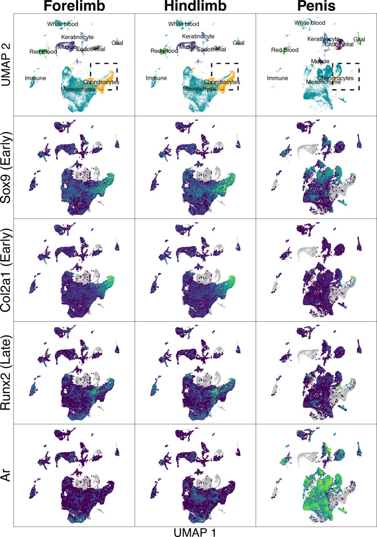
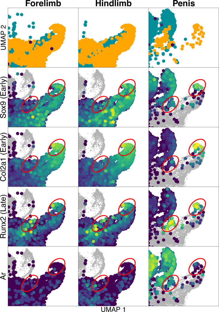
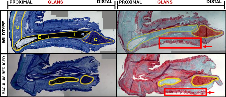
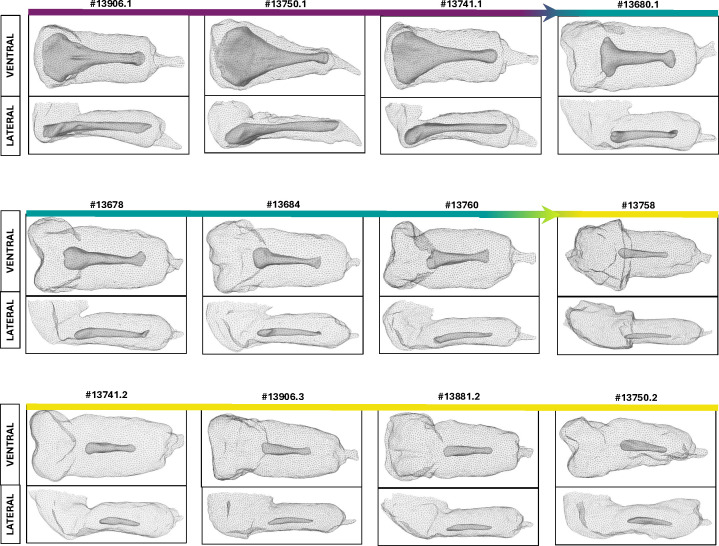
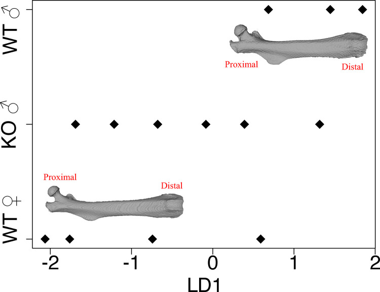
The baculum, a bone in the penis of many mammal species, shows an astonishing level of morphological divergence between species. Despite hundreds of years of interest, biologists have been unable to directly test its function. The goal of the current study is to uncover molecular details that could allow selective disruption of the baculum while allowing normal sexual differentiation and skeletal development. We compare patterns of androgen receptor binding and single cell gene expression in the developing penis, forelimbs and hindlimbs of mice. We identified chondrocytes in all three tissue types, but those from the developing penis show several unique features, including a population of chondrocytes that express both Runt-related transcription factor 2 (Runx2) and Androgen receptor (Ar). By combining a Runx2-Cre allele with a floxed Ar allele in mice, we selectively knocked out androgen signaling in late chondrocytes, resulting in a range of defects in baculum morphology. Males with the most disrupted bacula were unable to copulate, and their bacula appears to be disconnected from the corpus cavernosum muscle. Our study provides insights into the diversity of molecular mechanisms leading to bone and offers the first opportunity to directly test the function of the baculum.
Copyright: © 2025 Ghione et al. This is an open access article distributed under the terms of the Creative Commons Attribution License, which permits unrestricted use, distribution, and reproduction in any medium, provided the original author and source are credited.
Conflict of interest statement
The authors have declared that no competing interests exist.
Figures
Similar articles
-
Sexual Harassment and Prevention Training.2024 Mar 29. In: StatPearls [Internet]. Treasure Island (FL): StatPearls Publishing; 2025 Jan–. 2024 Mar 29. In: StatPearls [Internet]. Treasure Island (FL): StatPearls Publishing; 2025 Jan–. PMID: 36508513 Free Books & Documents.
-
The Black Book of Psychotropic Dosing and Monitoring.Psychopharmacol Bull. 2024 Jul 8;54(3):8-59. Psychopharmacol Bull. 2024. PMID: 38993656 Free PMC article. Review.
-
Role of Kctd13 in modulating AR and SOX9 expression in different penile cell populations.Andrology. 2025 Jul;13(5):1201-1212. doi: 10.1111/andr.70005. Epub 2025 Jan 29. Andrology. 2025. PMID: 39888193
-
Systemic pharmacological treatments for chronic plaque psoriasis: a network meta-analysis.Cochrane Database Syst Rev. 2021 Apr 19;4(4):CD011535. doi: 10.1002/14651858.CD011535.pub4. Cochrane Database Syst Rev. 2021. Update in: Cochrane Database Syst Rev. 2022 May 23;5:CD011535. doi: 10.1002/14651858.CD011535.pub5. PMID: 33871055 Free PMC article. Updated.
-
Systemic pharmacological treatments for chronic plaque psoriasis: a network meta-analysis.Cochrane Database Syst Rev. 2017 Dec 22;12(12):CD011535. doi: 10.1002/14651858.CD011535.pub2. Cochrane Database Syst Rev. 2017. Update in: Cochrane Database Syst Rev. 2020 Jan 9;1:CD011535. doi: 10.1002/14651858.CD011535.pub3. PMID: 29271481 Free PMC article. Updated.
References
-
- Dines JP, Mesnick SL, Ralls K, May-Collado L, Agnarsson I, Dean MD. A trade-off between precopulatory and postcopulatory trait investment in male cetaceans. Evolution. 2015;69(6):1560–72. - PubMed
-
- Kahrl AF, Johnson MA, Cox RM. Rapid evolution of testis size relative to sperm morphology suggests that postcopulatory selection targets sperm number in Anolis lizards. Journal of Evolutionary Biology. 2019;0–2. - PubMed
MeSH terms
Substances
Grants and funding
LinkOut - more resources
Full Text Sources
Research Materials
