. 2025 Jul 16;21(7):e1013254.
doi: 10.1371/journal.pcbi.1013254. eCollection 2025 Jul.

Impact of ligand binding on VEGFR1, VEGFR2, and NRP1 localization in human endothelial cells

Affiliations

Impact of ligand binding on VEGFR1, VEGFR2, and NRP1 localization in human endothelial cells

Sarvenaz Sarabipour et al. PLoS Comput Biol. .

Abstract

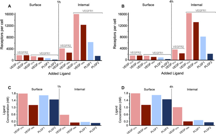
The vascular endothelial growth factor receptors (VEGFRs) bind to cognate ligands to facilitate signaling pathways critical for angiogenesis, the growth of new capillaries from existing vasculature. Intracellular trafficking regulates the availability of receptors on the cell surface to bind ligands, which regulate activation, and the movement of activated receptors between the surface and intracellular pools, where they can initiate different signaling pathways. Using experimental data and computational modeling, we recently demonstrated and quantified the differential trafficking of three VEGF receptors, VEGFR1, VEGFR2, and coreceptor Neuropilin-1 (NRP1). Here, we expand that approach to quantify how the binding of different VEGF ligands alters the trafficking of these VEGF receptors and demonstrate the consequences of receptor localization and ligand binding on the localization and dynamics of signal initiation complexes. We include simulations of four different splice isoforms of VEGF-A and PLGF, each of which binds to different combinations of the VEGF receptors, and we use new experimental data for two of these ligands to parameterize and validate our model. We show that VEGFR2 trafficking is altered in response to ligand binding, but that trafficking of VEGFR1 is not; we also show that the altered trafficking can be explained by a single mechanistic process, increased internalization of the VEGFR2 receptor when bound to ligand; other processes are unaffected. We further show that even though the canonical view of receptor tyrosine kinases is of activation on the cell surface, most of the ligand-receptor complexes for both VEGFR1 and VEGFR2 are intracellular. We also explore the competition between the receptors for ligand binding, the so-called 'decoy effect', and show that while in vitro on the cell surface minimal such effect would be observed, inside the cell the effect can be substantial and may influence signaling. We term this location dependence the 'reservoir effect' as the size of the local ligand reservoir (large outside the cell, small inside the cell) plays an integral role in the receptor-receptor competition. These results expand our understanding of receptor-ligand trafficking dynamics and are critical for the design of therapeutic agents to regulate ligand availability to VEGFR1 and hence VEGF receptor signaling in angiogenesis.


Conflict of interest statement

The authors have declared that no competing interests exist.

Figures

Fig 1
Fig 1. Diagram of molecular interactions of the computational model.
Cellular biophysical and biochemical reactions between VEGFR1, VEGFR2 and NRP1 receptors, extracellular matrix components and VEGF121a, VEGF165a, PLGF1 and PLGF2 ligands. The receptors can homodimerize (at a lower level than that induced by ligands), and unlike VEGFR2, VEGFR1 can form a complex with NRP1 in the absence of ligands [45]. PLGF1 binds to VEGFR1 in the absence or presence of NRP1 binding to VEGFR1. VEGF165a binds to VEGFR1, VEGFR2 and NRP1 and the matrix proteins. VEGF165a can only bind to NRP1 or matrix, not both simultaneously. PLGF2 and VEGF165a can only bind to VEGFR1 when VEGFR1 is not bound to NRP1. VEGF165a can bind to NRP1 and VEGFR2 simultaneously. During trafficking, surface protein complexes (monomeric, dimeric, or higher order) can be internalized (rate constants denoted kint). Early endosomal (“Rab4a/5a”) receptors can be degraded (rate constant kdeg), recycled (rate constant krec4), or transferred to the Rab11a compartment (rate constant k4to11) which leads to an additional recycling pathway (rate constant krec11). New surface receptors (monomers) are produced at rate kprod. Each of the rate constant values can be different for the different receptors. Reaction rates and species concentrations are detailed in S1–S18 Tables.
Fig 2
Fig 2. Experimental measures of whole-cell and surface levels of VEGFR1, VEGFR2, and NRP1 in HUVECs.
A, B, Western blot of total HUVEC lysates treated as indicated for 0.25–4 hours with 50 µg.mL-1 of VEGF165a (A) or PLGF1 (B) and stained for VEGFR1, VEGFR2 and NRP1. Representative of n = 3 replicates. C, D, Quantification of whole cell receptor levels shown in A,B. E, F, Western blot of Biotin labeling assay to measure surface and internal VEGFR1, VEGFR2, and NRP1 levels in HUVECs; for 1 hour and 4 hours with 50 µg.mL-1 of VEGF165a or PLGF1 and stained for VEGFR1, VEGFR2 and NRP1. Representative of n = 3 replicates. Western blot showing the effect on VEGFR1, VEGFR2 and NRP1 levels of depletion of both Rab4a and Rab11a for 1 hour and 4 hours ligand treatment; Representative of n = 3 replicates. Control conditions in E and F include nontargeting siRNA (siNT). Reagents used for the experiments are detailed in S19 Table. G, H, Quantification of whole cell receptor levels shown in E, F. The trend of reduced whole cell VEGFR2 following VEGF165a administration observed in A and C is consistent with significant differences (by single factor ANOVA analysis, *, p < 0.01; **, p < 0.0001) for surface and whole cell VEGFR2 levels at 1 hour and 4 hours post-VEGF165a addition compared to no ligand addition, observed in E-H.
Fig 3
Fig 3. Ligand treatment - here, VEGF165a - alters VEGFR2 distribution but not VEGFR1 or NRP1 distribution.
A-C, total (ligated and unligated) receptor levels on the cell surface, inside the cell, and combined (whole cell) following VEGF165a treatment. The experimentally-observed decline in surface and whole cell levels, simultaneous with a stable internal level, is consistent with a VEGF-induced three-fold increase in VEGFR2 internalization (V.R2.kint), compared to the unligated receptors, lines represent 8x, 4x, 3x, 2x, 1x, D-F, total (ligated and unligated) receptor levels on the cell surface, inside the cell, and combined (whole cell) following VEGF165a treatment. Using the increased VEGFR2 internalization as a baseline, changes to ligand-induced degradation of VEGFR2 (V.R2.kdeg) do not improve the fit to experimental observations, lines represent 5x, 2x, 1x, 0.5x, 0.2x, G-I, the simulated results with enhanced VEGFR2 internalization shows a match between the predicted and experimentally changes in surface receptor levels for VEGFR1, VEGFR2, and NRP1. J-L, Experimental and simulated surface to internal ratios for VEGFR1, VEGFR2 and NRP1 in the absence of ligands and under 1h and 4h ligand treatment conditions (normalized to the unligated values). See S2–S7 Figs.
Fig 4
Fig 4. Distribution of ligated and active VEGFR1 and VEGFR2 receptors following addition of one ligand.
A-C, Cell surface (A), internal (B), and whole cell (C) VEGFR2 following 50 ng.ml-1 VEGF121a or VEGF165a, D-F, cell surface (D), internal (E), and whole cell (F) VEGFR1 following 50 ng.mL-1 VEGF121a, VEGF165a, PLGF1 or PLGF2. V165 represents VEGF165a, V121 represents VEGF121a, P1 represents PLGF1, and P2 represents PLGF2.
Fig 5
Fig 5. Distribution of ligands and of active ligated VEGF receptors.
A, B, Levels of active VEGFR1 and VEGFR2 on the cell surface and internally, following 1 hour (A) or 4 hour (B) treatment with 50 ng.mL-1 of VEGF165a, VEGF121a, PLGF1, or PLGF2. C, D, Concentration of extracellular ligand (“surface”) and intracellular ligand (“internal”) following 1 hour (C) or 4 hour (D) treatment with 50 ng.mL-1 of VEGF165a, VEGF121a, PLGF1, or PLGF2. See the dose-dependent impacts on these levels in S13 and S14 Figs.
Fig 6
Fig 6. Impact of VEGFR1 on whole cell VEGFR2 ligation by VEGF165a.
A, changes in VEGFR2.VEGF165a.VEGFR2 levels under varying VEGF165a concentrations B, changes in VEGFR2.VEGF165a.VEGFR2 in the absence of VEGFR1 levels under varying VEGF165a concentrations C, percent change in VEGFR2.VEGF165a.VEGFR2 levels under loss of VEGFR1 molecules and under varying VEGF165a concentrations. When VEGFR1 is removed, whole cell VEGFR2.VEGF165a.VEGFR2 levels change.
Fig 7
Fig 7. Impact of VEGFR1 on A,B, Surface and C,D, Internal VEGFR2 ligation by VEGF165a.
Changes in VEGFR2.VEGF165a.VEGFR2 levels under varying VEGF165a concentrations. Change in VEGFR2.VEGF165a. VEGFR2 levels under loss of VEGFR1 molecules and under varying VEGF165a concentrations. When VEGFR1 is removed, surface VEGFR2.VEGF165a.VEGFR2 does not change but internal VEGFR2.VEGF165a.VEGFR2 levels change over a range of VEGF165a concentrations.
Fig 8
Fig 8. PLGF-VEGF competition: impact on active ligand-bound receptor complexes.
Predicted level of active VEGFR.ligand.VEGFR complexes, following two hours of ligand treatment at different doses for PLGF1 and VEGF165a, across the whole cell: VEGF-bound VEGFR2 (top), VEGF-bound VEGFR1 (middle), PLGF-bound VEGFR1(bottom).
Fig 9
Fig 9. Differences in VEGFR1, VEGFR2 and NRP1 trafficking in the absence of ligands (steady state) and presence of ligands in HUVECs.
A, VEGFR1, VEGFR2, NRP1 are constitutively trafficked in the absence of ligands in HUVECs. B, VEGFR2 internalization is faster as a result of VEGF165a treatment. PLGF treatment has no impact on VEGFR1, VEGFR2 or NRP1 trafficking in HUVECs. The size of the arrow represents the amount of receptor movement. The size of the receptor represents the expression level relative to other receptors shown here.

Similar articles

References

    1. Bautch VL. VEGF-directed blood vessel patterning: from cells to organism. Cold Spring Harb Perspect Med. 2012;2(9):a006452. doi: 10.1101/cshperspect.a006452 - DOI - PMC - PubMed
    1. Carmeliet P. Mechanisms of angiogenesis and arteriogenesis. Nat Med. 2000;6(4):389–95. doi: 10.1038/74651 - DOI - PubMed
    1. Jeltsch M, Leppänen VM, Saharinen P, Alitalo K. Receptor tyrosine kinase-mediated angiogenesis. Cold Spring Harb Perspect Biol. 2013;5(9):a009183. - PMC - PubMed
    1. Annex BH. Therapeutic angiogenesis for critical limb ischaemia. Nat Rev Cardiol. 2013;10(7):387–96. doi: 10.1038/nrcardio.2013.70 - DOI - PubMed
    1. Annex BH, Cooke JP. New Directions in Therapeutic Angiogenesis and Arteriogenesis in Peripheral Arterial Disease. Circ Res. 2021;128(12):1944–57. doi: 10.1161/CIRCRESAHA.121.318266 - DOI - PMC - PubMed

MeSH terms

Substances

LinkOut - more resources